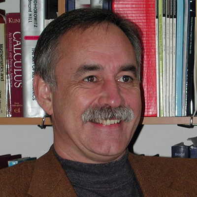
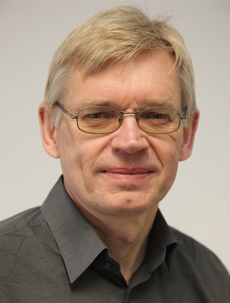
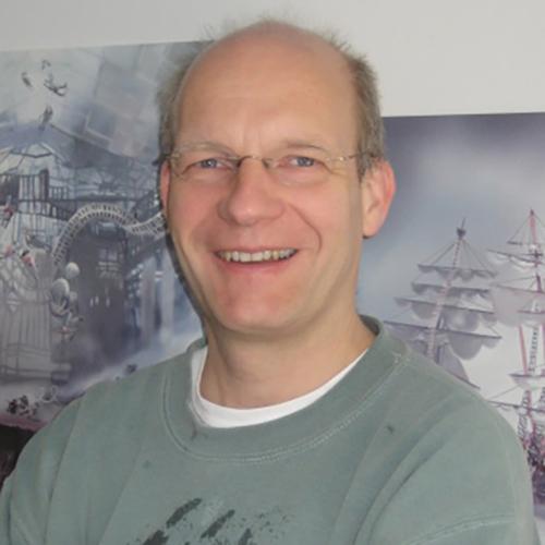
Istvan Mody is a Professor at the University of California, Los Angeles (UCLA) with appointments in the Department of Neurology and Department of Physiology . His research focuses on synaptic signaling in health and disease, including mechanisms of GABAergic transmission, calcium homeostasis, and their roles in neurological disorders such as epilepsy, Alzheimer's disease, Huntington's disease, stress, alcoholism, and postpartum depression. He utilizes advanced techniques like patch-clamp electrophysiology, neuroanatomical and immunohistochemical methods, and molecular biology in animal models and human brain tissue . Research Interests: Dr. Mody investigates the physiology, pharmacology, and pathology of synaptic transmission and extrasynaptic receptor activation , with a particular emphasis on GABA(A) receptors and their subunit-specific modulation. His work explores how disruptions in excitation-inhibition balance contribute to neurological diseases, including mechanisms of tonic inhibition , calcium signaling , and neurosteroid interactions . He also studies the effects of chronic stress and hormonal fluctuations on neural excitability and behavior. Publications Trends: Recent studies highlight his work on gamma oscillations in Alzheimer's models, microglial dynamics , and rehabilitation strategies for stroke. His lab develops optical tools like dqGEVI for neuronal activity monitoring and investigates human brain organoids to model network dysfunction in epilepsy and intellectual disability. Laboratory Location: 635 Charles Young Dr S, Los Angeles, CA 90095, United States.
Daan Brinks is an Assistant Professor at Delft University of Technology in the Department of Imaging Physics within the Faculty of Applied Sciences. He leads the Brinks Lab, which operates at the intersection of physics, biochemistry, optics, mathematics, and nanofabrication, focusing on developing novel imaging tools for neuroscience applications. His research spans both fundamental biophysics and practical biomedical applications, with significant collaborations including Erasmus MC. Faculty of Applied Sciences, Delft University of Technology Department of Imaging Physics (ImPhys) Brinks Lab leader Founding member of BIOlab (Biomedical Intervention Optimization lab) Lead of a convergence Health and Technology Consortium Dr. Brinks' academic journey began with an MSc in Molecular Nanophotonics from the University of Twente (2002-2007), followed by a PhD at ICFO Institute Barcelona (2007-2012). He then completed prestigious fellowships at Harvard University as a Rubicon Fellow (2012-2014) and HMMI Fellow (2014-2017) before joining TU Delft as an Assistant Professor in 2017. His research interests center on voltage imaging techniques to monitor neural activity, optogenetics for neural control, nonlinear optical microscopy for enhanced resolution, and AI applications in bioimaging . The lab develops tools to transduce information in neurons into detectable photons, addressing questions from biophysical principles to behavioral consequences and from subcellular compartments to complete organisms. Current projects include Voltage nanoscopy using plasmonic enhancement, Absolute Voltage Imaging through fluorescence lifetime measurements, Multiphoton Voltage Imaging for deep tissue applications, and advanced image analysis with machine learning. The publications reveal a strong focus on developing novel optical tools for neuroscience, particularly genetically encoded voltage indicators and plasmonic enhancement techniques. His work bridges physics, molecular biology, and neuroscience, with applications ranging from fundamental understanding of neural circuits to cancer cell identification. The research shows progression from fundamental physics (early career) to increasingly applied neuroscience and biomedical applications (recent work), with publications in top journals including Nature, Science Advances, and Nature Biomedical Engineering. Rubicon Fellow (2012-2014) HMMI Fellow (2014-2017) Publications in Nature, Science Advances, Nature Biomedical Engineering Media coverage in major outlets including Delta TU Delft and Trouw Dr. Brinks actively mentors students and researchers, with his lab welcoming enthusiastic students, PhD candidates, and postdocs interested in multidisciplinary projects at the junction of optics, molecular biology, and neuroscience. His research has received external funding through fellowships and likely additional grants supporting his lab's operations. The Brinks Lab collaborates extensively with both academic and medical institutions, particularly evident in the cancer cell research with Erasmus MC. The lab maintains strong physical infrastructure including advanced microscopy systems and nanofabrication capabilities, supporting their work in voltage imaging, plasmonics, and single-cell analysis. They have developed several hardware and software interfaces for automated interaction with excitable tissues and model dynamics in hybrid systems, reflecting their interdisciplinary approach to neuroscience questions.
Professor Ahmed Abdelfattah is the Robert J. and Nancy D. Carney University Assistant Professor of Brain Science and Assistant Professor of Engineering at Brown University. His work focuses on developing molecular tools to study brain function through bioengineering and chemical approaches. He leads the Abdelfattah Lab at the Neuroscience Department and Carney Institute for Brain Science. His research integrates electrophysiology, fluorescence imaging, protein engineering, and genetic methods to visualize neural circuits and their functions. Key areas include voltage imaging, optogenetics, and fluorescent protein development for in vivo applications. The lab’s tools enable real-time observation of electrical and chemical signals in the nervous system, addressing gaps in understanding memory and behavior formation. Notable contributions include chemigenetic voltage and calcium indicators, far-red fluorescent sensors, and all-optical neurophysiology techniques. His work bridges molecular biology and advanced imaging to decode neural activity at unprecedented resolution. Advising and grants are central to his academic role, though specific student names or grant details are not provided in available texts. The lab actively collaborates across disciplines to advance neurobiological research tools.
William N. Ross, Ph.D., is a Professor of Physiology at New York Medical College (NYMC), holding joint appointments in both the Graduate School of Biomedical Sciences and the School of Medicine. With a distinguished career spanning several decades, Dr. Ross has established himself as a pioneer in the field of neurophysiology, particularly in the development and application of optical imaging techniques for studying neuronal function. Dr. Ross received his Ph.D. in Physics from Columbia University, followed by postdoctoral training in Physiology at Yale University and Neurobiology at Harvard University. His educational background in high energy physics provided a strong quantitative foundation that he successfully transitioned to neuroscience research, where he has made numerous groundbreaking contributions. Dr. Ross's research focuses on the development and application of optical imaging techniques to study neuronal signaling, particularly in dendrites, axons, and spines. His laboratory was the first to apply voltage sensitive dye imaging to dendrites, to apply calcium imaging to CNS dendrites, and to use sodium imaging to examine neurons in slices. They pioneered high speed CCD imaging synchronized with electrical recording and were the first to describe Ca 2+ waves in dendrites and spontaneous Ca 2+ spark-like events. Most recently, his team has developed techniques for simultaneous imaging of sodium and calcium changes in extremely small neuronal structures like nodes of Ranvier and dendritic spines. Analysis of Dr. Ross's recent publications reveals a consistent focus on the development and refinement of optical imaging techniques for studying ionic dynamics in neurons. His work spans multiple scales, from molecular mechanisms of voltage indicators to cellular and subcellular signaling processes. The research demonstrates a progression from foundational studies of calcium signaling to increasingly sophisticated investigations of sodium-calcium interactions in specific neuronal compartments. This trend reflects both technological advances in imaging capabilities and a deepening understanding of the complex interplay between different ionic signals in neuronal computation. Grass Faculty Fellow, Marine Biological Laboratory (2007-2008) Dean's Distinguished Research Award - NYMC (1998) Fogarty Senior International Fellow (1993-1994, 1984-1985) Irma T. Hirschl Career Scientist (1981-1985) Dr. Ross has served on various committees including the TAP committee and as Chair of the Alternative Pathways committee at NYMC. His research has been supported by multiple prestigious funding mechanisms including Fogarty International Fellowships and the Irma T. Hirschl Career Scientist award, demonstrating sustained recognition of the significance and quality of his work by major funding agencies. Dr. Ross's laboratory has been at the forefront of developing and applying optical imaging techniques to study neuronal function. His group's work has consistently pushed the boundaries of what can be measured in living neurons, particularly in subcellular compartments like dendritic spines. The lab's technical innovations have enabled new discoveries about the role of calcium and sodium signaling in neuronal computation, with implications for understanding both normal brain function and neurological disorders.
Vladislav Verkhusha serves as Research Director at the University of Helsinki's Department of Anatomy and Supervisor for the Doctoral Programme in Biomedicine. His research integrates biomedicine, protein engineering, and advanced imaging techniques to develop optogenetic tools and biosensors. Research Focus: Primary interests include optogenetics, near-infrared fluorescent proteins, and biomedical imaging technologies. His work enables high-sensitivity deep-tissue visualization and manipulation of cellular processes, with applications in neuroscience and synthetic biology. Publication Trends: Recent articles (2024-2025) focus on near-infrared imaging probes, genetically encoded biosensors, and optogenetic manipulation. Key themes include protein engineering for enhanced imaging depth and precision in biological systems. Grants & Projects: Verkhusha akatemihanke 24-28 (Academy of Finland: €600,000, 2024-2028) Jane ja Aatos Erkon säätiö/Verkhusha (€626,001, 2023-2025) Magnus Ehrnrooth 2023/Vlad Verkhusha (2023-2024)
Ganesh Vasan is an Assistant Professor at the University of Minnesota's Department of Orthopedic Surgery. His research focuses on neurophysiology, biophysics, and marine biology, particularly investigating neural dynamics, voltage imaging techniques, and bioluminescent phenomena in marine organisms. He holds a PhD and has contributed to advancements in genetically encoded voltage indicators and high-resolution imaging technologies. Key research areas include dopamine-mediated memory interactions, olfactory bulb signaling, and deep-sea bioluminescence. His work bridges neuroscience and engineering, with applications in understanding neural circuitry and developing innovative imaging tools. Notable contributions include studies on neuronal voltage dynamics and the first evidence of bioluminescence on hydrothermal vents. Publications span 2011–2024, emphasizing interdisciplinary approaches. Awards are not explicitly listed, but his research has been published in high-impact journals. No grants or advising roles are detailed in the provided text. His department affiliation suggests involvement in orthopedic research, though specific clinical applications are not elaborated here.
Dr. François St-Pierre is an Associate Professor in the Department of Neuroscience at Baylor College of Medicine and an Adjunct Faculty member in Electrical and Computer Engineering at Rice University. He is a McNair Scholar at Baylor and leads the St-Pierre Lab, dedicated to developing tools for monitoring and perturbing biological systems. His research focuses on synthetic biology, voltage imaging, brain connectivity, and high-throughput screening methods. Education: BA and MA from the University of Cambridge, PhD from MIT, and postdoctoral training at Stanford University. His lab is located in the Texas Medical Center, the world's largest medical complex, and collaborates with institutions like Rice University. Research Interests: Engineered proteins, gene circuits, voltage imaging technologies, optogenetics, and novel screening methods. Key tools include genetically encoded voltage indicators and advanced microscopy techniques. Awards: McNair Scholar (Baylor College of Medicine) Labs/Teams: St-Pierre Lab at Baylor College of Medicine Lab Website: stpierrelab.com Publications emphasize voltage indicator development, optogenetic tools, and neural circuit imaging, with a focus on enabling high-resolution, in vivo studies of brain activity.
Marco Locarno is a Lecturer in the Department of Cell Biology, Neurobiology and Biophysics at Utrecht University's Faculty of Science. He teaches courses in the Molecular and Biophysical Life Sciences Bachelor program including Advanced Light Microscopy and Bioimaging, Biological Physics, Cell Biology, and Neurobiology of behavior. His research background focuses on enhancing Genetically Encoded Voltage Indicators (GEVIs) through plasmonic enhancement and non-linear excitation. With expertise in light microscopy and nanoparticles, his work bridges biophysics and neurobiology. He employs evidence-based teaching techniques grounded in constructivism, emphasizing critical thinking development through active learning and self-reflection. Dr. Locarno holds a University Teaching Qualification (UTQ/BKO) and Laboratory Animal Science certification. His educational philosophy integrates modern technologies like AI while preserving the human elements of learning such as emotional engagement and ethical considerations. He previously served as a PhD Candidate at TU Delft (2020-2024), Teaching Assistant at Università di Genova (2018-2019), and Student Tutor at the same institution (2017-2018).
Chelsea Goulton is a Research Fellow at the School of Psychology, University of New South Wales (UNSW Sydney). Her research spans neuroscience, neuropharmacology, and neurology, with a focus on seizure mechanisms, neuronal inhibition, and neuroprotective strategies. Core research areas: Epilepsy, KCC2 transporter modulation, GABAergic inhibition, and neurotechnology development Key studies: Investigation of ketamine's effects on BDNF, pharmacological preconditioning for neuroprotection, and optical voltage indicator validation Contact: c.goulton@unsw.edu.au
Mark J. Schnitzer is the Anne T. and Robert M. Bass Professor at Stanford University, with primary faculty appointments in the Departments of Biology, Applied Physics, and Neurosurgery within the School of Humanities and Sciences . He co-directs Stanford's Cracking the Neural Code Program and holds affiliations with the Bio-X , Wu Tsai Neurosciences Institute , and multiple graduate programs. Research focuses on neural circuit dynamics and optical imaging innovations for studying learning, memory, and motor behaviors in awake animals Develops high-resolution fluorescence microscopes and miniaturized imaging systems for clinical translation Scientific contributions include: 2019 Nature Methods Method of the Year for miniature fluorescence microscope HHMI Investigator (2008) NIH Director's Pioneer Award (2007) Allen Distinguished Investigator Award (2010) Teaching roles include: Advanced Imaging Lab in Biophysics (APPPHYS 232/BIO 132) Introduction to Biophysics (APPPHYS 205/BIO 126) Multiple independent study and graduate research courses Laboratory affiliations span biomedical engineering , neuroscience , and molecular imaging initiatives at Stanford.
Daan Brinks serves as a Researcher in the Department of Molecular Genetics at Erasmus Medical Center (Erasmus MC), Erasmus University Rotterdam, where he develops optical imaging technologies for cellular analysis. His work bridges biophysics, molecular biology, and biomedical engineering to address challenges in neuroscience and oncology. His core research focuses on: Voltage imaging via engineered proton-pumping rhodopsins Single-cell sequencing and transcriptomics in cancer heterogeneity Near-infrared fluorescence applications Real-time optical tagging of cellular phenotypes Proton transport mechanisms in microbial systems Nanostructured array fabrication for neuronal growth studies Recent publications (2022-2025) reveal a trajectory toward miniaturized imaging systems and genetically encoded sensors, with high-impact work in Nature Biomedical Engineering demonstrating 17 citations. His voltage imaging innovations show particular promise for neurological disorder research, as highlighted in 26 media appearances across 26 news outlets and social platforms. Dr. Brinks maintains an extensive collaboration network evidenced by multi-institutional publications, including partnerships with researchers in cancer biology (e.g., Chien, Hardillo) and optics engineering (e.g., Meng, Ganapathy). His team's press coverage on neurological disorders indicates translational relevance of fundamental imaging research.
Jeffrey Lopez-Rojas is an Assistant Professor in the Department of Psychology at the University of Wisconsin–Milwaukee (UWM), with strong affiliations to the Neuroscience and Clinical Psychology programs. He is actively recruiting graduate students for the Clinical PhD, Neuroscience PhD, and Health MS programs, indicating an active research and mentoring role. His research focuses on the neural basis of social cognition, particularly the role of the lateral entorhinal cortex and hippocampal CA2 circuit in social memory. Using advanced techniques such as optogenetics, pharmacogenetics, and in vivo neuronal recordings, his lab investigates how multisensory social signals are integrated and processed in the brain. His work has significant implications for understanding and treating neuropsychiatric and neurodegenerative disorders characterized by social deficits. The recent publications highlight a consistent trajectory in neuroscience, particularly in hippocampal function, synaptic plasticity, ion channel regulation, and neural circuit dynamics. Key themes include social memory, dendritic excitability, calcium and potassium channel function, and structural plasticity in the dentate gyrus and hippocampus. His work bridges molecular, cellular, and systems-level neuroscience. Dr. Lopez-Rojas has contributed to high-impact journals such as Neuron , Nature Communications , and EMBO Journal , demonstrating a strong publication record. While no specific scientific awards are listed in the provided text, his research output suggests recognition in the neuroscience community. He advises graduate students in clinical, neuroscience, and health-related programs and likely secures external funding to support his experimental work, though specific grants are not mentioned. His lab employs a multidisciplinary approach, combining behavioral assays with cutting-edge neurophysiological and molecular tools, positioning his team at the forefront of systems neuroscience research. His laboratory focuses on the entorhinal-hippocampal network, utilizing transgenic models, circuit tracing, and functional imaging to dissect the mechanisms of social memory and pattern separation. The team’s work contributes to broader efforts in understanding cognitive decline and social dysfunction in brain disorders.
Peter Hegemann is a Professor specializing in optogenetics and neuroengineering. His research focuses on developing molecular tools for precise control of neuronal activity, with applications in neuroscience and physiology. He collaborates extensively with institutions such as Humboldt-Universität zu Berlin (implied from project context) and international partners. Research Interests: Hegemann's work spans optogenetic actuator engineering, ion channel modulation, and optical control systems. Key areas include: Design of potassium/calcium-selective channelrhodopsins All-optical electrophysiology techniques Bidirectional neural control systems Low-light optogenetic inhibition Cross-species tool validation (C. elegans to mammals) Recent publications (2021-2023) demonstrate a consistent focus on developing next-generation optogenetic tools. These include WiChR (potassium-selective inhibitor), CapChR (calcium-permeable channels), BiPOLES (dual-color actuator), and closed-loop voltage clamps. The research emphasizes molecular engineering, photophysics, and physiological validation in neuronal/cardiac systems.
Daisuke Shimaoka is a Research Fellow at Monash University's Physiology department. His research focuses on neural circuit dynamics, visual processing, and brain-wide activity patterns. He leads and collaborates on projects including an NHMRC-funded initiative developing Digital Mirror Device (DMD) technology for neural circuit manipulation, and a study on brain-wide activity patterns' simplicity. His work integrates neurophysiological techniques with computational modeling. Active Projects: 2023-2025 (DMD system development) and 2025-2029 (brain connectivity analysis) Key research areas include: Functional and anatomical connectomics Arousal state effects on sensory processing Neural mechanisms of consciousness and sleep-wake transitions Optical imaging techniques for behaving animals Major contributions include defining visual cortex organization in mice and primates, elucidating bilateral neural activity impacts, and developing genetic tools for neural circuit interrogation. His work has been cited over 300 times across 11 publications.
Dieter Jaeger is a Professor in the Department of Biology at Emory University, leading the Jaeger Lab at the Rollins Research Center. His research focuses on computational and experimental neurophysiology to study motor control and neural networks in the basal ganglia, cerebellum, and motor thalamus. He holds a Ph.D. from the University of Michigan (1990). Research Interests: Combining neurophysiology with computer simulations to investigate neural networks involved in motor tasks and Parkinson’s disease. Key areas include cerebellar nuclei dynamics, thalamocortical signaling, and optogenetic manipulation of brain activity. Publications Highlight: Recent work (2023–2025) explores cortical voltage imaging, astrocyte complexity, and neurovascular coupling, advancing understanding of neural circuit dysfunction in disease. Contact: Email djaeger@emory.edu , Office: Rollins 2129.