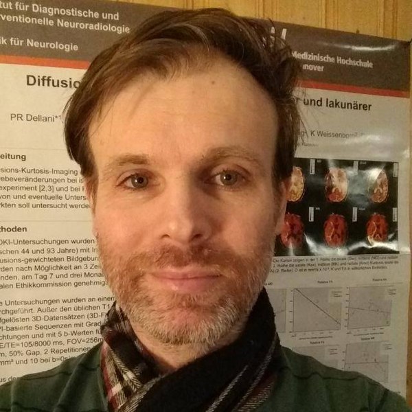
Dr. Paulo R. Dellani serves as a Postdoctoral Researcher at the Research Center Jülich, affiliated with the Institute of Neuroscience and Medicine (INM-1), where he investigates structural and functional brain organization. His work integrates advanced neuroimaging techniques, artificial intelligence, and clinical neuroscience to explore brain architecture, connectivity, and pathology. His research interests encompass: Novel brain mapping methodologies Clinical neuroanatomy and genomic imaging Big data analytics for neural systems Diffusion tensor imaging and fiber tract analysis Neurodegenerative disorder biomarkers Recent publications demonstrate a strong focus on translational neuroscience, including MRI-based diagnostics for vestibular disorders, Alzheimer's disease biomarkers, and computational approaches to white matter tractography. Common themes include validation of imaging protocols, neuropathology characterization, and clinical application of quantitative neuroimaging techniques. Dr. Dellani contributes to the Bernstein Coordination Site and participates in teaching activities at universities. His laboratory develops tools and services supporting brain research, including the Julich-Brain atlas project.
Dr. Valentin Beyer is a Tenure Track Research Fellow at the Aston Institute for Membrane Excellence (AIME), Aston University. Previously, he held roles as a Research Associate at KU Leuven and a Research Fellow at the University of Warwick. His research focuses on polymer science, processing of semicrystalline materials, and biodegradable polyesters. Education: PhD in Polymer Science from Queen Mary University of London (2020), MSc in Chemical Biology from Karlsruher Institut für Technologie (2016). He is a member of the Royal Society of Chemistry. Research interests include advanced polymer synthesis, sustainable materials, and applications in environmental biotechnology. Recent work explores flocculation techniques for microalgae harvesting and novel polymer characterization methods using NMR spectroscopy.
Valencia Koomson is an Associate Professor at Tufts University across three departments: Electrical and Computer Engineering (since 2011), Computer Science (since 2023), and Tisch College (since 2020). She previously served as a VLSI research engineer at USC/ISI and held visiting roles at MIT, Boston University, and Rensselaer Polytechnic Institute. Ph.D., University of Cambridge (2003) M.Phil., University of Cambridge (2000) M.Eng. and B.S., MIT (1999 and 1998) Her research focuses on silicon-based mixed-signal VLSI systems for biomedical imaging and optical wireless communication, with additional work in analog signal processing and nanoscale system integration across visible to millimeter-wave frequencies. Recent publications highlight her innovations in fdNIRS-tDCS brain stimulation systems , microfluidic cellular connectivity platforms , and ChromaSense wearable health sensors that address skin-tone bias in medical devices. She has secured multiple NSF grants including a CAREER award, plus NIH and industry funding. Scientific Awards: NSF CAREER Award Marshall Scholar IEEE Senior Member MLK Jr. Visiting Professor Fellowship (MIT) Two U.S. patents issued She leads the Advanced Integrated Circuits and Systems Lab at Tufts, which develops nanoscale systems spanning 60GHz RF to optical frequencies for medical and communication applications.
Dr. Tony Cafolla is an Emeritus Associate Professor at Dublin City University's School of Physical Sciences. He has held this position since 1992, with prior research roles at the University of Manchester and University of Wales, College of Cardiff. He is affiliated with the HEA-funded National Centre for Sensor Research since 1999. Education: B.Sc. in Experimental Physics, Trinity College Dublin (1975) M.Sc. in Nuclear Instrumentation Physics, Trinity College Dublin (1977) Ph.D. in Solid State Physics, University of Virginia (1985) Research Interests: Electronic and chemical properties of semiconductor surfaces Photoelectron spectroscopy using synchrotron radiation Surface structural studies via I-V Low Energy Electron Diffraction (LEED), Scanning Tunnelling Spectroscopy (STS), and optical probes Development of advanced materials for sensor applications (e.g., graphene, ceria thin films) Recent Research Trends: His work spans nanomaterial characterization (e.g., MoS₂ flakes, ZnO nanostructures), surface functionalization (e.g., bromine-functionalized molecules), and interdisciplinary applications in robotics and biomedical coatings. Publications emphasize experimental techniques like STM/STS and synchrotron-based spectroscopy. Awards: None explicitly listed. Grants/Advising: No specific grants or advisees mentioned, though his involvement with the National Centre for Sensor Research indicates collaborative research activity. Labs/Teams: Active in the National Centre for Sensor Research, focusing on sensor technology and material innovation.
Professor Markus Barth is a Full Professor in Biomedical Engineering at the University of Queensland (UQ), specializing in MRI physics and medical imaging. He holds roles as Deputy Head of School (Research) in the Faculty of Engineering, Architecture and Information Technology, Director of the National Imaging Facility – Queensland Node, and member of the ARC College of Experts. His academic journey includes a Doctorate in Technical Physics from Vienna University of Technology (1999) and prior appointments at the Medical University Vienna, Donders Institute, and Erwin L. Hahn Institute. His research focuses on advancing MRI techniques for high-resolution anatomical and functional imaging, including susceptibility-weighted imaging (SWI), quantitative susceptibility mapping (QSM), and ultra-high field MRI applications. He has pioneered methods to enhance MRI sensitivity for neurodegenerative diseases, cancer, and cognitive neuroscience studies. Barth’s work integrates cutting-edge technologies like deep learning and parallel transmission to improve image quality and clinical utility. His contributions include developing open-source tools (e.g., QSMxT) and large-scale datasets (e.g., HumanBrainAtlas) for reproducible neuroimaging research. Education: Bachelor of Technical Physics, Vienna University of Technology (1995) Doctorate in Technical Physics, Vienna University of Technology (1999) Research Interests: Optimizing MRI for ultra-high field applications, neuroimaging of brain function and connectivity, SWI/QSM for disease markers, and translational imaging techniques for clinical diagnostics. His work addresses topics like layer-specific fMRI, brain-computer interfaces, and decoding neural activity. Grants & Awards: ARC Future Fellowship (2014), leadership in national imaging initiatives, and contributions to MRI software frameworks. Labs/Teams: Leads the Ultra-high Field Human MR Research program at UQ’s Centre for Advanced Imaging and collaborates with interdisciplinary teams in biomedical engineering and neurology. His lab develops novel MRI protocols and open-source tools for global research impact.
Dr. Shajan Gunamony is the Head of MRI RF Engineering at the University of Glasgow's School of Psychology & Neuroscience. His work focuses on advancing MRI technology, particularly in RF engineering, coil design, and high-field MRI systems. He leads projects involving parallel transmit arrays, SAR management, and neurovascular imaging at 7T and higher field strengths. Grants & Awards: UKRI National facility for 11.7T human MRI (2024–2028) Wellcome Trust 7T Neurovascular Imaging Development (2021–2023) EU Living Lab Precision Medicine (2020–2025) Research Interests: Dr. Gunamony's research emphasizes cutting-edge MRI technology, including RF coil optimization, high-field imaging applications, and clinical translation. His contributions span from hardware development to biophysical modeling, with a focus on improving spatial resolution and safety in neuroimaging. Teams: Collaborates with interdisciplinary teams including physicists, clinicians, and engineers. Supervises research in MRI RF systems and leads the Glasgow MRI engineering group.
Cristina Ramona Cudalbu Moldovan is a Senior Lecturer and Researcher at École Polytechnique Fédérale de Lausanne (EPFL), where she serves as Head of Unit at the Center for Biomedical Imaging (CIBM), Animal Imaging and Technology (AIT) section. She is affiliated with the School of Basic Sciences and contributes to teaching in the Doctoral Program in Physics (EDPY) and the School of Physics (SPH). Her work integrates advanced MRI and MRS techniques to study brain metabolism and neurological complications of liver disease. Institution: École Polytechnique Fédérale de Lausanne (EPFL) School: School of Basic Sciences Department: CIBM - MRI Technologies Research Section Roles: Head of Unit, Senior Lecturer, Researcher Her research focuses on in vivo magnetic resonance spectroscopy (MRS) and imaging at ultra-high magnetic fields (up to 14.1T), with applications in hepatic encephalopathy, glioma, brain energy metabolism, and neurochemical profiling. She investigates metabolic alterations in animal models of liver disease, brain tumors, and neurodegenerative conditions, often employing multimodal imaging approaches such as 1 H, 31 P, and 13 C MRS, as well as PET and DTI. Her work emphasizes translational neuroscience and methodological innovation in spectral quantification and data acquisition. The most recent publications demonstrate a strong trend toward understanding metabolic dysregulation in hepatic encephalopathy, particularly focusing on ammonia toxicity, oxidative stress, and therapeutic interventions like urease inhibition and creatine supplementation. She also contributes significantly to methodological consensus in MRS, promoting standardization across research centers. Her articles span high-impact journals in neuroscience, radiology, and metabolism, reflecting interdisciplinary collaboration and technical rigor. She has supervised several PhD students, including Jessie Julie Mosso, Veronika Rackayová, and Dunja Simicic, whose theses reflect the lab’s focus on brain metabolism in liver disease and glioma. She is actively involved in expert task forces and collaborative networks such as ENIGMA, shaping best practices in neuroimaging. While no specific scientific awards are listed in the provided text, her leadership in consensus guidelines and high-impact publications underscores her recognition in the field. Her academic background includes a PhD from University Claude-Bernard Lyon 1, France, and a Master of Science in Biophysics and Medical Physics from University Babes-Bolyai, Romania. She has been continuously active at EPFL since 2007, progressing from postdoctoral researcher to senior leadership in imaging research. She leads a research unit at CIBM-AIT, which provides translational MR neuroimaging and spectroscopy services and training, emphasizing preclinical ultra-high-field MRI systems.
Bernard Lanz is a Researcher at the CIBM-AIT (Animal Imaging and Technology) unit within École Polytechnique Fédérale de Lausanne (EPFL) , with concurrent Lecturer roles in the EDPY-ENS and SB-SPH-ENS teaching programs. His work integrates Magnetic Resonance Spectroscopy (MRS) , Positron Emission Tomography (PET) , and mathematical modeling to study brain energy metabolism , neuroglial interactions , and metabolic flux dynamics in health and disease. Research Interests : Neuroenergetics : Quantifying glucose and lactate metabolism in rodent models of liver disease and cancer. Metabolic Imaging : Developing hyperpolarized C-13 glucose techniques for real-time metabolic tracking. Brain-Liver Axis : Investigating metabolic disruptions in hepatic encephalopathy via in vivo PET-MRS. Publication Trends : Recent work focuses on ultrahigh-field MRI , FLASH radiotherapy effects , and cross-disease metabolic comparisons (Alzheimer’s, glioblastoma, depressive disorders). Collaborative studies emphasize methodological innovation in MRS quantification and neurochemical modeling . Teaching & Supervision : Co-teaches MRI Practicals and Translational MR Neuroimaging courses. Advises PhD student Siviglia Alessio . Labs & Teams : Affiliated with the CIBM center and EPFL’s Swiss School of Physics units, contributing to interdisciplinary research in biomedical imaging .
Assoc. Prof. Dr. Filiz Celebi serves as an Associate Professor in the Department of Medicine at Yeditepe University Faculty of Medicine, Istanbul, Turkey, with expertise spanning clinical oncology and advanced medical imaging. Her research focuses on critical breast cancer domains: Neoadjuvant therapy optimization and vitamin D supplementation effects Lymphedema prediction via bioimpedance monitoring AI integration in diagnostic breast ultrasound Ovarian function preservation during chemotherapy Advanced PET/MRI applications in cancer diagnostics Recent publications demonstrate strong interdisciplinary trends, particularly the fusion of artificial intelligence with radiology for improved diagnostic accuracy and investigations into long-term quality-of-life outcomes for breast cancer survivors. Dr. Celebi has supervised multiple theses and led research projects as documented in her institutional profile, though specific awards are not detailed in available sources.
Mauro Riccò is an Associate Professor in the Department of Physics and Earth Sciences at the University of Parma, Italy, where he holds the chair of General Physics 2 for Engineering Management and serves as Director of the Materials Science and Technology program. Since 2005, he has been a confirmed Associate Professor in Physics of Matter (SSD FIS/03), following 14 years as a researcher at the University of Parma's Faculty of Engineering. Professor Riccò's academic background includes: PhD in Physics (1985-1988) from University of Parma Degree in Physics (1978-1984) with highest honors (110/110 cum laude) His research focuses on carbon nanostructures with three primary applications: hydrogen storage materials, superconducting fullerides, and battery electrode materials. Riccò's laboratory pioneered studies on alkali metal cluster-intercalated fullerenes achieving 6% hydrogen storage by weight, demonstrated defect-induced magnetism in graphene, and discovered superionic conductivity in fulleride-based electrolytes. His work combines advanced characterization techniques including solid-state NMR, μSR spectroscopy, neutron scattering, and SQUID magnetometry. Analysis of his publication history shows consistent output in materials physics with recent expansion into biomedical applications. His 2024-2025 publications reveal strategic diversification into virology and biosensors while maintaining core expertise in carbon nanomaterials. This interdisciplinary shift demonstrates his ability to apply fundamental physics knowledge to emerging societal challenges. Professor Riccò has secured significant research funding through: NEST-EU project "Ferrocarbon" (2005-2008) as international coordinator Swiss National Science Foundation Sinergia project "Hycarbo" (2010-2013) IRSES-EU project "MagNonMag" (since 2011) Multiple national research projects in Italy He founded and directs the Carbon Nanostructures Laboratory (CNL) since 1991, maintaining active international collaborations. The laboratory's research spans chemical synthesis of nanostructures, characterization techniques, and practical applications in energy storage, with recent expansion into biomedical materials as evidenced by his 2025 sericin electrode publication.
Oleksandr Misiats is an Assistant Professor at the Courant Institute of Mathematical Sciences, New York University. His research focuses on stochastic partial differential equations, mathematical physics, and materials science, with applications in thin film dynamics, superconductivity models, and biological systems. Research Areas: Stochastic PDEs, Materials Science, Mathematical Biology Affiliation: Courant Institute of Mathematical Sciences, New York University His recent publications explore nonlinear and stochastic perturbations in physical models, including thin film equations, bidomain models with random noise, and Ginzburg-Landau superconductivity frameworks. These works emphasize energy minimization, asymptotic behavior, and invariant measures in complex systems. While no formal awards or student advisement records appear in the provided data, his scholarly output demonstrates expertise in both deterministic and stochastic mathematical modeling across diverse scientific domains.
Dr. Babak Shadgan is an Assistant Professor in the Department of Orthopaedics, Faculty of Medicine, and Lecturer at the School of Biomedical Engineering, all at the University of British Columbia. He also holds research roles at the Vancouver Prostate Centre and Vancouver General Hospital, with affiliations to ICORD, the Peter Wall Institute, and the Canadian National Transplant Research Program. Education: M.D., Tehran Azad University M.Sc. in Sports Medicine, University of London Ph.D. in Muscle Biophysics, University of British Columbia Fellowship in Diffused Optical Tomography, MIT/Harvard Postdoctoral Fellowship in Clinical Biophotonics, University of British Columbia Dr. Shadgan’s research focuses on clinical biophotonics and translational applications of near-infrared spectroscopy (NIRS) for monitoring tissue oxygenation and hemodynamics in acute spinal cord injury, reconstructive surgery, and sports medicine. His work spans neuroprotection, musculoskeletal health, and non-invasive diagnostics. Recent articles highlight NIRS for spinal cord monitoring, muscle spasm analysis, and cardiac arrest detection. His lab has also explored blood flow restriction exercise for spinal cord injury rehabilitation and wearable sensor design. Scientific Awards: MSFHR Scholar Award (2018) Peter Wall Institute Wall Solutions Award (2020) US DoD Clinical Trial Award (2021) MITACS Postdoctoral Fellowship (2020) Dr. Shadgan supervises a multidisciplinary team in his clinical biophotonics lab, including PhD and Master’s students. His grants include funding from the Canadian Institutes of Health Research, the Michael Smith Foundation, and the US Department of Defense.
Saad Jbabdi is Professor of Biomedical Engineering at the University of Oxford, holding a Wellcome Trust Senior Research Fellowship. He serves as Head of Diffusion Analysis at FMRIB (Centre for Functional MRI of the Brain) within the Nuffield Department of Clinical Neuroscience and is a Lecturer in Engineering at St Hilda's College. His work pioneers advanced neuroimaging techniques for systems-level brain analysis. Professor Jbabdi's research spans diffusion MRI , brain connectivity modelling , and microstructure analysis . His lab develops mathematical frameworks for diffusion-weighted imaging while comparing in-vivo, ex-vivo, and histological techniques across humans and non-human primates. Key initiatives include human-macaque comparative anatomy, novel MR pulse sequence development, dynamic spectroscopy analysis, and individual variation modelling in brain function. Recent publication trends reveal intense focus on multi-modal integration of MRI, microscopy, and spectroscopy for connectome mapping. His group drives standardization in tractography (XTRACT protocols), cross-species neuroimaging, and computational models of neurodegeneration like tau progression. Work bridges engineering physics, neuroscience, and clinical applications in vision loss and neurodegenerative disorders. Scientific recognition includes: Wellcome Trust Senior Research Fellowship Royal Netherlands Academy of Arts and Sciences (KNAW) membership Funding support comes from: Wellcome Trust : Core fellowship and lab infrastructure Medical Research Council (MRC UK) : Projects on brain connectivity and microstructure He leads the FMRIB Diffusion Analysis group within Oxford's WIN Analysis Group, directing a team including Abivardi, Cottaar, Eichert, Howard, Li, Rafipoor, and Zheng. The lab leverages Oxford's advanced imaging facilities for projects like the BigMac dataset (macaque brain imaging) and Developing Human Connectome initiatives.
Dibash Basukala is a Research Fellow at the Sloan Kettering Institute , Memorial Sloan Kettering Cancer Center, affiliated with the Medical Physics Department . His work focuses on advanced MRI techniques for breast cancer assessment. Research Focus : Functional MRI, hypoxia imaging, diffusion-weighted imaging, and multisite reproducibility studies Key Collaborations : Katja Pinker-Domenig, Sunitha Thakur, Arka Bhowmik, Panagiotis Kapetas Publications : 2 journal articles and 1 review (2024–2025) in Journal of Magnetic Resonance Imaging and Frontiers in Oncology His research addresses technical validation of MRI biomarkers and clinical translation of functional imaging techniques for breast tumor characterization.
Paul Sijens is a Researcher at the University of Groningen , affiliated with the Faculty of Medical Sciences and the BRIDGE (Basic and Translational Research and Imaging Methodology Development in Groningen) program. His career spans academic and clinical research in Magnetic Resonance Imaging (MRI) and NMR spectroscopy , with key contributions to oncology, neurochemistry, and breast imaging. Education: PhD in 31P NMR spectroscopy of tumors from Utrecht University (awarded October 24, 1988). Professional Experience: Led MR Spectroscopy at Daniel den Hoed Cancer Center (1991-2000), postdoctoral work at Cleveland Clinic Foundation (1989-1991) funded by NIH. His research focuses on MRI applications in breast cancer screening , phenylketonuria , and tumor metabolism , integrating deep learning and radiomics to enhance diagnostic precision. Recent publications highlight inter-individual variability in brain phenylalanine transport and AI-driven lesion localization in breast imaging. Notable scientific awards include the EPOS Magna Cum Laude Award (2017) and the Koning Albert Gedenkpenning from FWO Vlaanderen (2017). He serves as an editor for the Journal of Magnetic Resonance Imaging (since 2023) and on editorial boards for radiology journals. Collaborations span institutions like Cleveland Clinic Foundation and European Radiology teams.