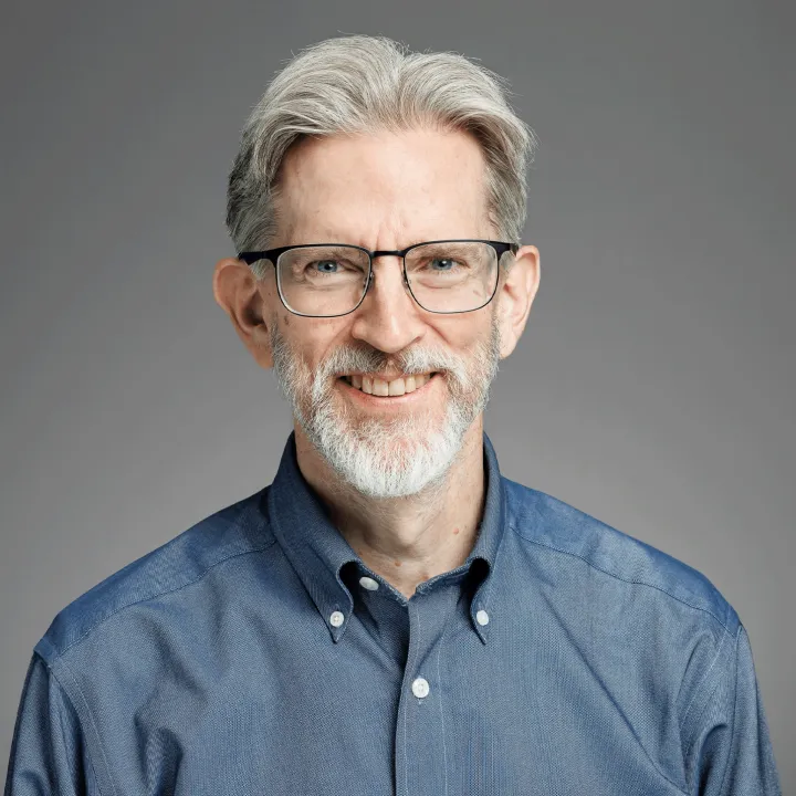
Professor Guy Williams is a leading academic at the University of Cambridge with a focus on imaging science and clinical neurosciences, affiliated with Downing College and the Wolfson Brain Imaging Centre . Holding a PhD in Physics from his initial Natural Sciences degree, he specializes in nuclear magnetic resonance (NMR) and MRI techniques for brain imaging. Education: BA, PhD in Physics His research centers on non-invasive imaging of brain structure and function, particularly in traumatic brain injury (TBI) and dementia. His work involves developing novel MRI pulse sequences and advanced data analysis algorithms, including AI-based diagnostic tools. He leads studies on white matter integrity post-trauma, longitudinal dementia assessment, and applications of MRI in disorders of consciousness and addiction. Recent publications highlight collaborations in traumatic brain injury outcomes, AI-guided dementia prediction, and neuroimaging of post-COVID cognitive deficits. His team's work on ultra-high field laminar fMRI and distortion correction methods has advanced clinical neuroscience applications. Key techniques include diffusion tensor imaging (DTI), 7 Tesla MRI, and positron emission tomography (PET/MR). His research spans from basic NMR physics to clinical translation, with a strong emphasis on multi-site studies and real-world diagnostic implementation.
Denise Head is Professor of Psychological & Brain Sciences and Associate Chair at Washington University in St. Louis, with an additional appointment as Associate Professor in Radiology. Her research integrates cognitive neuroscience and neuroimaging to study cognitive aging and Alzheimer's disease. PhD, University of Memphis MS, University of Memphis BS, University of New Orleans Her research focuses on age-related cognitive changes and their neural underpinnings. Key areas include spatial navigation deficits in aging, the role of lifestyle factors (exercise, sleep, stress) in brain aging, and interventions to support cognitive function in older adults. She uses virtual reality, mobile eye-tracking, and neuroimaging techniques such as fMRI and DTI. The recent publications highlight a consistent trajectory in cognitive neuroscience and aging research, with emphasis on neuroimaging biomarkers, structural brain changes, and cognitive performance in normal and pathological aging. Her work bridges psychology, neurology, and radiology, contributing to early detection and understanding of Alzheimer's disease. Scientific Awards: No awards listed in the provided text. Dr. Head advises graduate students and leads a research lab focused on cognitive aging, though specific student names are not listed. Her lab investigates mediators of brain aging and develops methods to support spatial navigation in older adults. While specific grants are not mentioned, her ongoing research and recent publications suggest active external funding. She leads a research team in the Department of Psychological & Brain Sciences, utilizing advanced neuroimaging and behavioral methods to study aging and dementia. The lab integrates real-world and virtual experimental designs to understand spatial cognition and brain health in older populations.
Lawrence H. Staib is a Professor of Biomedical Engineering at Yale University, with additional academic appointments in Electrical & Computer Engineering and Radiology & Biomedical Imaging. He holds a Ph.D. from Yale University and specializes in automated medical image analysis, including techniques like model-based segmentation, nonrigid registration, and diffusion tensor imaging (DTI). His research focuses on applications in neuroscience, cardiology, and cancer imaging, emphasizing machine learning and functional MRI analysis. His key contributions include advancements in white matter tractography via anisotropic wavefront evolution, real-time neural tract parcellation (Fasciculography), and noise reduction in diffusion tensor fields. Staib is a Fellow of the American Institute for Medical and Biological Engineering (2015), recognizing his impactful work in medical imaging technologies. Staib's research also encompasses statistical deformation models, perturbation-based shape analysis, and 3D deformable models for volumetric segmentation. He has developed patented 3D ultrasound computed tomography systems (USPTO #6878115, 7025725). His work bridges clinical needs with computational methods, addressing challenges in image registration, structural connectivity analysis, and medical robotics.
Dr. Chris A. Flask is a Professor at the Case Western Reserve University School of Medicine, with joint appointments in Radiology, Pediatrics, and Biomedical Engineering. He serves as Co-Director of the Imaging Research Core and Associate Director of the Medical Scientist Training Program, while also contributing to the Cancer Imaging Program at the Case Comprehensive Cancer Center. Research Focus Quantitative Magnetic Resonance Imaging (MRI) MRI Physics and Pulse Sequence Design Lung Imaging in Cystic Fibrosis Kidney Imaging in Polycystic Kidney Disease, Sickle Cell Disease, and Diabetic Nephropathy Liver Imaging for Inflammation and Fibrosis Scientific Recognition Distinguished Investigator Award 2023 from The Academy for Radiology and Biomedical Imaging Research Reviewer with Distinction for Magnetic Resonance in Medicine Semi-Finalist for ISMRM Young Investigator Award 2013 His recent publications focus on pH-responsive imaging agents, MR fingerprinting techniques, and applications in pediatric and adult diseases. Dr. Flask's work bridges technical MRI innovation with clinical translation across multiple organ systems.
Moo Chung is a Professor of Biostatistics and Medical Informatics at the University of Wisconsin-Madison, affiliated with the School of Medicine and Public Health. He holds additional appointments in the Department of Statistics and the College of Letters and Science. Chung earned his Ph.D. in Mathematics and Statistics from McGill University under Professors Keith J. Worsley and James O. Ramsay. His research focuses on computational neuroimaging, leveraging MRI, fMRI, and DTI to study brain dynamics through topological and geometric methods. Key areas include persistent homology, brain network analysis, and statistical modeling of high-dimensional imaging data. He has pioneered methods like hyper-network construction and exact topological inference for paired brain networks, addressing computational challenges in analyzing large-scale neuroimaging datasets. Chung’s work integrates advanced mathematical techniques such as Hodge Laplacian, spectral graph theory, and Wasserstein distances to analyze brain connectivity. He has secured NIH funding for projects like the Brain Initiative (2017-2020) and current grant MH133614 (2023), focusing on geometric data analysis and topological dynamics. His contributions include three books on brain imaging and network analysis, with ongoing research on topological data analysis applications. Awards: Vilas Associate Award (2013-2014), NIH Brain Initiative Award, Editor's Award (2011) Labs/Teams: Leads brain imaging workshops globally, including Seoul National University (2024) and POSTECH (2024), and organizes conferences like ISBI and MICCAI special sessions. Grants: NIH EB022856 (Brain Initiative), MH133614 (2023), and collaborations with institutions like Vanderbilt University and University of Chicago. Chung actively mentors students and postdocs in biomedical data science, offering fellowships through the CIBM program. His group maintains a Google mailing list for brain image analysis discussions and hosts regular seminars on methodological advancements.
Mojtaba Zarei is a researcher at the Department of Clinical Research, Faculty of Health Sciences, University of Southern Denmark, with additional affiliations at Odense University Hospital (OUH) and Karolinska Institutet (KI). His primary research unit is the Neurology Research Unit in Odense, focusing on advanced neuroimaging techniques and their applications in neurological and sleep disorders. Dr. Zarei's research spans multiple domains within neuroscience, with particular expertise in Positron Emission Tomography (PET), Diffusion Tensor Imaging (DTI), and cognitive function assessment. His work frequently addresses Alzheimer's Disease, Parkinson's Disease, and insomnia disorders, utilizing both clinical and computational approaches. His fingerprint analysis shows strong activity in neuroscience (100% for PET), diffusion tensor imaging (66%), cognitive function (45%), and Alzheimer's Disease (40%). His recent publications reveal a clear trajectory toward integrating multimodal imaging techniques with machine learning approaches for improved diagnosis and understanding of neurological conditions. The work on OPETIA (Odense-Oxford PET Image Analysis) demonstrates his contribution to developing standardized tools for neuroimaging analysis. His research increasingly bridges computational methods with clinical neuroscience, as evidenced by his work on image stitching algorithms and machine learning applications for insomnia classification. Dr. Zarei actively collaborates with researchers across multiple institutions, with notable external collaborations visible on the international network map. His work has been mentioned by peer review sites, picked up by news outlets, and shared across social media platforms, indicating growing impact in his field. Within his research unit of Neurology in Odense, Dr. Zarei appears to be part of a multidisciplinary team working at the intersection of clinical neurology, advanced imaging, and computational analysis, contributing to both methodological development and clinical applications of neuroimaging techniques.
Peter J. Basser is a leading research scientist at the National Institutes of Health (NIH), specifically within the Eunice Kennedy Shriver National Institute of Child Health and Human Development (NICHD), where he heads the Section on Quantitative Imaging and Tissue Sciences (SQITS). His work bridges physics, engineering, and neuroscience to develop non-invasive MRI methods for probing tissue microstructure, particularly in the brain. His educational background is not explicitly mentioned, but his scientific achievements reflect deep training in biophysics and medical imaging. He earned his Ph.D. and has built a career at NIH as a principal inventor of key neuroimaging technologies. Basser's research focuses on quantitative imaging and tissue sciences , especially using diffusion MRI to study brain structure and function. He pioneered Diffusion Tensor MRI (DTI) , Streamline Tractography , and advanced methods like MAP MRI , CHARMED , and AxCaliber , enabling in vivo measurement of axon diameters and microstructural features previously accessible only through histology. His work aims to translate these tools into clinical use for diagnosing developmental disorders, trauma, and neurodegeneration. The 15 most recent articles reflect a consistent focus on developing novel MRI biomarkers, particularly through diffusion and relaxometry methods. They explore water exchange, restriction, glymphatic clearance, latency connectomes, and cortical microstructure, demonstrating a trajectory toward in vivo MRI histology and precision imaging for pediatric and neurological applications. Scientific Awards: National Academy of Engineering (NAE), Inducted 2020 National Academy of Inventors (NAI) Fellow, 2024 Eduard Rhein Technology Award, 2021 ISMRM Gold Medal, 2008 ISMRM Lauterbur Lecturer, 2020 American Society of Neuroradiology Honorary Member, 2019 Victor M. Haughton Award, 2017 ISMRM Fellow, 2010 AIMBE Fellow Best Paper Award, Frontiers in Physics, 2023 Basser leads a dynamic research group that mentors postdoctoral fellows and trainees, many of whom have received prestigious awards. His lab has secured significant grants from the NIH BRAIN Initiative, NICHD, USUHS, and the Bill & Melinda Gates Foundation. The SQITS lab develops open-source software tools like TORTOISE , dmritool , and HI-SPEED , which are widely used in the neuroimaging community. The lab collaborates with institutions such as Uniformed Services University and participates in major initiatives like the Human Placenta Project and the Human Connectome Project. Basser’s vision is to transform clinical MRI scanners into quantitative scientific instruments for precision medicine and large-scale brain mapping. Labs and Teams: Section on Quantitative Imaging and Tissue Sciences (SQITS), NICHD, NIH Neuropathology-Neuroradiology Integration Core (with USUHS) Advanced Translational Neuroimaging Research & Development Core Diffusion – Data Processing Center (DPC)
Dr Erica Dall'Armellina serves as a Senior Lecturer at the School of Medicine, University of Leeds, and holds an Honorary Consultant Cardiologist position. She leads research in quantitative cardiac magnetic resonance imaging with international recognition in cardiovascular MRI techniques. Her academic background includes: Medical degree and Cardiology specialization, University of Trieste, Italy 2-year Cardiac MR Fellowship, Wake Forest University, USA DPhil in CMR applications for acute myocardial infarction, University of Oxford (2012) Her research focuses on clinical translation of quantitative CMR for ischemic heart disease, particularly diffusion tensor imaging (DTI) to predict cardiac remodeling post-infarction and applications in acute coronary syndromes. Current projects target DTI biomarkers for hypertrophic cardiomyopathy and myocardial infarction outcomes. Scientific recognition includes: British Heart Foundation Intermediate Clinical Research Fellowship (2013, extended 2018) She supervises PhD/MRes students and has trained numerous UK/international clinicians. Major grants include BHF FS/13/71/30378 on DTI remodeling prediction and Heart Research UK RG 2668/18/20 on IVIM DTI in hypertrophic cardiomyopathy. Active in Leeds Institute of Cardiovascular and Metabolic Medicine, Biomedical Imaging Science group, and Multidisciplinary Cardiovascular Research Centre, she collaborates on advanced imaging translation.
Chiara Begliomini is an Associate Professor at the Department of General Psychology, University of Padova. Her research focuses on neural mechanisms underlying action planning and execution, particularly using functional MRI (fMRI) to investigate motor control, handedness effects, and cerebellar contributions to cognition. She explores cognitive and motor functions across the lifespan, with a special emphasis on aging populations and neurological disorders. Her work integrates neuroimaging techniques such as VBM and DTI to study brain structure-function relationships. Key research topics include cerebellar laterality, the impact of cognitive reserve on neurodegeneration, and the neuropsychological consequences of post-COVID-19 infection. She also investigates behavioral and neural correlates of binge eating, impulse control, and sensorimotor integration in grasping movements. Publications highlight interdisciplinary approaches, combining fMRI with kinematic analyses and neuromodulation techniques. Her studies often address clinical populations, such as individuals with mild cognitive impairment, anorexia nervosa, and cerebellar hypoplasia. Her research portfolio reveals a strong focus on translational neuroscience, bridging basic science and clinical applications. Ongoing work explores cerebellar-cortical interactions, the role of handedness in motor networks, and the neurobiological basis of agency and intentionality.
Associate Professor Fatima Nasrallah is an Associate Professor and Principal Research Fellow at the Queensland Brain Institute (QBI), University of Queensland (UQ). Her research focuses on functional neuroimaging and brain injury mechanisms, particularly traumatic brain injury (TBI) and its link to neurodegenerative diseases like dementia. She leads a lab investigating multimodal imaging techniques to map structural, functional, metabolic, and molecular changes post-TBI, linking these to behavioral outcomes and biomarkers. Education: PhD in neurochemistry from the University of New South Wales (2009). Postdoctoral work at Singapore Bioimaging Consortium (2009–2012), followed by roles at the Clinical Imaging Research Center and QBI since 2015. Appointed as a Motor Accident and Injury Commission Fellow in 2015. Active in clinical and preclinical TBI research, translational medicine, and neuroimaging innovation. Research Interests: Her work spans basic and clinical neuroscience, emphasizing early diagnosis of TBI biomarkers and neuroimaging advancements. Key areas include: functional MRI, diffusion tensor imaging, quantitative susceptibility mapping, and biomarker discovery. Her lab explores the pathophysiological pathways connecting TBI to Alzheimer's disease and other dementias. Recent Article Themes: Recent publications highlight advancements in TBI biomarkers, neuroinflammation profiling, preclinical imaging standards, and MRI techniques for rodent models. Work also addresses clinical applications, such as pediatric TBI prediction and stroke rehabilitation via robotic devices. Awards: Recognized with the Motor Accident and Injury Commission Fellowship (2015), supporting her TBI research. Advising & Grants: Supervises PhD students (e.g., Linfeng Liu, Junyan Lyu) and collaborates on projects like the PREDICT-TBI trial (multicenter TBI outcome prediction). Leads teams in biomarker development, imaging innovation, and translational studies. Labs & Teams: Heads her independent research group at QBI, collaborating with institutions like the Singapore Bioimaging Consortium and international ISMRM networks. Engages in cross-disciplinary efforts to bridge preclinical and clinical TBI research.
Halima Chahboune is the Executive Director of the Carol and Gene Ludwig Program at Yale School of Medicine, leading research in neuroimmunity and dementia. She holds a PhD in Biomedical Engineering from Université Claude Bernard Lyon 1 and completed postdoctoral training at Yale in Radiology and Biomedical Engineering. Her career includes roles as Assistant Director at the Center for Research on Interface Structures and Phenomena (CRISP) and the Center for the Physics of Biological Function (CPBF). Her research focuses on advancing multi-modal Magnetic Resonance Imaging (MRI) techniques for applications in neuroscience, molecular imaging, and regenerative medicine. Key interests include neurodevelopmental disorders, tissue engineering, and translational imaging strategies. Her work bridges engineering and medicine, with publications in journals like Magnetic Resonance in Medicine and Stroke . Her research trends emphasize interdisciplinary approaches, leveraging MRI innovations to address challenges in regenerative medicine, neurodegenerative diseases, and developmental neuroscience. While no specific awards are listed, her contributions to imaging methodologies have been widely cited. She collaborates with prominent researchers such as Douglas Rothman and Flora Vaccarino, advancing studies on brain development and injury mechanisms. Halima’s work is affiliated with Yale’s Ludwig Program for Neuroimmunity in Dementia and the Neuroscience department, reflecting her commitment to integrating advanced imaging with biomedical research. Her office is located at 100 College Street, New Haven, CT.
University of California, Los AngelesUnited States
Barbara Knowlton is a Professor and Vice Chair in the Department of Psychology at the University of California, Los Angeles (UCLA), with affiliations to the Behavioral Neuroscience Division and the Brain Research Institute. She earned a B.A. in Psychology from Johns Hopkins University and a Ph.D. in Neuroscience from Stanford University, followed by postdoctoral work in Psychiatry at UC San Diego. Her research focuses on cognitive neuroscience, memory systems, learning mechanisms, and their implications in schizophrenia and aging. Recent publications highlight her work on value-directed remembering, procedural learning, age-related memory differences, and neural correlates of decision-making. She employs advanced neuroimaging techniques (e.g., high-resolution fMRI, DTI) and neurostimulation (tDCS) to investigate memory encoding, reward processing, and brain connectivity. Key themes include the interaction between cognitive and emotional systems, habit formation, and interventions to enhance memory retention through targeted stimulation of prefrontal and medial temporal regions. Selected Research Grants: 2022: NIH grant for "Causal Role of Prefrontal Cortex in Value-Directed Memory Encoding" Techniques: High-resolution 7T fMRI Transcranial Direct Current Stimulation (tDCS) Diffusion Tensor Imaging (DTI) Collaborations: Prof. Susan Bookheimer (UCLA) Prof. Itzhak Fried (UCLA Neurosurgery) Dr. Adam Ekstrom (UCLA)
University of California, Los AngelesUnited States
James McCracken is a tenured Professor of Psychiatry and Biobehavioral Sciences at UCLA's David Geffen School of Medicine. As Director of the Division of Child and Adolescent Psychiatry at the Semel Institute for Neuroscience and Human Behavior, he leads the NIMH-funded Research Center 'Translational Research to Enhance Cognitive Control' focusing on novel treatments for cognitive deficits in child psychiatry. His research spans pharmacogenomics, neurodevelopmental disorders (ASD, ADHD, OCD), and autonomic neurophysiology. Key affiliations: Center for Autism Research and Treatment (CART), ADHD Brain Research Institute, Society for Neuroscience Research themes: Pharmacologic interventions for neurodevelopmental disorders, white matter sex differences, EEG biomarkers, and genome-wide association studies Leadership roles: Principal Investigator for multiple NIMH grants, editorial board member for the Journal of Child and Adolescent Psychopharmacology His recent publications (2023-2025) emphasize precision medicine in ASD, OCD genetics, and pupillary response as a neurophysiological marker. Awards include the APA Young Psychiatrist Research Award and listings in Best Doctors and Top Doctors databases. Scientific Awards American Psychiatric Association Young Psychiatrist Research Award Best Doctors in America America's Top Doctors Publications Over 150 peer-reviewed articles across child psychiatry Leading research in pharmacogenomics and neuroimaging
Martijn Froeling serves as an Assistant Professor at University Medical Center Utrecht, actively contributing to the Precision Imaging research group within the High Field division. His work bridges advanced MRI technology development with clinical applications targeting critical health domains including brain cancer, circulatory health, dementia, and musculoskeletal disorders. His academic foundation includes: Master's in Biomedical Engineering from Eindhoven University of Technology (July 2009) PhD in Diffusion Tensor Imaging of the human forearm from Amsterdam University Medical Center and Eindhoven University of Technology (October 2012) Dr. Froeling's research centers on pioneering quantitative MRI methodologies, with specialized expertise in Diffusion Tensor Imaging (DTI) across multiple organ systems (brain, peripheral nerves, muscle, kidney, heart). He drives innovation in 7T MRI hardware development—including specialized coils for multi-nuclei imaging—and conducts clinical studies focused on neuromuscular diseases. His QMRITools software platform for Mathematica enables sophisticated quantitative MRI analysis, directly supporting his mission to 'see the unseen' for advancing clinical diagnostics in cancer, cardiovascular disease, stroke, and MSK conditions. Analysis of his 2024-2025 publications reveals a cohesive research trajectory: DTI applications dominate clinical studies (hamstring injuries, fasciculation mapping), while parallel technical work advances ultra-high-field hardware (double-tuned coils for ²H/³¹P imaging). This dual focus on clinical impact and technological innovation demonstrates his commitment to translating engineering breakthroughs into tangible medical solutions. No scientific awards were documented in the provided materials. While the text confirms Dr. Froeling's role in supervising PhD work (evidenced by his PhD completion under prominent supervisors), no current students or specific grant funding details are explicitly mentioned in the source material. He operates within the High Field group at University Medical Center Utrecht, leading the Precision Imaging research initiative. This team specializes in developing cutting-edge MRI hardware (particularly for 7T systems), maintaining the QMRITools analysis platform, and executing clinical trials targeting neuromuscular pathologies alongside broader applications in oncology and cardiovascular medicine.
Dr. Tsang-Wei Tu is an Associate Professor in the Department of Radiology at Howard University College of Medicine. He is a key faculty member of the Molecular Imaging Laboratory and the Howard University Imaging Core, where he leads research in advanced neuroimaging techniques. His educational background includes a Ph.D. in Mechanical Engineering and Materials Science from Washington University in St. Louis (2011) and postdoctoral training in Radiology and Imaging Sciences at the National Institutes of Health (2016). Dr. Tu's research focuses on molecular and functional imaging to understand traumatic brain injury and other neurological disorders. His work emphasizes bridging radiological findings with underlying brain pathophysiology. Key areas include neuroimaging, diffusion tensor imaging (DTI), chemical exchange saturation transfer (CEST) MRI, blood-brain barrier disruption, and spinal cord injury . He teaches courses such as Medical Imaging Technology (MPHS 502) and Topics in Anatomy & Physiology (BIOL 501). The recent publications show a strong trend in using multimodal imaging (MRI, PET, MRS) in preclinical models to study brain injury, neuroinflammation, and regenerative therapies. His work frequently involves pulsed focused ultrasound, microbubbles, and stem cell delivery, with a focus on safety, efficacy, and mechanistic insights. Dr. Tu has not been publicly recognized with scientific awards in the provided text. He actively mentors students and researchers in the Molecular Imaging Laboratory, though specific advisees are not listed. His research is supported by extensive collaborations, particularly with scientists at the National Institutes of Health. Ongoing work appears to focus on imaging biomarkers for cerebral metabolic depression, BBB modulation, and regenerative medicine applications. Dr. Tu is affiliated with the Molecular Imaging Laboratory and the Howard University Imaging Core, which provide advanced imaging resources for preclinical and translational research.