Prof. Dr. Stephanie E. Combs is a leading academic in Radiation Oncology at Technische Universität München (TUM), holding the position of Professor and Chair of the Department of Radiation Oncology. She also serves as the Dean of the TUM Faculty of Medicine since 2022. Her expertise spans highly conformal radiation therapy techniques (e.g., IMRT/IGRT/ART, proton, and carbon ion therapy), with a focus on brain and skull base tumors, pediatric oncology, gastrointestinal oncology, and biomarker-driven therapies. Prof. Combs studied medicine in Heidelberg and the United States, completed postdoctoral training in Heidelberg, and became Vice Chair of Radiation Oncology there before joining TUM in 2014. She has held leadership roles, including heading the TUM Senate from 2019 to 2022. Her research emphasizes precision medicine, radiation biology, and translational radiotherapy innovations. Her awards include the Basic/Translational Senior Science Award (2019), Robert Janker Award (2012), and multiple honors from German and international radiation oncology societies. Her work bridges clinical practice and research, with contributions to guidelines on target delineation for glioblastoma and skull base tumors. Prof. Combs’ publications focus on advancing radiation techniques, radiomics, and personalized therapy, with over 300 peer-reviewed articles. She leads initiatives in education, including a Master of Science program in Radiation Biology, and collaborates internationally in translational research.
Joanna Pylvänäinen is a Project Researcher at the Faculty of Science and Engineering, Åbo Akademi University, specializing in Biochemistry and Cell Biology. She is affiliated with the InFLAMES flagship program (Innovation Ecosystem based on the Immune System) focusing on health-related solutions. Her research interests include: Bioimage analysis Live cell imaging Quantitative cell biology Software development for life science applications Flow dynamics Cancer cell biology Image analysis software Cell tracking Microfluidics 4D microscopy The research articles and publications cover a broad range of topics in image analysis, software development for life sciences, cell tracking, and microfluidics applications in biological research. The datasets she has contributed to focus on these same areas, showing a consistent research trajectory in computational biology and advanced imaging techniques. Joanna is active in developing tools and platforms for life scientists, with a particular focus on making bioimage analysis more accessible and on reducing technical challenges in live cell imaging. She is also interested in addressing phototoxicity issues and improving workflow efficiency in quantitative cell biology research. Her work includes contributions to educational tools like Ocul-AR for microscopy learning, Fast4DReg for microscopy data registration, and CellTracksColab for cell tracking data analysis. She is involved in the development of TrackMate 7 for integrating state-of-the-art segmentation algorithms into tracking pipelines.
Dr. Kurt Schmoller is a Group Leader at Helmholtz Munich, heading the Schmoller Lab within the Institute of Functional Epigenetics (IFE). His research focuses on understanding how cells regulate their size, maintain organelle homeostasis during growth, and adjust protein composition according to cell size. The lab employs interdisciplinary approaches combining quantitative biology, live-cell microscopy, AI-based image analysis, and mathematical modeling. Dr. Schmoller received his Diploma in Biophysics from TU München (2004-2008), followed by PhD studies at TU München in Andreas Bausch's lab (2008-2012), where he studied the mechanics of in vitro reconstituted cytoskeletal networks. He then pursued postdoctoral research at Stanford University with Jan Skotheim (2012-2017), developing his interest in cell size regulation. Since 2017, he has led the 'Cell Size and Organelle Control' research group at Helmholtz Munich. His research spans four main areas: Maintenance and Adaptation of Cell Size, Histone Homeostasis, Mitochondrial DNA Maintenance, and AI for live-cell imaging data analysis. Using model organisms including budding yeast (S. cerevisiae) and green algae (C. reinhardtii), his lab investigates fundamental cellular processes that are broadly conserved across eukaryotes. His work has significant implications for understanding diseases like cancer where cell size regulation is often disrupted. Analysis of his recent publications reveals a strong focus on the relationship between cell size and various cellular processes, with particular attention to histone regulation, mitochondrial DNA homeostasis, and the development of AI tools for image analysis. His work bridges molecular biology, biophysics, and computational approaches to address fundamental questions in cell biology. Dr. Schmoller leads a diverse research team including postdocs, doctoral researchers, and technicians working across multiple projects. His lab has developed notable tools such as Cell-ACDC, an open-source software for bioimage analysis that has been adopted by multiple laboratories. The lab maintains strong collaborations across institutions and participates in the broader 'Epigenetics at Helmholtz Munich' initiative. The Schmoller Lab operates from the Neuherberg Campus (Building 35.26 / Room 014) and maintains an active presence through their lab website, BioRxiv, and GitHub repositories. Their work receives support from Helmholtz Munich's infrastructure and resources, including access to advanced microscopy facilities and computational resources for AI-based image analysis.
Michaël Unser is a Full Professor at École Polytechnique Fédérale de Lausanne (EPFL) in the School of Engineering , leading the Biomedical Imaging Laboratory . He serves as Academic Director for Imaging at EPFL and contributes to cross-departmental teaching in Microengineering , Mathematics , and Life Sciences Engineering . His research spans Image Processing , Medical Imaging , Wavelets , and Spline-based Modeling , with a focus on multiresolution analysis and single-molecule localization microscopy . He has mentored over 30 PhD students and supervised numerous research projects. Recent publications highlight advancements in super-resolution microscopy , deep learning integration , and inverse problem solving for biomedical imaging. His work emphasizes mathematical rigor and open-source software development for accessible bioimaging tools. IEEE Technical Achievement Award (2008) IEEE EMBS Career Achievement Award (2020) Three ERC Advanced Grants (FUNSP, GlobalBioIm, FunLearn) As Academic Director for Imaging , he leads EPFL's cross-disciplinary imaging initiatives. His teaching includes Fundamentals of Image Analysis and Signals and Systems courses.
Haohan Wang is an Assistant Professor at the University of Illinois Urbana-Champaign's School of Information Sciences, with affiliations to the Carl R. Woese Institute for Genomic Biology and the National Center for Supercomputing Applications. His research focuses on developing trustworthy machine learning methods for computational biology and healthcare applications , emphasizing robustness , causality , and interpretability in vision-based models. Recent research trends in his work include: Large Language Model interactions with biomedical challenges (GenoAgent, GenoTex) Adversarial security in language and vision models (Guard, Jailbreakzoo) Genomic data analysis through robust machine learning frameworks (Precision Lasso, Kernel Mixed Models) Interactive toolkits like Robustar for data annotation and model training Scientific awards include recognition as Baidu's Top 50 AI+X Rising Young Scholars (2022), Best Paper Honorable Mention at WSDM 2023, and Broad Institute's Next Generation status (2019). Current projects explore AI-made scientists for biomedical discovery and Robustar development for GUI-based robust vision learning.
Fatima Boukari is an Associate Professor in Computer Science within the Division of Physics, Engineering, Mathematics and Computer Sciences at Delaware State University. Her research bridges artificial intelligence, deep learning, and mathematical modeling to develop robust solutions for biomedical engineering, cell biology, and agricultural technology challenges. Education: B.Sc. in Computer Science Engineering from University of Annaba, Algeria Dual M.Sc. degrees in Computer Systems Architectures and Parallel Computing from Algeria-Glasgow Ph.D. in Mathematics & Physics from Delaware State University Dr. Boukari's research centers on foundational Deep Learning architectures and mathematical modeling applied to biomedical diagnostics and cell dynamics analysis. Her work in reinforcement learning and transfer learning enhances decision-making for autonomous systems, while her cognitive modeling research decodes human perception using EEG data. She pioneers multi-modal distributed learning systems that maintain privacy across heterogeneous sensor networks, addressing critical gaps in military ISR applications. Her recent publications reveal a strong trajectory toward spectral data analysis for medical diagnostics and AI-driven cognitive modeling , with increasing emphasis on trustworthy AI solutions for healthcare and environmental sustainability. The consistent focus on cell segmentation/tracking algorithms demonstrates her commitment to advancing biomedical image analysis. Scientific Awards: No scientific awards, prizes, or fellowships listed in available information Dr. Boukari has mentored over 40 undergraduate and 2 graduate students from underrepresented STEM backgrounds. Her active research portfolio includes: NSF CISE grant for biomolecular detection using physics-informed machine learning Air Force RITA/UARC project on neuroscience computational modeling Air Force project building robust multi-modal distributed learning systems DE-CTR ACCEL project for COVID-19 respiratory disease diagnosis NSF grant for Delaware and Mid-Atlantic Data Science Corps Research scientist role in AI-CLIMATE National AI Research Institute She leads the Applied Interdisciplinary Data Science (AIDA) Laboratory and serves as Project Lead for the CAST E-IoT Center's four agricultural research thrusts. As Team Lead of the 1890 Working Group on Artificial Intelligence, she drives initiatives addressing climate change resilience and food security through responsible AI development.
Dr. Hannah Mitchell is a Lecturer at Queen's University Belfast's School of Mathematics and Physics, affiliated with the Intelligent Autonomous Manufacturing Systems and Mathematical Sciences Research Centre. She specializes in spatial data analysis, Hidden Markov models, and survival analysis, with research focusing on single-molecule imaging and statistical modeling. Key Research Areas: Spatial data analysis, Hidden Markov models, reversible jump MCMC for changepoint detection in imaging Recent Publications: Advanced statistical methods for FLImP super-resolution imaging and photobleaching correction Awards: 1st Prize for oral presentation at international conference (2024) Her work bridges computational statistics and biomedical imaging, developing techniques to improve imaging accuracy and efficiency. She actively supervises PhD students and contributes to peer review activities for journals.
Victor Hatini is an Associate Professor in the Department of Developmental, Molecular and Chemical Biology at Tufts University School of Medicine. His research focuses on understanding the mechanisms controlling epithelial morphogenesis, particularly using the Drosophila retina as a model system. He investigates the roles of adhesion molecules like Sidekick (Sdk) in coordinating contractile and protrusive forces during tissue organization. His work has revealed critical insights into tricellular adherens junction dynamics and their role in developmental processes. Education: PhD, Cornell University, 1996 MSc, Weizmann Institute of Science, 1991 BSc, Hebrew University, 1989 Research Interests: Dr. Hatini studies how adhesion molecules and cytoskeletal networks regulate epithelial tissue organization. His lab explores pulsatile mechanical cycles in epithelial cells, focusing on Sidekick’s dual roles in modulating actomyosin contraction and actin branching. Current work emphasizes the formation and function of tricellular junctions, with implications for understanding diseases involving tissue disorganization. Teaching & Mentoring: Dr. Hatini contributes extensively to graduate education at Tufts, directing core courses such as Cell Behavior and Molecular Cell Biology of Development. He has mentored 18 graduate students, 5 postdoctoral fellows, and 5 technicians, many of whom have pursued successful careers in academia, industry, and medicine. He also serves on NIH study sections supporting early-career researchers. Awards: Tufts Collaborates Award (2014) Grants: Active funding includes NIH grants investigating the WAVE regulatory complex in epithelial morphogenesis (2018–2023) and prior support for cellular interactions in patterning (2004–2009). Labs & Teams: His laboratory at Tufts focuses on integrative approaches combining genetics, live imaging, and computational modeling to study epithelial dynamics. Collaborations emphasize bridging basic research with clinical relevance in tissue repair and disease.
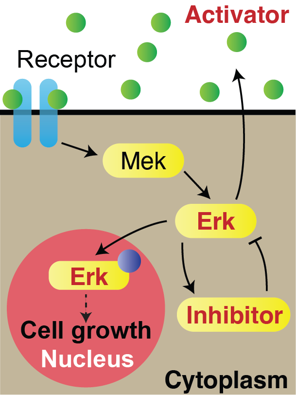
Alessandro De Simone is an Assistant Professor in the Department of Genetics and Evolution at the University of Geneva. His research group investigates the physical principles of vertebrate regeneration using zebrafish scales as a primary model system, focusing on how cells coordinate growth and patterning during tissue repair. Research interests center on the integration of biochemical signaling and mechanical forces in regeneration. Key areas include: Erk activity waves controlling tissue size/shape Mechano-chemical coupling in cell proliferation Coordination of bone formation with cell growth Quantitative live imaging of signaling dynamics Computational modeling of morphogenesis Publications demonstrate a strong interdisciplinary focus, with recurring themes in regeneration signaling waves, live imaging of developmental processes, and mathematical modeling of tissue dynamics. Recent work emphasizes Erk/JNK wave propagation in zebrafish models and stem cell-based approaches to human development. Scientific awards and recognitions include: SNSF Eccellenza Professorial Fellowship (PCEFP3_202776) ERC Starting Grant (Gradients, waves and nematics: quantitative perspectives on regeneration) De Simone advises PhD students and oversees research projects on regeneration mechanisms. Current PhD advisees include Konrad Marx, Coline Coudeville, and Oriane Foussadier. External funding supports investigations into signaling dynamics and tissue patterning. The laboratory, located in Sciences III building, includes postdoctoral researchers, PhD students, and technical staff specializing in live imaging, molecular biology, and zebrafish genetics. Team members collaborate on projects bridging experimental biology with computational approaches.
Ala'a Al-Habashna is an Adjunct Professor at Carleton University's School of Computer Science within the Faculty of Engineering and Design. He holds a PhD from Carleton University and focuses on advanced wireless communication systems, 5G/6G networks, machine learning applications, and discrete-event simulation. His research integrates AI-driven solutions for network optimization, spectrum management, and smart urban infrastructure analysis. Education: PhD in Computer Science, Carleton University Research Interests: 5G/6G Networks, Machine Learning for Communications, Cognitive Radio Systems, IoT Integration, and Urban Modeling via Computer Vision Focus on resource allocation algorithms, channel modeling, and AI-driven network architectures Key Article Trends: Recent work emphasizes AI integration in next-gen networks (e.g., RIS-assisted MIMO, GenAI-6G), 5G energy efficiency, and urban analytics using street-view imagery. Earlier contributions include DEVS-based simulation frameworks for fire modeling and D2D video streaming optimization. Collaborations & Funding: Active partnerships include Ericsson Canada for projects like Channel Reconstruction for LTE/NR and Spectrum Sharing with Machine Learning . A funded PhD position is available in Wireless Communication and 5G Networks requiring expertise in Mathematical Optimization. Labs & Teams: Engaged in Ericsson-Carleton Partnership initiatives and leads interdisciplinary teams addressing 6G challenges. Research leverages Carleton's advanced wireless and AI facilities.
Kevin Lin is an Assistant Professor in the Department of Biostatistics at the University of Washington, joining in Fall 2023. His research focuses on developing statistical methods for analyzing single-cell data to uncover cellular mechanisms in diseases like Alzheimer’s and immune resistance. He holds a PhD from Carnegie Mellon University and completed postdoctoral training at the University of Pennsylvania. His work bridges computational methods with biological questions, emphasizing matrix factorization, network modeling, and changepoint detection. Education: PhD in Statistics & Data Science (Carnegie Mellon University, 2020) Previous Role: Postdoctoral Researcher at University of Pennsylvania (Wharton Statistics) Research interests include high-dimensional data analysis, statistical genetics, and single-cell RNA-Seq. Notable contributions include methods like Tilted-CCA for multimodal data integration and eSVD-DE for cohort-wide differential expression analysis. His work has been recognized with awards such as the Wikimedia Foundation Research Award (2023). Lin’s lab collaborates on projects involving endolysosomal dysfunction in Alzheimer’s, yeast cell division dynamics, and lineage-aware machine learning. Key achievements include over 20 publications in journals like Nature Biotechnology , PNAS , and Biometrics . He advises on grants related to single-cell technologies and serves on the EDI subcommittee for mental health initiatives at UW. Outside academia, Lin enjoys zumba and cooking.
Jin U. Kang is the Jacob Suter Jammer Professor of Electrical and Computer Engineering at Johns Hopkins University, with a joint appointment in the Department of Dermatology at the Johns Hopkins School of Medicine. He is affiliated with the Kavli Neuroscience Discovery Institute and the Laboratory for Computational Sensing and Robotics (LCSR). His research focuses on optical imaging, sensing, and robotic systems for biomedical applications, including OCT-based surgical tools and image-guided interventions. Kang has pioneered real-time 4D OCT systems and autonomous robotic devices for microsurgeries, such as retinal and vascular procedures. His work also involves developing smart surgical tools through startups like LIV Med Tech Inc. Education: B.S. in Physics (Western Washington University, 1992); M.S. and Ph.D. in Optical Science and Electrical Engineering (University of Central Florida, 1993–1996). Prior to JHU, he worked at the U.S. Naval Research Laboratory. Research interests include fiber optic sensors, medical imaging systems, and surgical robotics. His recent work emphasizes real-time OCT-guided procedures, autonomous robotic surgery, and innovations in corneal and retinal surgeries. Over 20 patents and key awards such as the ONR Young Investigator Award reflect his contributions. Scientific Awards: Recipient of the ONR Young Investigator Award, Australian Institute of Advanced Studies Fellowship, NASA Faculty Fellowship, and Brain Korea Distinguished Faculty Fellowship. He is a Fellow of the Optical Society of America, SPIE, and the American Institute for Medical and Biological Engineering. Advising & Grants: Kang’s research is supported by grants and collaborations with industry (e.g., LIV Med Tech). He has advised numerous projects on surgical robotics and imaging systems, though specific student names are not listed here. His work spans academic and translational efforts, including ventures to commercialize medical technologies. Labs & Teams: Active in the Kavli Neuroscience Institute and LCSR. His lab develops cutting-edge imaging and robotic tools, with recent focus on autonomous systems for microsurgery and intraoperative guidance.
Andrew John Campbell is a Knowledge Exchange Fellow in the Department of Electronic & Electrical Engineering at the University of Strathclyde's Faculty of Engineering. With a Doctor of Engineering in Advanced Manufacturing, Dr. Campbell specializes in applying image processing and hyperspectral imaging techniques to solve complex measurement challenges across nuclear decommissioning, aerospace, and industrial manufacturing contexts. His research bridges academic innovation with practical industrial applications, particularly in environments where traditional measurement approaches are impractical or hazardous. Doctor of Engineering, Advanced Manufacturing Dr. Campbell's research focuses on developing advanced image analysis methodologies for diverse applications. His primary expertise lies in hyperspectral imaging for nuclear fuel characterization during decommissioning processes, where his work enables remote analysis of radioactive materials through specialized techniques like short-wave infrared imaging through leaded glass. He has also applied similar methodologies to space object identification, granular material analysis in foundry applications, and biomedical contexts such as platelet analysis. His approach consistently combines cutting-edge image processing algorithms with domain-specific knowledge to address real-world industrial challenges. Analysis of Dr. Campbell's recent publications (2019-2025) reveals a clear research trajectory from fundamental image analysis techniques toward increasingly specialized applications in high-consequence environments. The 2024-2025 publications demonstrate a strong emphasis on nuclear decommissioning applications, with multiple papers addressing the characterization of nuclear fuel debris through multimodal spectroscopy. This work represents a significant advancement in the field, enabling safer and more efficient nuclear site cleanup. His publications span multiple high-impact venues including IEEE conferences and scientific journals, reflecting the interdisciplinary nature of his work. Best Technical Paper in IIF Journal Award (Non-Ferrous Category) - February 2025 Dr. Campbell has secured significant research funding through multiple competitive grants, serving as Principal Investigator for the "Spectroscopy Suite for Direct Low Waste Measurement for All States of Matter" project funded by Sellafield Limited (January-March 2024), and as Co-investigator on several other substantial projects including the HSI for Granular Material project funded by Fraunhofer UK Research Limited (2023-2024). His funding portfolio spans nuclear, aerospace, manufacturing, and biomedical applications, demonstrating the versatility of his image processing expertise. He has supervised doctoral research as evidenced by his role in the thesis "Image processing for the analysis of TI-6AL-4V microstructures" and has presented research at professional venues including a 2020 presentation on "Automated tracking of mitochondrial motility using image processing and epifluorescence microscopy". Dr. Campbell works within research groups focused on advanced imaging applications, collaborating extensively with researchers including Paul Murray, Jaime Zabalza, and Stephen Marshall. His laboratory work suggests capabilities in hyperspectral imaging, spectral analysis, and advanced image processing techniques. He appears to be part of Strathclyde's nuclear technology research initiatives and contributes to the Universities' Nuclear Technology Forum. His fingerprint analysis indicates significant contributions to image processing (100%), space object identification (87%), and microstructure analysis (65%), reflecting the interdisciplinary nature of his research program.
Mona Garvin is a Professor in the Department of Electrical and Computer Engineering at the University of Iowa's College of Engineering. She also holds researcher positions at the Iowa Institute for Biomedical Engineering and the Iowa Initiative for Artificial Intelligence , blending engineering principles with medical imaging applications. Education PhD in Biomedical Engineering, The University of Iowa (2008) MS in Biomedical Engineering, The University of Iowa (2004) BSE in Biomedical Engineering (2003) BS in Computer Science (2003) Research Focus Her work specializes in ophthalmic image analysis , leveraging machine learning and graph-theoretic approaches for 3D segmentation of retinal structures. Current projects involve differentiating optic disc pathologies (papilledema, NAION) using OCT and enhancing retinal blood flow analysis through computational models. Professional Affiliations Institute of Electrical and Electronics Engineers (IEEE) Society of Photographic Instrumentation Engineers (SPIE) Association for Research in Vision and Ophthalmology (ARVO) American Society for Engineering Education (ASEE) Innovative Contributions Developed tools like AxoNet 2.0 and eyeFusion for automated retinal segmentation and visual field quantification. Her team explores deep learning solutions for OCT analysis, latent variable modeling in retinal thickness patterns, and radiation effects on retinal structures.
Dongfang Li is a Research Fellow in the Department of Neurosurgery at Yale University's School of Medicine, specializing in magnetically-driven microrobotics for neurosurgical applications and brain cancer therapy. His research focuses on: Microrobot design and magnetic actuation Image-guided navigation in dynamic biological environments Swarm control for targeted drug delivery Degradable materials for precision cancer treatment Magnetic hyperthermia-chemotherapy integration Analysis of his 15 most recent publications (2019-2025) reveals a cohesive trajectory advancing microrobot technology for brain interventions. Key innovations include OCT-guided in vivo navigation, torque-driven microswimmers, vessel-targeting nanoclovers, and rotating-field control systems. His work bridges robotics, nanotechnology, and neurosurgery to enable non-invasive, cell-level therapeutic precision with significant implications for glioblastoma treatment.