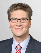
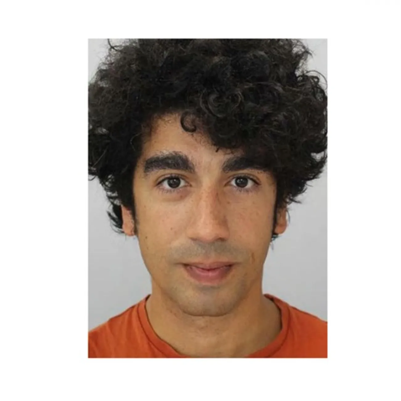
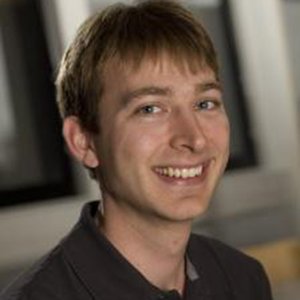
Prof. Dr. Franz Pfeiffer is a full professor at the Chair of Biomedical Physics within the Department of Physics at the Technical University of Munich (TUM) . He has served as director of the Munich School of BioEngineering since 2016. His research focuses on translating advanced X-ray physics concepts to biomedical imaging and clinical applications, particularly for early cancer and osteoporosis diagnostics. Research Interests: X-ray phase-contrast and dark-field imaging, synchrotron instrumentation, CT reconstruction algorithms, and medical imaging technology. Awards: Alfred Breit Prize (2017) ERC Advanced Grant (2016) Leibniz Prize (2011) National Latsis Prize (2010) ERC Starting Grant (2009) His work bridges fundamental X-ray physics with clinical translation, involving collaborations with radiologists, engineers, and medical researchers. Recent publications emphasize AI integration in CT, dark-field chest radiography, and spectral imaging applications.
Daniel Berthe is a Researcher affiliated with the Department of Physics at the Technical University of Munich , operating under the Faculty of Medicine . His work focuses on advanced X-ray imaging techniques such as grating-based phase-contrast and spectral X-ray imaging , with applications in medical diagnostics and dentistry . Specializes in photon counting detectors and dark-field imaging Collaborates with institutions like the Munich Institute of Biomedical Engineering Develops simulation frameworks for improving panoramic dental imaging His research spans breast-CT , mammography , and bone mineral density estimation , leveraging grating-based X-ray phase-contrast for enhanced tissue visualization. Recent publications highlight his contributions to non-destructive testing in industrial contexts and self-supervised denoising algorithms in imaging. Presentations include talks at the SpecXray 2024 conference in Geneva and the IMXP 2023 in Munich on spectral X-ray applications in dental imaging.
Dr. Siul Ruiz is a Lecturer at the University of Southampton, affiliated with the Bioengineering Group. His research focuses on physical processes in soils and biological systems, including solid/fluid mechanics, mass/energy transport, and imaging techniques like X-ray computed tomography (XCT) and neutron radiography. He develops mathematical models to study soil biomechanics, biofilm dynamics in plants, and the impact of fertilisers on crop nutrition. Current projects include quantifying soil biomechanics via X-ray diffraction and modeling olive tree resistance to Xylella fastidiosa. Funded by the Royal Society and BBSRC, his work bridges applied mathematics, mechanical engineering, and environmental science. Education: MSc in applied mathematics and mechanical engineering (focus on soft robotics). Research Interests: Soil-plant interactions, biofilm modeling, and biophysical constraints in ecological systems. His recent publications explore topics like phosphate removal mechanisms in soil, Xylella fastidiosa biofilm spread in olive trees, and high-throughput analysis of plant stem structures. He supervises two PhD students in Engineering and the Environment. Dr. Ruiz aims to extend biomechanical quantification techniques for broader applications, leveraging interdisciplinary approaches.
Jef Vandemeulebroucke is a researcher at the Department of Electronics and Informatics , Vrije Universiteit Brussel (VUB) , specializing in medical imaging, computer vision, and augmented reality applications in healthcare. His work bridges artificial intelligence with radiology and biomechanics , focusing on automated segmentation, predictive modeling, and real-time surgical navigation systems. Research interests include: Medical image analysis for disease prognosis (e.g., COVID-19 severity , neurosurgical drains ) Development of MedShapeNet , a 3D medical shape dataset for computer vision Augmented reality systems in orthopedic and neurosurgical interventions AI-driven fluorescence endoscopy and dynamic CT for joint kinematics Key trends in his 140+ publications emphasize deep learning , image registration , and 4D-CT applications . Supervised theses include brain age prediction and chest radiography automation. Active in 38 projects (e.g., AI-NIMO , TumorScope ), he collaborates with institutions like the Universitair Ziekenhuis Brussel (UZB) and FWO (Fund for Scientific Research-Flanders).
Bertrand VIGNERON is a Professor at the French School of Public Health (EHESP), teaching in the Institute of Management. His career spans biomedical engineering leadership roles at CH Elbeuf (1999-2015) and contractual engineering at CHU Amiens (1997-1999). He specializes in medical logistics, information systems, and project management, with a focus on Agile methodologies. Education: General Engineering Diploma (ENIB, 1996), Specialized Master's in Biomedical Equipment (UTC/EHESP, 1997), EEA License (University of Lille, 1993) Research interests include hospital technical platforms (operating rooms, imaging), healthcare information systems, medical supply chain optimization, and health safety protocols. He has developed international training programs in medical logistics across Ivory Coast, Congo, Algeria, Lebanon, Mongolia, and Vietnam. Key publications cover topics such as business intelligence in healthcare, endoscope sterilization, and radiology equipment digitalization. He has also contributed to national biomedical engineering guidelines and delivered oral presentations at major French medical engineering conferences.
Martin Bech serves as Senior Lecturer at Lund University's Department of Clinical Sciences within the Medical Radiation Physics division and holds a concurrent postdoctoral position at Technical University Munich's Physics department E17 since 2009. He is an active LINXS Fellow contributing to the Integrative Pharmacology and Drug Discovery Working Group 3 and Opportunities in Imaging initiatives. His academic credentials include: PhD from Niels Bohr Institute, University of Copenhagen (2009) Master of Physics from Niels Bohr Institute, University of Copenhagen (2006) Bachelor's degree from Niels Bohr Institute, University of Copenhagen (2004) Bech's research pioneers advanced X-ray methodologies for biomedical applications, specializing in grating interferometer-based phase contrast and dark-field imaging to visualize soft tissues previously undetectable with conventional radiography. His work bridges physics and clinical medicine through Micro-CT development using both synchrotron radiation and laboratory X-ray tubes, enabling high-resolution tissue analysis without contrast agents. This research directly addresses critical diagnostic challenges in cancer detection and vascular disease. His publication trajectory demonstrates rapid methodological evolution from foundational grating interferometry (2008) to compact light source adaptation (2009) and clinical translation via standard X-ray tubes (2009), reflecting a strategic shift toward accessible medical imaging technologies. Recognition includes: Carlsbergs Mindelegat for Brygger I.C. Jacobsen Scholarship (2005) As a LINXS Fellow, Bech actively shapes Scandinavia's neutron and X-ray research infrastructure through thematic working groups and young researcher initiatives, though specific grant details and advisory roles remain undocumented in source materials. His laboratory operations at Lund integrate synchrotron and laboratory-scale imaging systems to advance preclinical diagnostic frameworks.
Ruben Pauwels is an Associate Professor in the Department of Dentistry and Oral Health at Aarhus University, Denmark. As the strategic research coordinator of the 'Intelligent Systems' research theme, his work focuses on integrating artificial intelligence (AI) and deep learning into clinical workflows, particularly in medical imaging, radiation protection, and dental data science. His research spans applications like image enhancement, automated segmentation, lesion detection, and risk assessment for treatment planning, with a strong emphasis on cone-beam computed tomography (CBCT) and medical physics in dentistry. His educational background includes a PhD and MSc, and he actively contributes to interdisciplinary research involving multiple departments. Teaching activities align with his expertise, covering digital workflows and novel technologies in dental practice. Key research areas include biomedical image processing, machine learning in odontology, and biophysics. He has authored 118 publications, with recent work focusing on AI-driven medical imaging solutions and radiation protection standards. Notable contributions include the European consensus on patient contact shielding and organ-specific deep learning models for radiation dose calculation. Received awards such as the Bagger-Sørensen Young Researcher Award (2024) and ECMP Best Radiation Protection Presentation (2022). Active in professional networks: EMRA, EFOMP, and ITU/WHO/WIPO initiatives on AI in healthcare. Supervised students like B. N. de Freitas and R. J. Gonçalves da Motta in projects involving digital twin technology and mandibular canal labeling.
Tina Dorosti is a researcher at the Technical University of Munich , affiliated with the TUM Faculty of Medicine and the Department of Physics . Her work focuses on applying artificial intelligence to medical imaging, particularly in CT and X-ray technologies. Research Interests: Tina specializes in AI-driven medical imaging solutions, with emphasis on machine learning for disease detection, dark-field X-ray imaging, and spectral X-ray imaging. Her projects address challenges in low-dose imaging, artifact reduction, and lung volume quantification. Publications: Her recent work (2025) includes optimizing CNNs for COPD detection in CT scans, enhancing lung tumor imaging with sparse sampling, and developing deep learning methods for lung volume estimation from chest radiographs. Earlier studies (2024-2021) explore hemorrhage detection, artifact correction, and bone segmentation in clinical imaging. Awards: Cover image of the Radiology: Artificial Intelligence July 2025 issue Collaborations: Tina collaborates with Prof. Franz Pfeiffer and colleagues at the Chair of Biomedical Physics, contributing to interdisciplinary projects in radiology, oncology, and respiratory disease diagnostics.
Dr. Glenn Myers is a Research Fellow in the Department of Materials Physics at Australian National University (ANU), where he works within the X-ray tomography and applications research group. His work focuses on advanced imaging techniques, particularly in the field of X-ray tomography and computed tomography. He collaborates extensively with researchers across multiple institutions and has made significant contributions to the development of imaging methodologies for both scientific and industrial applications. Dr. Myers' research interests span X-ray tomography, computed tomography, micro-CT imaging, image reconstruction algorithms, and beam hardening correction techniques. His work addresses fundamental challenges in imaging physics, including photon statistics, spectral information extraction, and dose reduction in imaging procedures. He has developed novel approaches for motion correction, alignment, and high-fidelity imaging of complex materials, particularly additively manufactured metal components. His research bridges theoretical physics with practical applications in materials science and non-destructive testing. Analysis of Dr. Myers' publication record reveals a strong focus on advancing X-ray imaging technologies, with particular emphasis on ghost imaging techniques, neutron imaging applications, and methods for improving image quality and reducing radiation dose. His work demonstrates consistent innovation in imaging methodology over the past decade, with applications spanning medical imaging, materials characterization, and environmental science. The interdisciplinary nature of his research connects physics, engineering, and materials science to solve complex imaging challenges. Key Collaborators: Andrew Kingston, Wilfred Fullagar, Shane Latham, Glenn Sheppard Research Group: X-ray tomography and applications group Primary Applications: Materials characterization, non-destructive testing, medical imaging
Dr Joanna Long is a Lecturer in Physiotherapy at Queen Margaret University (QMU), part of the Division of Nursing, Biomedical Sciences, Pharmacy, Physiotherapy and Radiography (DNBSPPR). She joined QMU in 2021 and teaches on undergraduate and postgraduate Physiotherapy programs, focusing on respiratory care and developing entrepreneurial skills. Her clinical background includes roles in London, Glasgow, Edinburgh, and the Scottish Borders, with a specialization in dizziness and preventing hospital admissions via Emergency Department collaborations. She holds a PhD from the University of Edinburgh (2022), where her research explored point-of-care diagnostics for tuberculosis in low-resource settings, alongside entrepreneurship and healthcare innovation modules. Education: - BSc (Hons) Biomedical Science, University of Sheffield - PhD in point-of-care diagnostics, University of Edinburgh Research Interests: Rehabilitation, dizziness, global health equity, and belonging in healthcare. She employs both quantitative and qualitative research methods and is a full member of the Centre for Health, Activity and Rehabilitation Research at QMU. Affiliations: - Chartered Society of Physiotherapy - Health and Care Professions Council Teaching Highlights: - Leads respiratory physiotherapy education - Integrates entrepreneurship into healthcare training
Megan K. Mills, MD , is an Associate Professor and Associate Vice Chair for Clinical Operations at the University of Utah's Department of Radiology and Imaging Sciences. She specializes in Musculoskeletal Imaging and serves as Chief of the Musculoskeletal section. Education: B.S. in Business Administration from Utah State University MD from University of Utah Residency in Diagnostic Radiology at University of Utah, School of Medicine Fellowship in Musculoskeletal Radiology at University of Colorado Research Interests : Dr. Mills focuses on innovating training techniques for medical students and radiology residents, particularly in windowing and perceptual training for diagnostic imaging. Her work spans musculoskeletal disorders, image-guided procedures, and orthopedic radiology. Recent Publications : Her research covers musculoskeletal imaging, pain management via radiofrequency ablation, AI integration in diagnostics, and educational methodologies. Key areas include hip stability assessment, foot and ankle imaging, and improving diagnostic accuracy through perceptual training. Professional Contributions : Dr. Mills is board-certified by the National Board of Medical Examiners and leads in clinical operations within her department. She has co-authored reviews on postoperative imaging, pediatric orthopedics, and stress injuries in athletes.
George Dedes is a Senior Lecturer at the Department of Medical Physics within the Faculty of Physics at Ludwig-Maximilians-Universität München. His research focuses on proton/ion radiography, computational treatment planning, second cancer risk assessment in proton therapy, ion range monitoring, and deep learning applications in medical physics. He also contributes to radiation protection initiatives under the CALA framework. Proton and ion beam therapy optimization Radiation dosimetry and Monte Carlo simulations Medical physics education Teaching responsibilities include 'Computational Methods in Medical Physics', 'Monte Carlo Applications Seminars', and a lab course on dosimetry. He maintains an open-access habilitation manuscript and publishes in biomedical journals via PubMed.
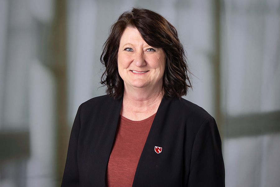
Lisa Bartenhagen is a Professor and Charles R. O'Malley Endowed Chair in the Department of Clinical, Diagnostic, & Therapeutic Sciences at the University of Nebraska Medical Center (UNMC). She serves as Program Director for the Radiation Therapy program and chairs the department, roles she has held since 2000. Her academic background includes a BS in Biology (University of Nebraska-Lincoln, 1990), BS in Radiography/Radiation Therapy (UNMC, 1993), and MS in Radiation Science Education (Midwestern State University, 2005). She is ARRT-certified in Radiography and Radiation Therapy. Her research focuses on educational methodologies in healthcare, including flipped-classroom models, e-learning platforms, and interprofessional collaboration. She also investigates patient education in oncology and occupational health risks for healthcare workers exposed to radiation. Notable work includes studies on redox dysregulation in radiation-exposed professionals and anxiety mitigation strategies for head and neck cancer patients. Bartenhagen's contributions to medical education reform include pioneering the use of virtual learning environments and 3D printing in radiation therapy training. Her programmatic leadership has emphasized quality improvement in oncology care and adherence to accreditation standards like ASTRO's APEx. She maintains active roles in UNMC's College of Allied Health Professions and collaborates with clinical and academic teams across the institution. Endowed Position: Charles R. O'Malley Endowed Chair Key Contributions: Developed Therapy Physics Education in a Virtual Learning Environment and Flipped-Classroom Medical Physics Course Labs/Teams: Oversees the Radiation Therapy Program and collaborates with the Department of Clinical, Diagnostic, & Therapeutic Sciences on interdisciplinary initiatives.
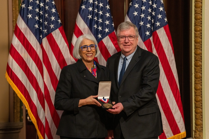
Eric R. Fossum is the John H. Krehbiel Sr. Professor for Emerging Technologies at the Thayer School of Engineering at Dartmouth College. He serves as Vice Provost for Entrepreneurship and Technology Transfer and Director of Dartmouth's PhD Innovation Program. As one of the world's leading experts in solid-state image sensors, he invented the CMOS active pixel sensor technology that revolutionized digital imaging in smartphones, medical devices, and automotive systems. His work has earned him numerous accolades, including the National Medal of Technology and Innovation (2025) and the Queen Elizabeth Prize (2017). His research interests focus on: Solid-state image sensors (CCDs, CMOS active pixel sensors, Quanta Image Sensors) Advanced imaging systems and on-chip processing New applications for image sensors in medicine, security, and space Dr. Fossum's recent publications demonstrate significant advancements in: Photon-counting sensors for low-light applications High-speed imaging for microscopy and radiography Backside-illuminated and sub-diffraction-limit pixel designs Quantum random number generation using sensor technology Infrared spectral extension of CMOS sensors His scientific awards include: National Medal of Technology and Innovation (2025) Queen Elizabeth Prize for Engineering (2017) IEEE Andrew S. Grove Award (2009) Induction into National Inventors Hall of Fame (2011) Emmy Award for Technology & Engineering (2021) Doctor of Science, Honoris Causa from Trinity College (2014) As an entrepreneurial leader, Dr. Fossum has: Co-founded Gigajot Technology with former PhD students Previously led Photobit and Siimpel Corporations Active participant in technology transfer initiatives at Dartmouth Founder and Past President of the International Image Sensor Society
Shadi Ebrahimian is affiliated with Yale School of Medicine's Radiology & Biomedical Imaging department. Their research focuses on AI-driven advancements in medical imaging, radiation safety protocols, and health equity in academic radiology. Key work includes developing AI algorithms for diagnostic quality control, optimizing CT radiation doses, and analyzing pulmonary embolism prognostics using CT features. Expertise: AI in Radiology, CT Imaging, Radiation Dosimetry, Medical Image Analysis Key Projects: Radiologist-trained AI models, national CT dose standards in Brazil, motion artifact detection Research interests span AI integration into clinical workflows, improving diagnostic accuracy through machine learning, and addressing disparities in academic medicine. Recent studies explore predictive analytics for kidney stone composition and pulmonary embolism severity using CT radiomics. Advising and grants: No formal students listed, but collaborates on multi-institutional studies across 4 continents. Active in global radiology initiatives addressing imaging quality and radiation safety in diverse clinical settings. Labs/Teams: Part of Yale Radiology & Biomedical Imaging's AI and medical imaging research groups, contributing to interdisciplinary teams developing clinical AI solutions without data scientists.