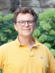
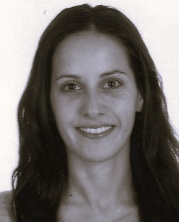
Eden Fussner-Dupas is an Assistant Professor in the Department of Biochemistry and Molecular Biology at the University of British Columbia's Faculty of Medicine. With a background in biochemistry education from the University of Toronto (BSc, MSc, PhD), her work bridges structural biology and nuclear organization research. Department: Biochemistry and Molecular Biology University: University of British Columbia Academic Rank: Assistant Professor Her research focuses on nuclear architecture and chromatin structure , employing advanced techniques like electron spectroscopic imaging to explore DNA-RNA-protein interactions in genome regulation. Recent publications highlight her contributions to understanding chromatin fiber density in pluripotency, heterochromatin reorganization during cell reprogramming, and phase transitions in membraneless organelles. Contact: Biological Sciences Building, Room 3119 | Phone: (604) 822-8260 | Email: eden.fussner@ubc.ca
Dr. Susanne Dunker is a Working Group Leader and Deputy Department Head at the Department for Physiological Diversity at the Helmholtz Centre for Environmental Research (UFZ) and the German Center for Integrative Biodiversity Research (iDiv) in Leipzig, Germany. Her research bridges aquatic and terrestrial ecosystems with a focus on biodiversity assessment using advanced imaging techniques. Dr. Dunker's research interests span water quality, phytoplankton diversity, cyanobacterial blooms, air quality, pollination, and pollen diversity. She has pioneered the use of multispectral imaging flow cytometry for environmental monitoring, enabling high-throughput analysis of phytoplankton communities and pollen diversity. Her work integrates machine learning with biological analysis to develop innovative monitoring strategies for both aquatic and aerial ecosystems, with applications in understanding biodiversity dynamics, ecosystem services, and human health impacts. Her publication record demonstrates a strong focus on developing and applying imaging flow cytometry techniques across multiple domains. The research shows a clear trajectory from aquatic systems (phytoplankton analysis) to terrestrial systems (pollen analysis), with increasing integration of machine learning and deep learning approaches. Her work has significant implications for environmental monitoring, biodiversity assessment, and understanding the impacts of environmental change on ecosystem functioning. UFZ Technology Transfer Award (2019) UFZ Sustainability Challenge Award (2022) Dr. Dunker leads multiple research projects including PollenNet, OrthoDiv, ProtistQuant, PlanktAI, and SedimentAI, demonstrating her leadership in interdisciplinary research. Her work on reducing plastic in laboratories shows her commitment to sustainable research practices. She serves as a reviewer for numerous high-impact journals and participates in important scientific commissions including the German VDI/DIN mirror committee on automated pollen monitoring. Her laboratory utilizes advanced imaging flow cytometry and deep learning approaches to analyze biodiversity across different ecosystems. The iCyt platform she co-leads provides integrative cytomics support for biodiversity research. Her team's work connects microscopic biological processes with ecosystem-level functions, creating a bridge between cellular traits and environmental monitoring applications.
Herbert Kruitbosch is a researcher specializing in machine learning and computer vision for biomedical applications, with active contributions to cell segmentation, gait analysis in chronic pain conditions, and medical image diagnostics. His work bridges computational methods and clinical research through interdisciplinary collaborations. Research interests center on Machine Learning, Computer Vision, and Medical Image Analysis, with specific expertise in annotation-free segmentation techniques for biological imaging and machine learning models for chronic low back pain assessment. His methodologies frequently leverage convolutional neural networks and synthetic data generation to address challenges in bioimage informatics and pain management. Recent publications reveal a strong trend toward applying transfer learning and deep neural networks to biomedical imaging problems, particularly in cell segmentation (2024) and yeast cell analysis (2022), alongside clinically focused studies on gait patterns in chronic pain patients (2021-2022). These works demonstrate consistent integration of computer vision techniques with medical diagnostics, emphasizing practical applications in healthcare settings.
Ali Pezeshki is a Professor at Colorado State University with joint appointments in the Department of Electrical and Computer Engineering and the Department of Mathematics . His research spans statistical signal processing , machine learning , optimization , and applied harmonic analysis , with applications to radar, bioimaging, and network systems. B.Sc. and M.Sc. in Electrical Engineering from University of Tehran (1999, 2001) Ph.D. in Electrical Engineering from Colorado State University (2004) He has served on the editorial board of IEEE Access (2012-2018) and received the IEEE SP Society Best Young Author Award (2011). His students include PhD candidates like Yifan Yang and alumni such as Pooria Pakrooh (Qualcomm) and Wenbing Dang (Argo AI). His recent publications focus on greedy optimization strategies , submodular functions , adaptive radar , and single-pixel imaging . Keywords across his work include Compressed Sensing , Network Learning , and Statistical Estimation .
Michael Unser is a Full Professor at EPFL’s School of Engineering and Academic Director of EPFL’s Center for Imaging in Lausanne, Switzerland. His research focuses on biomedical imaging, applied functional analysis, sampling theory, wavelets, splines, and computational bioimaging. Current Position: Full Professor, EPFL School of Engineering Academic Role: Director of EPFL Center for Imaging Key Research Areas: Inverse problems, sparse stochastic processes, machine learning for imaging Dr. Unser’s research bridges mathematical theory with practical biomedical imaging applications. He has pioneered the use of splines for image processing, developed advanced regularization frameworks for inverse problems, and contributed to computational bioimaging techniques like SMLM and optoacoustic tomography. His recent work explores connections between deep learning and classical kernel methods through variational formulations. He has published over 400 journal papers and authored the seminal book on sparse stochastic processes. His scientific contributions have been recognized with multiple awards including IEEE and EURASIP Technical Achievement Awards, five IEEE-SPS Best Paper Awards, and three consecutive ERC Advanced Grants (FUN-SP, GlobalBioIm, FunLearn). He has served on editorial boards for leading journals and founded the IEEE SPS Bio Imaging and Signal Processing Technical Committee.
Esther van t Veld is a Researcher and Teacher at the Centre for Cell Imaging (CCI) , part of the Faculty of Veterinary Medicine at Utrecht University . Her work focuses on fluorescence microscopy and advanced light microscopy techniques, including confocal microscopy, TIRF microscopy, live cell imaging, and super-resolution microscopy. She also serves as a Contact Person for Occupational Health & Safety and Environment . The CCI facility, where she is based, provides expertise in histology specimen analysis and high-end fluorescence microscopy, operating within a ML-II biosafety environment. Her role involves both research and education, supporting users through equipment access and technical guidance. Contact details: Androclus Building, Yalelaan 1, Room O.163, 3584 CL Utrecht, Netherlands. Phone: +31 30 253 7704.
François Lagugné-Labarthet is a Professor in the Department of Chemistry at Western University , where he has been since 2007. He holds a Canada Research Chair in Nanoscience (2008-2018) and serves as the Scientific Director of Western's Nanofabrication Facility . His research bridges Nanoscale Spectroscopy , Plasmonics , and Advanced Microscopy to explore materials and biomaterials at the nanoscale. Education: PhD in Physical Chemistry, Université Bordeaux (1998); Postdoc at Queen's University (2000-2004) His work focuses on Tip-Enhanced Raman Spectroscopy (TERS) , Surface-Enhanced Raman Spectroscopy (SERS) , and Nanofabrication to surpass the diffraction limit of light. Key projects involve 2D materials (e.g., MoS 2 , WS 2 ), ultrasensitive detection platforms , and single-cell chemical analysis . His lab designs plasmonic fractal structures for multi-resonant optical applications. Recent trends in his publications include TERS for amyloid studies , fractal plasmonics , and gold nanoraspberry SERS sensors . His group collaborates with industry partners like SCATR Inc. on street drug detection and applies machine learning to analyze nanoscale data. Scientific Awards: CNRS Bronze Medal (2004) Canada Research Chair in Nanoscience (2008-2018) He mentors graduate students in Nanochemistry and Physical Chemistry , including Zeiad Saad (MSc), Lorena Veliz (PhD), and Cédric Lambin (PhD). His lab trains students in Nanofabrication , Optical Microscopy , and Plasmonic Modelling .
Kevin Braeckmans is a Full Professor at Ghent University's Faculty of Pharmaceutical Sciences, Department of Pharmaceutics (FW01). He leads the Bio-Photonic Research Group , BIOMARKED , and Ghent Research Group on Nanomedicines , with a focus on photoporation and laser-induced nanobubble technologies. Research areas include Medical Nanotechnology , Nanophotonics , and Non-Viral Gene Delivery . His work spans Intracellular Delivery of macromolecules, Photoporation for immunotherapy, and Optical Molecular Microscopy methods. Over 15 doctoral researchers, including Amin Ahmad , Baihao Huang , and Chloë De Clercq , are under his supervision. Funding sources include the Special Research Fund , FWO , and European Framework Programmes . Key technologies developed involve Polydopamine Nanosensitizers , Photothermal Nanofibers , and STED Nanoscopy integration.
Dr. Lars Palsson is an Associate Professor in the Department of Chemistry at Durham University, specializing in chemical physics and biophysics. His research focuses on the application of optical spectroscopy and microscopy for investigations of biological systems and organic materials for display applications, molecular electronics, and sensing of the biological environment. His work is highly interdisciplinary, involving collaborations with colleagues in synthetic chemistry and cell biology. His primary research interests include chemical physics and biophysics with emphasis on optical spectroscopy. Palsson investigates how the dielectric medium modulates excited states using time-resolved emission techniques, develops luminescence imaging methods for bio-active materials, and pursues circularly polarized luminescence spectroscopy for studying biological systems and molecular recognition. His work spans from fundamental photophysical studies to practical applications in sensing and bioimaging. Analysis of his recent publications reveals a strong focus on time-resolved fluorescence techniques, luminescent probes for biological applications, and molecular recognition. His research consistently bridges chemistry, physics, and biology, with particular emphasis on developing novel optical sensing methodologies and understanding excited state dynamics in complex environments. The publications demonstrate expertise in both fundamental photophysics and practical applications in bioimaging and sensing. Palsson's research involves significant interdisciplinary collaboration, particularly with colleagues in synthetic chemistry and cell biology. His work often involves developing novel optical techniques for biological applications and creating new luminescent probes for cellular imaging and sensing. The research has practical implications for developing new sensing technologies and understanding fundamental biological processes at the molecular level.
Hervé Waxin is a researcher at the Pasteur Institute in Paris, France, where he is affiliated with the Image Analysis Hub (UTechS PBI) within the Computer Science Resource and Research Center. He contributes to various research initiatives focused on bioimaging and microscopy technologies. Dr. Waxin's research interests span multiple areas of molecular biology and bioimaging, with a particular focus on: Bioimaging techniques and applications Molecular biology of cellular processes Microscopy technology development Genomics and epigenetics His work demonstrates expertise in applying advanced imaging techniques to biological research questions. Dr. Waxin has published research on yeast genetics and epigenetic mechanisms, showing his interdisciplinary approach bridging imaging technology with fundamental biological research. Dr. Waxin is actively involved in educational initiatives at the Pasteur Institute, contributing to multiple specialized courses including: Pasteur Course: Creation and Management of Databases Pasteur Course Vaccinology Pasteur Course Fundamental Immunology Pasteur Course Emerging Viruses Development and Plasticity of the Nervous System His collaborative work spans multiple research teams at the Pasteur Institute, demonstrating his integration within the institute's research ecosystem.
Professor Rudolf Allemann serves as Pro Vice-Chancellor, International at Cardiff University, with responsibility for the institution's international activities, partnerships, and student recruitment. He also provides overall leadership and management for the College of Physical Sciences and Engineering, setting strategic direction and overseeing academic performance and resource allocation. Professor Allemann's research focuses on enzyme catalysis mechanisms, particularly in terpene synthases and dihydrofolate reductase systems. His work integrates protein engineering, biocatalysis, and molecular dynamics to understand and manipulate enzyme function. Recent research explores water capture mechanisms in sesquiterpene synthases, photocontrol of protein function, and chemoenzymatic approaches to natural product synthesis. Analysis of his recent publications (2020-2025) reveals a strong emphasis on enzyme engineering for biocatalytic applications, with particular focus on terpene synthases and their catalytic mechanisms. His work combines computational modeling, protein engineering, and advanced kinetic analysis to unravel the molecular choreography of enzyme catalysis. The research has significant implications for synthetic biology, natural product synthesis, and understanding fundamental principles of enzyme function. Professor Allemann has made substantial contributions to understanding dynamic effects in enzyme catalysis, particularly in dihydrofolate reductase systems, and has pioneered approaches to enzyme engineering for biocatalytic production of terpenes and other natural products. His work spans multiple disciplines including biochemistry, chemical biology, and synthetic biology. As Pro Vice-Chancellor, International, Professor Allemann leads Cardiff University's global engagement strategy while maintaining an active research program. His leadership extends to strategic direction of the College of Physical Sciences and Engineering, where he oversees academic performance and resource allocation across multiple departments.
Dr. Angelo Amoroso serves as Senior Lecturer in Inorganic Chemistry and Director of Learning and Teaching at Cardiff University's School of Chemistry. With a career spanning over two decades since his 1998 appointment, he maintains an active research program while overseeing educational initiatives within the department. His dual role combines significant administrative responsibilities with continued scholarly contributions to the field of coordination chemistry. Dr. Amoroso earned his PhD from the University of Cambridge in 1992, working under B.F.G. Johnson and J. Lewis on high nuclearity osmium carbonyl clusters. His postdoctoral career included positions at the University of Bristol (1992-1995) with J.A. McCleverty and M.D. Ward studying Mo(NO) species, the University of Utah (1995-1997) with J.A. Gladysz investigating Re(NO) species, and the University of Nottingham (1997-1998) with M. Schröder modeling Ni/Fe hydrogenases. His research program focuses on developing novel imaging agents for stem cell implant tracking, with particular emphasis on ligand design for enhanced stability, bio-orthogonal reactivity, and tailored photophysical/electrochemical properties. The work bridges fundamental inorganic chemistry with practical medical applications, creating tools for visualizing transplanted cells directly in vivo. This research has evolved from basic coordination chemistry to increasingly sophisticated imaging applications, with recent work emphasizing dual-modal imaging capabilities. Analysis of his publication record reveals consistent focus on transition metal and lanthanide complexes for imaging applications, with growing emphasis on practical biomedical implementation. The research demonstrates strong interdisciplinary connections between coordination chemistry, molecular imaging, and regenerative medicine, particularly in developing tools for monitoring tissue repair processes. As Director of Learning and Teaching, Dr. Amoroso shapes the educational experience for chemistry students through courses including CH5202 (Reactivity And Properties Of The Elements And Their Compounds), CH2301 (Training in Research methods), CH4302 (Advanced Organometallic and Coordination Chemistry), and CH3403 (Bio-imaging Applications of Coordination Chemistry), effectively connecting theoretical principles with cutting-edge research applications.
Markus D. Herrmann is an Assistant Professor of Pathology at Harvard Medical School and serves as Director of Computational Pathology and Assistant Computational Pathologist at Massachusetts General Hospital (MGH). His lab operates from the Computational Pathology unit at MGH's Longfellow Building in Boston, MA. His research transforms qualitative histopathology into quantitative science using microscopy imaging, proteomics, and machine learning to develop image-based clinical diagnostics. Key interests include cancer biomarker discovery, computational diagnostic tests, and analyzing cellular interactions during tumorigenesis and metastasis through spatial molecular feature extraction. His 2014-2020 publications reveal consistent focus on computational pathology infrastructure (DICOM standards), multiplexed protein mapping, and AI-driven image analysis across oncology. Work spans quantitative cancer imaging informatics, digital pathology standardization, and high-content cell/transcriptome profiling, establishing him as a leader in translating computational methods to clinical pathology. As faculty, he mentors trainees within Harvard's ecosystem though specific students aren't listed. His research aligns with MGH Pathology's $19 million annual research portfolio, indicating substantial institutional support for computational pathology initiatives. He leads the Computational Pathology lab collaborating across MGH, Harvard, and clinical networks to develop multimodal imaging methods (immunofluorescence, electron microscopy, OCT) and clinical validation frameworks for AI-based diagnostic tools, emphasizing real-world implementation and performance monitoring.
Dr. Daniel Langenkämper is a researcher at the University of Bielefeld, affiliated with the Faculty of Engineering and the Center for Biotechnology (CeBiTec). He serves as a key member of the Biodata Mining Group, where he develops and applies advanced computational methods for marine biological data analysis. His office is located at UHG V10-107 with contact number +49 521 106-3678. Langenkämper's research focuses on the intersection of computer science and marine biology, with particular expertise in: Computer vision applications for marine ecosystem monitoring Deep learning approaches for diatom and coral classification Biodata mining from complex marine imagery Digital platform development for environmental monitoring systems Multi-sensor data analysis for marine infrastructure assessment His publication record shows consistent output through 2025, with recent work emphasizing expert-computer vision integration for coral status exploration and self-supervised learning techniques for diatom classification. The research demonstrates strong interdisciplinary collaboration across computer science, marine biology, and engineering disciplines, addressing critical challenges in marine environmental monitoring and infrastructure maintenance. His work contributes significantly to both theoretical advancements in image analysis and practical applications for marine conservation and industrial monitoring. Langenkämper actively participates in marine imaging workshops and contributes to the development of standardized image datasets for marine research. His work with the Biodata Mining Group at CeBiTec supports multiple research initiatives focused on transforming visual data into actionable ecological insights, particularly for deep-sea coral ecosystems and marine infrastructure maintenance.
Elda Fischi-Gómez is a Research Staff Scientist at the Center for Biomedical Imaging (CIBM), affiliated with EPFL and CHUV. She holds a BSc and MSc in Telecommunication Engineering from Polytechnic University of Catalonia (UPC, Spain) and a PhD in Electrical Engineering from EPFL (2015). Her research focuses on advancing MRI techniques by integrating software algorithms, MR physics/hardware, and neuroscience to improve understanding of brain development and disease. She has received notable grants including the SNFS Early-Postdoc Fellowship [SNF-P2ELP2_172286] and the EPFL-ETH Strategic Focal Area grant [PHRT-2018-425] for microstructure imaging in multiple sclerosis. Her work combines high-resolution dMRI acquisitions with advanced microstructural models and machine learning. As a member of the IEEE BioImaging and Signal Processing Technical Committee (BISP-TC), she chairs the Communications and Outreach subcommittee. She has served as a reviewer for ISBI, ISMRM, and conferences like ICASSP and ICIP, as well as high-impact journals such as Medical Image Analysis and Neuroimage: Clinical.