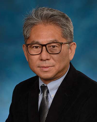
Dr. Shaojun Du is a Professor in the Department of Biochemistry and Molecular Biology at the University of Maryland School of Medicine and a faculty member at the Institute of Marine and Environmental Technology, University of Maryland. His research focuses on genetic and epigenetic mechanisms regulating muscle development, growth, and repair using zebrafish models. Education: PhD in Biochemistry and Molecular Biology (University of Toronto, 1993), Postdoctoral Fellowship in Genetics (Howard Hughes Medical Institute, University of Washington, 1993–1997) Dr. Du’s work explores the roles of molecular chaperones (e.g., Hsp90α1), histone modifiers (e.g., SmyD1), and fusogenic proteins (Myomaker/Myomixer) in myofibril assembly, sarcomere organization, and myoblast fusion. His lab employs Tol2 transposon, TALEN, and CRISPR technologies to generate transgenic and mutant zebrafish models. The scientific awards section is currently empty, but his research has been funded by the NIH (Grant 1R01AR072703-01A1, 2018–2025). His publications highlight breakthroughs in muscle cell identity specification via Hedgehog signaling and the critical role of post-translational modifications in protein stability. Dr. Du’s lab has developed over 40 zebrafish models to study muscle structural proteins, transcription factors, and microRNA networks, contributing to understanding neuromuscular diseases and advancing aquaculture applications.
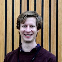
Dr. Oliver Plümper is an active researcher at Utrecht University's Faculty of Geosciences, specifically within the Earth Sciences department and the Structural Geology & Electron Microscopy group. His work spans multiple disciplines at the intersection of geology, chemistry, physics, and materials science, with a particular focus on understanding processes occurring at the nanoscale that influence large-scale geological phenomena. Plümper's research interests center around fluid-rock/mineral interaction, nano(geo)sciences, mineral physics, and rock deformation. He employs a multi-faceted approach that combines natural observations, experimental techniques, micro and nano-analytics, and numerical modeling to address fundamental questions in Earth sciences. His work particularly emphasizes how nanoscale processes in the Earth's interior can influence large-scale geological structures and phenomena, including mountain building, earthquake generation, and the carbon cycle. His current research portfolio includes several major projects: the ERC Starting grant 'nanoEARTH' investigating mineral-water interactions deep within the Earth; the Dutch Research Council Vidi project 'RELEASE' studying carbon cycling in subduction zones; the EU INFRAIA project 'EXCITE NETWORK' as co-Principal Investigator supporting access to advanced imaging facilities; the UIO-UU project 'serpAI' using artificial intelligence to study mantle rock alteration; and the UU-NIOZ project 'I-NANO' examining iron nanoparticles' effects on ocean biogeochemistry. These projects collectively demonstrate his focus on understanding how nanoscale processes influence planetary-scale phenomena. Plümper is a strong advocate for open science and transdisciplinary research, leading a diverse team of scientists from earth sciences, chemical engineering, mathematics, physics, and chemistry. He actively contributes to the Utrecht University Electron Microscopy Center and is involved in multiple initiatives promoting open access to analytical facilities. His collaborative approach is evident in his extensive publication record across high-impact journals including Nature Geoscience, PNAS, and Geology, with research spanning from fundamental mineral physics to applications in geothermal energy, raw material extraction, and carbon storage.
Thomas Maresca is an Associate Professor in the Biology Department at the University of Massachusetts Amherst, serving as Graduate Program Director for Molecular & Cellular Biology within the Interdisciplinary Doctoral Graduate Program (IDGP). His research laboratory investigates fundamental mechanisms of cell division with institutional affiliation to the university's life sciences division. His academic training includes: Ph.D. in Cell Biology from the University of California at Berkeley (2005) B.S. in Biology from the University of North Carolina at Chapel Hill (1999) Dr. Maresca's research centers on chromosome segregation mechanics during mitosis, employing Drosophila and Xenopus model systems with advanced live-cell imaging techniques. His laboratory specializes in quantifying kinetochore-microtubule interactions , error correction mechanisms , and force generation during cell division. Key approaches include FLIM-FRET biosensors, single-molecule tracking, and micromanipulation in egg extracts to dissect how intrinsically disordered proteins, motor complexes (particularly dynein), and kinase networks (Aurora A/B, MPS1) ensure mitotic fidelity. Current work focuses on spatiotemporal regulation of phosphorylation gradients and mechanical feedback at the kinetochore-microtubule interface. Analysis of his 15 most recent publications reveals a consistent trajectory in mitotic mechanics with increasing emphasis on intrinsically disordered proteins as mechanical regulators and subcellular signaling hubs . His work bridges molecular biophysics with cellular function, demonstrating how microtubule plus-ends act as physical signaling platforms and how kinase crosstalk establishes precise spatiotemporal control during anaphase. Recent methodological innovations include commercial FLIM-FRET adaptation and quantitative force measurements at kinetochores. As Graduate Program Director, Dr. Maresca oversees curriculum development and student mentorship for the Molecular & Cellular Biology track. His laboratory (located in Morrill Science Center IV, Room 436) maintains active research programs in kinetochore mechanics and mitotic regulation, supported by continuous publication output since 2000. Collaborative networks include structural biologists and biophysicists investigating mechanochemical transduction in division processes.
Prof. Wolfgang Rheinheimer is a Professor and Institute Director at the Institute for Ceramic Materials and Technologies , University of Stuttgart, since 2023. Previously, he held academic positions at RWTH Aachen (2022-2023), Forschungszentrum Jülich (Emmy Noether Group Leader, 2020-2022), TU Darmstadt (2020), and Karlsruhe Institute of Technology (2010-2017). Academic Rank: Full Professor Research Focus: Advanced sintering technologies, grain boundary engineering, defect chemistry, conductivity in ceramics, and microstructure evolution His research spans experimental and computational approaches to ceramic materials, with emphasis on field-assisted processing (electric/magnetic), grain boundary properties , and solid-state electrolytes . He has pioneered studies on flash sintering , cold sintering , and blacklight sintering mechanisms. Scientific Awards : Emmy Noether Fellowship (2020-2022) for establishing his independent research group His work integrates phase-field modeling with experimental characterization to optimize ceramic properties for energy applications (solid-state batteries, fuel cells) and structural uses. Collaborations include Robert Bosch GmbH and Purdue University (2018-2019 Visiting Professorship).
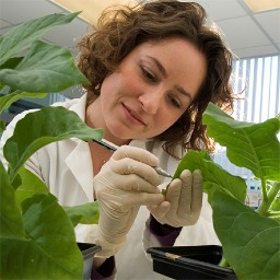
Verena Kriechbaumer is a Senior Lecturer in Biotechnology and Plant Sciences at the School of Biological and Medical Sciences, Oxford Brookes University . She is Deputy Director of the Oxford Brookes University Centre for Bioimaging and a leading expert in plant endoplasmic reticulum (ER) structure, membrane proteins, and auxin biosynthesis, utilizing biochemical techniques, high-resolution live cell imaging, and interdisciplinary approaches. Research Focus: Plant cell biology, ER architecture, auxin metabolic pathways, protein-membrane interactions, bioinformatics, and translational projects such as engineering plants to convert methane into biofuel. Key Techniques: FRET-FLIM, light sheet microscopy, single-particle tracking, and optogenetics. Publication Trends: Recent studies emphasize ER-membrane contact sites, organelle interaction networks, and the role of reticulons in viral trafficking and methane monooxygenase expression. Collaborative work spans physics, bioenergy, and industrial biotechnology. Scientific Awards: Fellowship from Korean Federation of Science and Technology Societies (2013) Santander Travel Fellowship (2018) Oxford Brookes Research Excellence Award (2020-21) Grants: Leverhulme Trust grant for "pMMO in plants" (2015-2017), STFC Harwell facility grants (2017-2021), BBSRC funding (2021-2026), and industry collaborations with Porton Biopharma Ltd. Labs & Teams: Leads the Endomembrane Structure and Function Group , collaborates with physicists at STFC Harwell Campus, and contributes to European Commission-funded projects like "Advanced Training for Next Generation Scientists in Spatio-Temporal Imaging."
Dr. Emily Gibson is an Associate Professor in the Department of Bioengineering at the University of Colorado School of Medicine. She holds a PhD from the University of Colorado Boulder (2004) and a BS from the Colorado School of Mines (1997). Her multidisciplinary research focuses on developing advanced optical technologies for neuroscience applications. Her primary research interests include: Development of implantable miniature microscopes for two-photon brain imaging in freely behaving animals Superresolution STED microscopy for subcellular imaging of protein dynamics Optical interfaces for neural modulation and sensing in central and peripheral nervous systems Applications in brain mapping, neural circuit analysis, and bioelectronic medicine Dr. Gibson's recent publications demonstrate a strong focus on neurophotonic tool development, including miniature microscopes, fiber-optic imaging systems, and superresolution techniques. Her work consistently applies these technologies to study neural coding, learning mechanisms, and neurodegenerative processes. She leads the Biophotonics Lab at CU Anschutz, which actively develops open-source neurophotonic tools. Current projects include BRAIN Initiative-funded work on voltage imaging and NSF-supported research on odor navigation. Her lab maintains active collaborations with neuroscientists and clinicians to translate optical technologies into neuroscience research and clinical applications.
Dr David Collins serves as the Mike Ashby Associate Professor in Materials Science at the Department of Materials Science and Metallurgy, University of Cambridge, and is a core member of the Rolls-Royce University Technology Centre (UTC) focused on advanced aerospace materials. His research spans superalloys , titanium alloys , and high-entropy alloys , with expertise in phase transformations , high-temperature deformation , and microstructural evolution . He pioneers advanced characterization techniques including in-situ synchrotron X-ray diffraction , electron backscatter diffraction (EBSD) , and three-dimensional X-ray diffraction (3DXRD) to investigate grain-scale stress interactions and failure mechanisms under extreme conditions. Analysis of his 2023-2025 publications reveals dominant trends in additive manufacturing validation , oxidation/corrosion resistance of superalloys , and grain-resolved mechanical behavior using coupled experimental-computational approaches. His work frequently addresses Rolls-Royce-relevant challenges in jet engine materials, particularly nickel-based superalloy performance under cyclic thermal-mechanical loading. No specific scientific awards for Dr Collins are mentioned in available departmental communications, though the Rolls-Royce UTC recently celebrated prizes for other researchers. Details regarding student supervision and grant funding are not publicly specified, but his position within the EPSRC- and Rolls-Royce-funded UTC indicates active involvement in large-scale collaborative research projects. As part of the Rolls-Royce UTC infrastructure, Dr Collins utilizes specialized facilities including electro-thermal mechanical testing rigs, cyclic oxidation test systems, and synchrotron beamline partnerships for real-time microstructural analysis during deformation and phase transformations.
Professor Kenneth Harris is a Distinguished Research Professor in the School of Chemistry at Cardiff University, specializing in the fundamental properties of solids and the development of advanced experimental techniques for materials characterization. His work bridges the gap between traditional crystallography and modern analytical methodologies, with a particular focus on overcoming limitations in structural analysis of complex materials. His research spans three primary interconnected themes: the development of techniques for determining crystal structures of organic solids directly from powder X-ray diffraction data; the advancement of in-situ solid-state NMR strategies for monitoring crystallization processes in real time; and the investigation of structural properties of anisotropic materials using polarized X-ray beam techniques, including the pioneering development of X-ray Birefringence Imaging (XBI). This work has significant implications for pharmaceutical development, materials science, and understanding biological crystallization processes. Analysis of his recent publications reveals a consistent trajectory toward increasingly sophisticated multi-technique approaches to materials characterization. His work increasingly integrates 3D electron diffraction, powder XRD, solid-state NMR, and computational methods like DFT calculations to solve previously intractable structural problems. A notable trend is the application of these methods to biologically relevant molecules (xanthine, riboflavin, L-tyrosine) and the development of techniques to monitor dynamic processes like crystallization and phase transitions in real time. His research demonstrates a shift from purely structural determination toward understanding the dynamic processes that govern material formation and transformation. Distinguished Research Professor title at Cardiff University Key contributor to the development of X-ray Birefringence Imaging Significant contributions to NMR crystallography methodologies Extensive publication record in top chemistry and materials science journals Professor Harris leads a research group focused on developing and applying cutting-edge techniques for materials characterization. His work has significant implications for pharmaceutical development, where understanding crystal structure and polymorphism is critical for drug efficacy and safety. His group has developed innovative approaches to monitor crystallization processes in real time, which has applications in both industrial manufacturing and understanding natural biomineralization processes. The group maintains strong collaborations with researchers across multiple disciplines, including physics, biology, and engineering, reflecting the interdisciplinary nature of modern materials science research.
Prof. Dr. Andreas Reiner is a faculty member in the Department of Cellular Neurobiology at the Faculty of Biology and Biotechnology, Ruhr University Bochum. His research focuses on glutamate receptor signaling, synaptic plasticity, and the development of optical techniques for studying receptor dynamics in the central nervous system. University: Ruhr University Bochum School: Faculty of Biology and Biotechnology Department: Cellular Neurobiology Email: andreas.reiner@ruhr-uni-bochum.de His work emphasizes the use of chemical photoswitches (photoswitchable ligands) for light-based activation/inhibition of ionotropic (iGluRs) and metabotropic (mGluRs) glutamate receptors, enabling precise optogenetic and pharmacological studies. Research also explores receptor desensitization, subunit occupancy, and structural diversity. Recent publications highlight advancements in photoswitchable tools (2023), structural analysis of kainate receptors (2021), and subunit-selective antagonists for NMDA receptors (2020). Earlier work (2013-2016) established foundational techniques for optogenetic control of glutamate receptors.
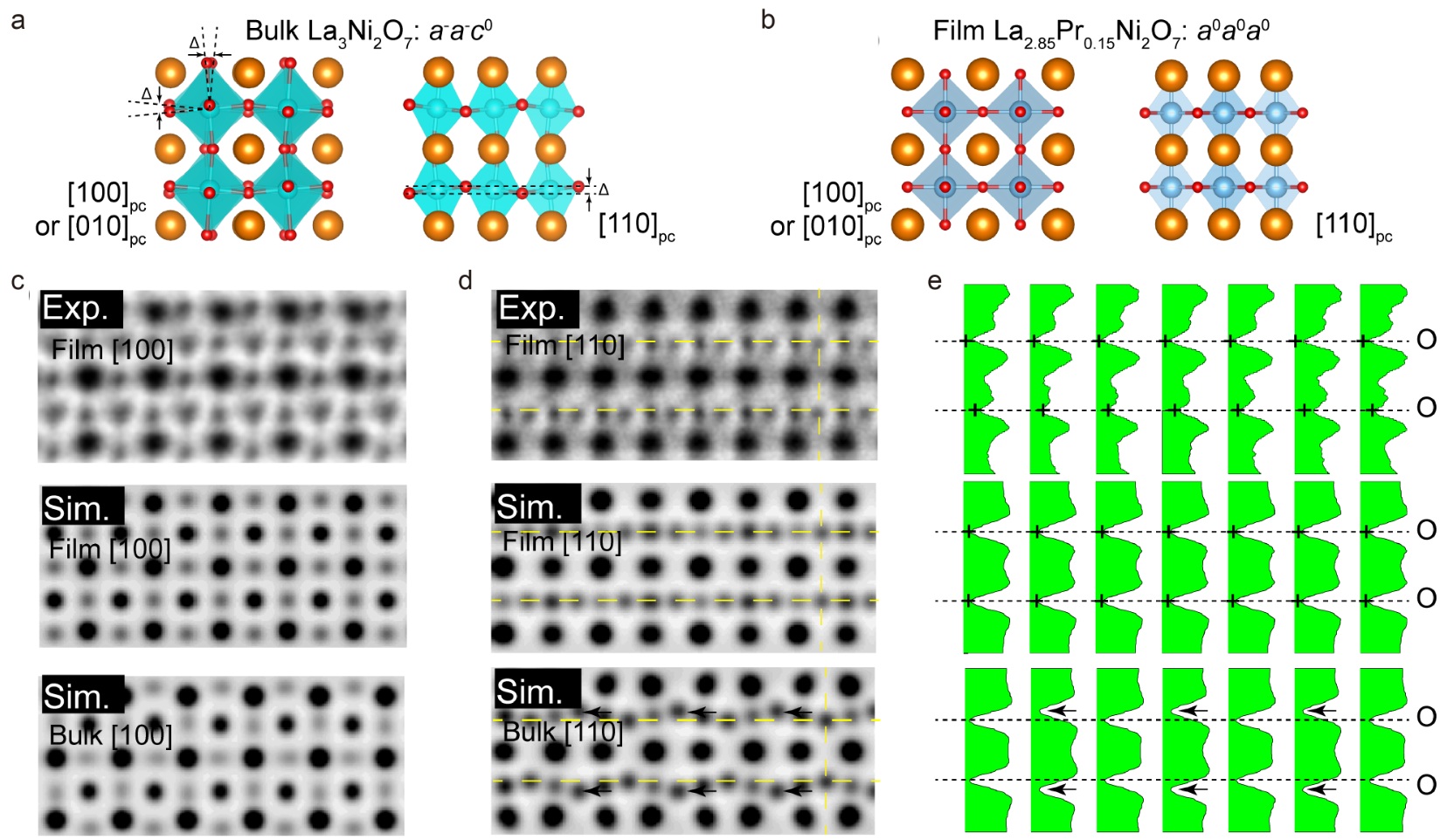
Prof. Junhao Lin is a tenured Full Professor at the Department of Physics, Southern University of Science and Technology (SUSTech). With a dual background in experimental and theoretical physics, he specializes in atomic-scale characterization of 2D materials, combining aberration-corrected STEM, EELS spectroscopy, and density functional theory calculations to study defect-structure-property relationships. Ph.D. (2015) from Vanderbilt University Postdoctoral work at National Advanced Institute of Science and Technology (Japan) under Kazu Suenaga Research Interests : Atomic-scale defect engineering in 2D materials In-situ high-resolution electron microscopy under external stimuli High-energy-resolution EELS for optical property characterization Quantum material structure-property correlations Publication Trends : Over 130 high-impact publications (Nature, Science, PRL) focusing on 2D material defects, phase transitions, and atomic engineering. Recent works explore nickelate superconductivity, magnetic phenomena in topological materials, and energy storage applications. Scientific Recognition : MIT Technology Review TR35 China (2021, Visionary category) Elsevier 2024 Highly Cited Researcher (China) Multiple international conference awards 8+ million RMB investment in glove-box interconnected lab systems Lab Overview : The Advanced Low-Dimensional Materials Laboratory at SUSTech (10+ members) integrates atomic-resolution STEM, in-situ TEM, and computational modeling. Collaborations span Oak Ridge National Lab, Peking University, and Singaporean/Japanese institutions.
Dr. Robert J. Usselman serves as an Assistant Professor of Chemistry in the College of Engineering and Science at Florida Institute of Technology , where he also acts as Deputy Director of Computational Research At Florida Tech (CRAFT) . His research bridges quantum physics and cellular biology, focusing on magnetic resonance, biophotonics, and redox biochemistry. Specializes in quantum biological clocks and ROS production mechanisms Leads a multidisciplinary lab with active undergraduate/graduate students Develops computational infrastructure for AI and quantum research Research interests include: Quantum-Classical Interface in Biological Systems Magnetic Field Effects on Cellular Redox Balance Autofluorescence Lifetime Microscopy ROS Dynamics in Cellular Metabolism Recent publications highlight quantum biological mechanisms in ROS production, magnetic field modulation of cellular processes, and multiphoton imaging techniques. His work integrates theoretical quantum biology with practical applications in bioenergetics and disease models. Dr. Usselman actively mentors students in biotechnology, biomedical engineering, and physics, while advancing collaborations in AI-driven computational research through CRAFT. His lab combines experimental and computational approaches to study redox signaling and quantum sensing in biological systems.
Mehmet Yaran is an Associate Professor in the Department of Biology at Gaziantep University's Faculty of Arts and Sciences. He has held this position since 2021, following previous roles as a Doctor Lecturer at the same university's Faculty of Arts and Sciences Department of Biology (2016-2021) and at the Islahiye Vocational School in various departments. Dr. Yaran completed his Doctorate in Biology at Gaziantep University (2009-2014), followed by a Master's degree in Biology from the same institution (2007-2009). His undergraduate education was in Biology Teaching at İnönü University's Faculty of Education, Department of Mathematics and Science Education (2002-2007). Dr. Yaran's research primarily focuses on entomology, with special emphasis on fruit flies (Diptera: Tephritidae) taxonomy, systematics, and morphology. His work extensively examines spermathecae morphology using scanning electron microscopy, wing shape analysis through geometric morphometrics, and regional fauna studies across various Turkish provinces. He has made significant contributions to documenting new species and records of Diptera in Turkey, particularly within the Tephritidae family, but also extending to other families like Ulidiidae and Pallopteridae. His research bridges traditional taxonomic methods with modern morphometric techniques to advance understanding of insect biodiversity and systematics in Turkey. Dr. Yaran's recent publications demonstrate a consistent focus on documenting and analyzing insect biodiversity in Turkey, with particular attention to fruit flies and related taxa. His work spans faunal surveys across multiple Turkish provinces, taxonomic descriptions of new species, morphological analyses using advanced techniques like geometric morphometrics and electron microscopy, and contributions to regional biodiversity databases. The pattern of his research shows increasing sophistication in analytical methods while maintaining a strong foundation in traditional taxonomic approaches. 2016 Public Publication Promotion from TUBITAK 2015 Public Publication Promotion from TUBITAK 2012 Public Publication Promotion from TUBITAK Dr. Yaran has supervised numerous graduate students, guiding research on various aspects of entomology including Pieridae fauna, fruit fly systematics, and flower fly taxonomy. His administrative roles have included serving as Head of Department (2016-2018), Vice Dean (2021), and Deputy Head of Department (2021). He has led and participated in multiple research projects funded by higher education institutions and other public organizations, focusing on entomological surveys and biodiversity documentation across various regions of Turkey. Dr. Yaran's work is closely associated with entomological research teams at Gaziantep University, collaborating extensively with colleagues like Murat Kütük, Vedat Görmez, and Mürşit Ömür Koyuncu on various projects documenting Turkish insect fauna. His research contributes significantly to the understanding of Turkey's entomological biodiversity, particularly in understudied regions.
Ruth E. Blake is a Professor of Earth & Planetary Sciences at Yale University, where she leads a research program focused on stable isotope geochemistry, geomicrobiology, and biogeochemical cycling of elements in diverse environments. Her work spans from deep-sea hydrothermal systems to volcanic hot springs and has significant implications for understanding Earth's history and potential extraterrestrial life. Dr. Blake's research interests center on stable isotope techniques, particularly oxygen isotopes in phosphate, to elucidate biogeochemical cycling of phosphorus, sulfur, carbon, and iron across geological time. Her laboratory conducts experimental geochemistry investigating phosphate-iron oxide interactions, nanogeochemistry studying carbon nanotubes and iron nanoparticles, and geobiology examining microbial communities in extreme environments like St. Lucia's volcanic hot springs. She has developed innovative methods for oxygen isotope ratio analysis of phosphate that are now widely used in the field. Analysis of her recent publications reveals a consistent focus on phosphate oxygen isotope applications across diverse systems - from deep biosphere phosphorus cycling to photochemical degradation mechanisms of organophosphorus compounds. Her work bridges geochemistry, microbiology, and analytical chemistry, with significant contributions to understanding phosphorus cycling in marine sediments, soil systems, and extreme environments. Dr. Blake actively mentors students and postdocs, with several researchers listed as student advisees (*) and postdoctoral advisees (†) in her publications. She has participated in major research initiatives including the Center for Dark Energy Biosphere Investigations (C-DEBI) and has sailed on Ocean Drilling Program Leg 201. Her outreach work with Amistad America includes creating educational programs engaging underrepresented high school students in marine biogeochemistry research. Her laboratory is equipped for analytical geochemistry and microbiological research, including molecular biology facilities, microscopy equipment, and access to stable isotope ratio mass spectrometers through the Earth System Center for Stable Isotopic Studies. Dr. Blake's interdisciplinary approach combines field studies, laboratory experiments, and advanced analytical techniques to address fundamental questions about biogeochemical cycles across Earth's history.
Professor Wei Wei is a distinguished researcher in the Department of Neurobiology at the University of Chicago, where he leads a lab focused on understanding the synaptic basis of neural computation in the retina. His work bridges cellular and systems neuroscience, with particular emphasis on how retinal circuits process visual information to detect motion and other visual features. Institution: University of Chicago Department: Neurobiology Research Focus: Retinal circuitry and visual processing Current Funding: Multiple NIH R01 grants Dr. Wei received his PhD in Neurobiology from Cold Spring Harbor Laboratory in 2008, followed by postdoctoral training at the University of California, Berkeley from 2008-2011. His educational background provided him with a strong foundation in both molecular/cellular neuroscience and systems-level approaches to neural circuit function. Wei's research primarily investigates how neural circuits in the retina are assembled to perform specific computations, with particular focus on motion detection. His lab leverages genetic tools that label specific retinal neuron types to target synapses of interest, using multiphoton microscopy, visual stimulation, electrophysiology, and molecular biology to characterize synaptic maturation and function. A key area of investigation involves starburst amacrine cells and their role in direction selectivity. His work has revealed how dendritic computations, synaptic plasticity, and circuit organization contribute to visual processing. Analysis of Wei's recent publications shows a clear progression from fundamental circuit mechanisms toward understanding more complex visual processing in naturalistic contexts. His work spans from cellular and synaptic physiology to systems-level circuit function, with a consistent focus on motion detection mechanisms. The research increasingly incorporates advanced imaging techniques and computational approaches to understand how retinal circuits transform visual inputs into neural representations. 2012 Whitehall Foundation Grant 2013 Sloan Research Fellowship 2013 E. Matilda Ziegler Foundation Grant 2014 Karl Kirchgessner Foundation Grant 2016 McKnight Scholar Award As Principal Investigator on multiple NIH R01 grants totaling millions of dollars, Wei directs a well-funded research program investigating the synaptic basis of motion detection in the retina. His current projects explore dynamic interactions between synaptic and intrinsic properties of starburst amacrine cells, circuit mechanisms for encoding naturalistic motion, and the developmental basis of motion detection circuits. The lab maintains strong collaborations with other researchers in visual neuroscience, as evidenced by co-authorship with scientists at multiple institutions. The Wei Lab operates within the University of Chicago's Department of Neurobiology, utilizing state-of-the-art facilities for retinal imaging and electrophysiology. The lab employs a multidisciplinary approach that combines genetic, optical, electrophysiological, and computational techniques to dissect retinal circuit function. Current research directions include investigating how short-term plasticity shapes circuit function, how dendritic computations contribute to visual processing, and how retinal circuits adapt to changing visual environments.
Sarah Butcher is a Professor of Microbiology at the University of Helsinki, affiliated with the Faculty of Biological and Environmental Sciences and the Molecular and Integrative Biosciences Research Programme. She serves as a supervisor for doctoral programmes in Integrative Life Science and Microbiology and Biotechnology. Her research focuses on structural virology, particularly using electron microscopy to study virus structure, assembly, and host interactions. She leads the Cryo-Electron Microscopy Facility and coordinates the FINStruct Structural Biology Finland infrastructure. Her research interests span structural virology with emphasis on picornaviruses , coronaviruses , and flaviviruses . She applies advanced techniques including cryo-electron microscopy , X-ray crystallography , and image processing to understand viral architecture, host-pathogen interactions, and mechanisms of viral entry and assembly. Her work has significant implications for antiviral drug development and vaccine design. Her recent publications reveal a strong focus on viral structure-function relationships, particularly in tick-borne encephalitis virus, coxsackieviruses, and SARS-CoV-2. Her research bridges structural biology with virology, producing insights into viral maturation, membrane interactions, and antiviral strategies. She has been instrumental in developing community standards for cryo-EM data validation and archiving. Alfred Kordelin Prize (2017) Maikki Friberg award (2013) Professor Butcher has supervised numerous doctoral and master's students and serves on multiple doctoral thesis committees. Her research has been supported by major competitive funding including Academy of Finland grants, Biocenter Finland, Sigrid Juselius Foundation, and European Commission projects. She currently leads the FINStruct infrastructure and the Instruct-ERIC Centre Finland. She directs the Cryo-Electron Microscopy Facility at the Institute of Biotechnology and coordinates the FINStruct Structural Biology Finland infrastructure, which provides state-of-the-art structural biology resources to researchers across Finland. Her team specializes in advanced cryo-EM techniques for studying complex biological structures, particularly viral particles and membrane proteins.