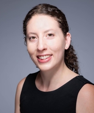
Lu Wei is an Assistant Professor of Chemistry at the California Institute of Technology and an Investigator at the Heritage Medical Research Institute. She holds a B.S. from Nanjing University (2010) and a Ph.D. from Columbia University (2015), joining Caltech in 2018. Research Areas: Optical spectroscopy, Biophysics, Bio-imaging, Chemical probe development Education: B.S. Nanjing University (2010), Ph.D. Columbia (2015) Research Interests include next-generation optical imaging techniques based on nonlinear vibrational spectroscopy for live-cell dynamics. Her work spans super-resolution label-free imaging , quantitative polyQ aggregate analysis in Huntington’s disease, Raman-guided pharmacometabolomics for melanoma, and environmental sensing in subcellular systems. Recent Publications highlight trends in vibrational thermometry (2025), high-speed bond-selective imaging (2025), and photochromic Raman microscopy (2023), with applications from single-molecule to cellular biology . 2024 Margaret Oakley Dayhoff Award 2023 NSF CAREER Award 2022 Sloan Research Fellowship 2021 Scialog Fellow Students : 5 Ph.D. graduates (Dr. Jiajun Du, Dr. Kun Miao, Dr. Xiaotian Bi, Dr. Li-En Lin, Dr. Dongkwan Lee) and current advisees including Adrian, RJ, Phil, Yulu, Berea, and Kwan. The lab has received grants from the Chan Zuckerberg Initiative, NSF, Curci Foundation, and Eli Lilly. Labs & Collaborations : The Wei Lab at Caltech collaborates with Karthikeyan (metabolic imaging) and Mazmanian Labs (microbiome studies). They host interdisciplinary teams in physical chemistry and chemical biology.
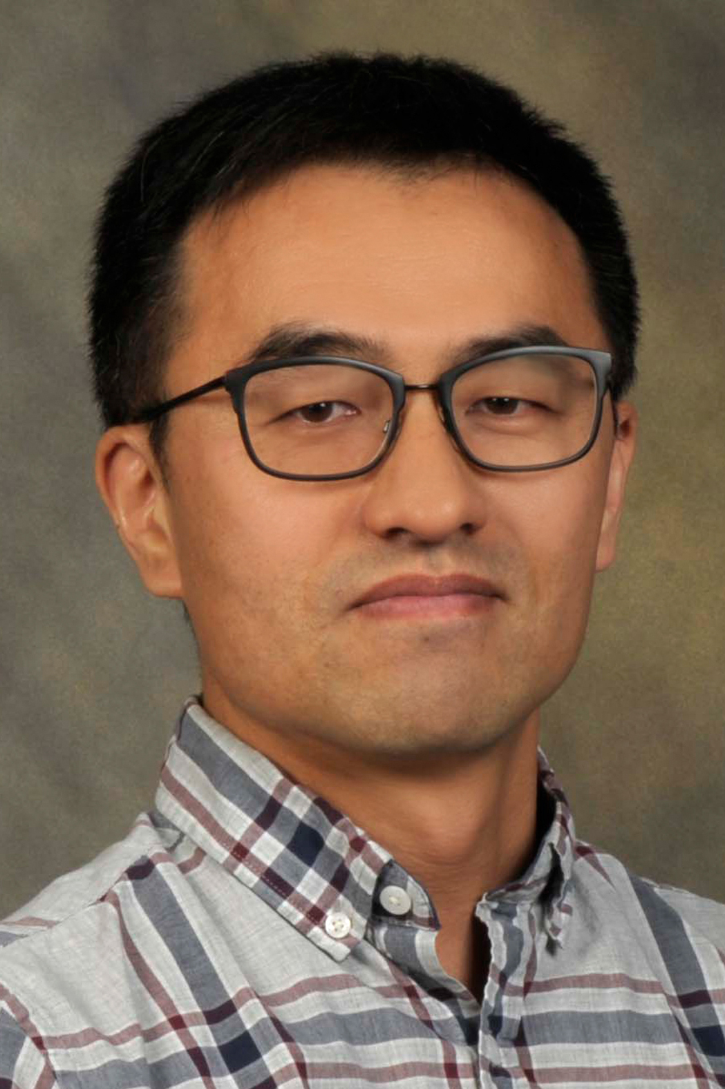
Yiming Li is an Associate Professor in the Department of Biomedical Engineering at Southern University of Science and Technology's School of of Engineering. His research focuses on cutting-edge 3D super-resolution imaging techniques and their biological applications, with expertise spanning optical instrumentation, theoretical optics, and advanced imaging algorithms developed during his postdoctoral work at EMBL and Yale University. Education: Ph.D. in Biophysics, Karlsruhe Institute of Technology (2010-2015) M.Sc. in Medical Physics, Heidelberg University (2009-2010) B.Eng. in Biomedical Engineering, Shanghai Jiao Tong University (2005-2009) Research Interests: Dr. Li specializes in developing advanced 3D super-resolution microscopy techniques with particular expertise in single-molecule localization microscopy , point spread function engineering , and real-time 3D imaging systems. His work bridges optical physics and biological applications, enabling nanoscale observation of cellular processes. His software earned first place in the SMLM Challenge 2016, the field's most prestigious software contest. Publication Trends: Dr. Li's research shows a clear progression from fundamental algorithm development to comprehensive imaging system design, with increasing emphasis on real-time 3D applications. His publications in Nature Methods, Nature Communications, and other high-impact journals demonstrate consistent innovation in super-resolution microscopy, particularly in point spread function calibration, aberration correction, and biological applications at the nanoscale level. Scientific Awards: National Overseas High-level Talents (Youth Program) (2020) Shenzhen Overseas High-level Talents Category B EMBL-EIPOD Marie Curie Postdoc Fellowship (2016-2019) Karlsruhe School of Optics and Photonics Fellowship (2010-2013) International Symposium on Biomedical Imaging Travel Grant (2013) Professional Activities: As a PhD supervisor at SUSTech, Dr. Li mentors graduate students in biomedical engineering. He serves as a reviewer for top journals including Nature Methods, Light: Science & Applications, and Optics Letters. His laboratory maintains active international collaborations with EMBL, Yale University, Oxford University, and Cambridge University, facilitating cross-institutional research in advanced imaging techniques. Research Laboratory: Dr. Li leads an active research group focused on next-generation imaging technologies, operating under the website https://li-lab-sustech.github.io/ . His laboratory combines theoretical optics, software development, and biological applications to push the boundaries of what's possible in optical microscopy for cellular and subcellular observation.
Peter Zijlstra is a Full Professor in the Department of Applied Physics at Eindhoven University of Technology (TU/e), leading the Molecular Plasmonics group. His research focuses on single-molecule sensing using plasmonic and nanophotonic approaches to study biomolecular interactions in complex environments. He is a core member of the Institute for Complex Molecular Systems at TU/e, collaborating across disciplines like chemistry, biomedical engineering, and mathematics. Education: MSc in Applied Physics, University of Twente (2005) PhD from Swinburne University of Technology (2009), studying plasmonic nanoparticles in optical data storage Postdoctoral fellowship at Leiden University under Prof. Michel Orrit Research Interests: Developing novel sensing concepts via nanophotonics and super-resolution microscopy. Key areas include plasmon-enhanced fluorescence, real-time biomolecular dynamics, and applications in cancer management. His work contributes to UN Sustainable Development Goals through advancements in biosensing technologies. Awards: 2013 NWO Vidi Award for research on plasmonic imaging of enzymes in living cells Teaching & Activities: Teaches courses like Advanced Optical Microscopy and Electromagnetism Supervised 32 academic works Contributed to conferences and editorial roles for journals like npj Biosensing Labs & Collaborations: Molecular Plasmonics group website: www.molecular-plasmonics.nl Marie Curie ITN SuperCol project: www.supercol.eu
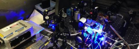
Andrea Pickel is an Assistant Professor at the University of Rochester, holding joint appointments in the Department of Mechanical Engineering, Materials Science, and the Institute of Optics, while also serving as a Scientist at the Laboratory for Laser Energetics (LLE). She received her PhD in Mechanical Engineering from UC Berkeley (2019) and a BS from Carnegie Mellon University (2014). Her research focuses on nanoscale heat transfer, leveraging luminescent materials and super-resolution imaging to address challenges in thermal management, catalysis, and energy systems. Education: PhD, Mechanical Engineering, UC Berkeley, 2019 BS, Mechanical Engineering, Carnegie Mellon University, 2014 Research interests include luminescence nanothermometry, single-nanoparticle imaging, and high-temperature thermal metrology. Her work integrates experimental methods like stimulated emission depletion (STED) imaging and operando spectroscopy to advance understanding of energy transport at the nanoscale. Notable awards include the NSF CAREER Award (2022), ACS PRF Doctoral New Investigator Award (2020), and Furth Fund Award (2021). She was also named a Scialog Fellow in 2024. Advancing thermal measurement techniques, her group collaborates across disciplines to tackle applications in carbon capture, battery technology, and plasmonic photocatalysis. Current projects emphasize developing dual-mode sensing tools for real-time thermal and chemical monitoring. Labs/Teams: Active at the Laboratory for Laser Energetics (LLE) and leads the Pickel Research Group in the Department of Mechanical Engineering.
Professor Daniel Gryko leads a prominent research group at the Institute of Organic Chemistry, Polish Academy of Sciences, specializing in advanced functional dyes and photochemistry. His work bridges fundamental organic synthesis with practical applications in bioimaging, molecular electronics, and nanomaterials. With over 150 publications and numerous high-impact grants, including an ERC Advanced Grant and multiple Horizon Europe projects, Gryko has established himself as a leader in the field of novel chromophore design. Gryko's research focuses on developing innovative fluorescent dyes with exceptional photophysical properties, particularly exploring fluorescence of nitroaromatics, two-photon absorption phenomena, and excited-state intramolecular proton transfer (ESIPT). His group specializes in several key structural platforms including corroles, diketopyrrolopyrroles, pyrrolo[3,2-b]pyrroles, dipyrrolonaphthyridinediones, porphyrins, and coumarins. Recent work has centered on creating strongly emitting helicenes, quadrupolar dyes with unique symmetry-breaking properties, and developing specialized fluorophores for super-resolution microscopy applications. Analysis of Gryko's recent publications reveals a strong emphasis on molecular design strategies for controlling photophysical behavior. His group frequently employs π-expansion techniques, heteroatom doping, and strategic substitution patterns to tune emission properties. A significant portion of their work focuses on overcoming traditional limitations in fluorophore design, such as the non-fluorescence of nitroaromatics, through innovative molecular architectures. Gryko has received prestigious recognition including an ERC Advanced Grant for the ARCHIMEDES project targeting NIR-II emission efficiency, multiple Horizon Europe grants, and the TEAM grant from the Foundation for Polish Science supporting development of fluorescent probes for super-resolution microscopy. His group's work has resulted in numerous publications in top-tier journals including Journal of the American Chemical Society , Chemical Science , and Angewandte Chemie . Professor Gryko actively mentors a diverse research team including PhD students, postdoctoral researchers, and collaborators worldwide. His group has secured substantial funding including Horizon Europe grants for PhotoBrane and APACE projects, ERC funding, and multiple Polish National Science Centre grants. Current projects focus on developing novel fluorescent probes for super-resolution microscopy, creating bio-mimetic sunlight-pumped lasers, and designing photo-switchable membranes for molecular separation. The Gryko group operates a well-equipped laboratory focused on organic synthesis and photophysical characterization. Their work spans from fundamental molecular design to practical applications in bioimaging and materials science. Recent expansions of their research program include development of probes for detecting SARS-CoV-2 proteases, demonstrating the group's ability to pivot toward addressing pressing societal challenges.
Kyu Young Han is an Associate Professor in Optics & Photonics at CREOL, The College of Optics and Photonics, University of Central Florida. His research focuses on developing advanced optical tools for biological and neuroscience applications, including super-resolution imaging (STED/GSD microscopy), label-free single-molecule imaging, and novel microscopy techniques. He holds a patent commercialized by Leica Microsystems and has received the 2020 NIH Maximizing Investigators’ Research Award (MIRA). Education: BS and PhD in Chemistry from Seoul National University (2004 and 2010). Postdoctoral work at the University of Illinois (2011–2016) and Max Planck Institute for Biophysical Chemistry (Germany), specializing in optical microscopy innovation. Research interests emphasize nanoscopy, biophotonics, and interdisciplinary applications in cell biology. His group explores nuclear structure in mammalian cells, DNA-protein interactions, and RNA imaging in live cells. Recent work includes optimizing imaging techniques like TIRF microscopy and integrating deep learning for faster, less damaging STED imaging. Publications span cutting-edge advancements in microscopy resolution, photobleaching reduction, and single-molecule analysis. He advises multiple PhD students and collaborates across disciplines, contributing to Parkinson’s disease research through imaging-driven molecular studies.
Dr. Xiaomin Liu is a Group Leader at the Max Planck Institute for Polymer Research's Department of Molecular Spectroscopy, specializing in optical super-resolution microscopy and laser technology. Her work focuses on developing advanced imaging techniques using nanomaterials and fluorescence dyes. PhD in Optical Engineering, Technical University of Denmark (2011) Postdoctoral Research, Technical University of Denmark (2011-2015) Postdoctoral Research, Max Planck Institute for Polymer Research (2015-2017) Her research explores fluorescent blinking mechanisms of nanomaterials and super-resolution imaging applications , leveraging laser systems and novel fluorescence dyes. Recent publications emphasize nanographene-based microscopy tools and room-temperature phosphorescence in advanced materials. Key trends in her work include: Development of broadband wavelength-tunable femtosecond fiber lasers Investigation of acid-induced fluorescence enhancement in nanographenes Engineering room-temperature phosphorescence via molecular charge-transfer effects Designing polarized emission materials through asymmetric center manipulation Her group collaborates with international researchers in laser technology and material science, supported by Danish FTP and NIH grants. She works with partners like Klaus Müllen, Mischa Bonn, and Andrey Turchinovich.
Peter Burke is a Professor of Electrical Engineering and Computer Science (joint appointments in Biomedical Engineering and Materials Science and Engineering ) at the Samueli School of Engineering, University of California, Irvine . His research bridges nanoelectronics with biotechnology , focusing on carbon nanotubes , graphene devices , and mitochondrial bioenergetics . He has received prestigious Young Investigator Awards from the Office of Naval Research and Army Research Office. Education: B.A. in Physics, University of Chicago (1992) Ph.D. in Physics, Yale University (1998) His work spans quantum electronics , high-speed semiconductor devices , and bio-nano interfaces . Recent publications highlight drone technology , mitochondrial electrical activity , and AI-driven nanoscale sensing . Research trends include terahertz spectroscopy , super-resolution imaging , and open-source medical devices like the NanoStat potentiostat . Scientific Awards Young Investigator Award, Office of Naval Research Young Investigator Program Award, Army Research Office As director of the BurkeLab , he develops nano-electronic interfaces for biological systems, including mitochondrial membrane potential assays and graphene-based biosensors . His lab's innovations in carbon nanotube arrays and scanning microwave microscopy have advanced bio-nano applications.
George M. Church is a Professor of Genetics at Harvard Medical School and affiliated with MIT, where he directs PersonalGenomes.org, providing open-access genomic, environmental and trait data. His laboratory focuses on transformative technologies for reading and writing 3D/4D biological structures with attention to ethics, safety, and equitable access. Church has co-initiated major scientific initiatives including the BRAIN Initiative (2011) and multiple Genome Projects (GP-Read-1984, GP-Write-2016, PGP-2005). Church's research spans multiple cutting-edge domains including genome engineering, synthetic biology, aging reversal, and space genetics. His lab pioneered foundational methods for direct genome sequencing, molecular multiplexing and barcoding in 1984, leading to the first genome sequence in 1994. His innovations contributed to nearly all next-generation DNA sequencing methods and companies. Current research directions include machine learning for protein engineering, tissue reprogramming, organoids, gene therapy, and in situ 3D DNA/RNA/protein imaging. His work bridges fundamental biology with therapeutic applications across diverse fields from Alzheimer's disease to de-extinction biology. Church's recent publications reveal a remarkable breadth of scientific inquiry, spanning from fundamental genome editing techniques to applications in aging research, neuroscience, and space biology. His work increasingly integrates artificial intelligence with biological systems, as seen in papers on machine-guided cell-fate engineering and automation of systematic reviews with large language models. His research maintains a strong translational focus, with numerous papers addressing therapeutic applications in cancer immunotherapy, gene therapy, and diagnostics. The consistent theme across his diverse publications is the development and application of transformative technologies to address fundamental biological questions and medical challenges. National Academy of Sciences (NAS) membership National Academy of Engineering (NAE) membership Franklin Bower Laureate for Achievement in Science Co-initiator of the BRAIN Initiative (2011) Director of multiple NIH Centers for Excellence in Genomic Science (2004-2020) Church directs numerous research centers including the NIH-CEGS, Personal Genome Project (PGP), Lipper Center for Computational Genetics, and Wyss Institute Synthetic Biology center. His laboratory has trained PhD students across multiple Harvard and MIT programs including Biophysics, BBS, Biomedical Informatics, ChemBio, Chemistry, SSQB, MCO, Virology, HST, EE/CS, Physics and Applied Math. His commercial impact is extensive through companies spanning medical diagnostics (Knome/PierianDx, Alacris, Nebula, Veritas) and synthetic biology/therapeutics (AbVitro/Juno, Gen9/enEvolv/Zymergen/Warpdrive/Gingko, Editas, Egenesis). Church also pioneered new privacy, biosafety, ELSI, environmental and biosecurity policies. The Church Lab operates across multiple research domains including molecular multiplexing, next-generation sequencing, nanopore technology, and genome engineering. The lab maintains strong connections with the Personal Genome Project, Wyss Institute, and multiple commercial ventures. Current research directions include the Spatial Atlas of Human Anatomy (SAHA), human skin rejuvenation via mRNA, and space genetics research through the Consortium for Space Genetics and BioAstra. The lab's mission focuses on transformative technologies for reading and writing 3D/4D structures at any scale, inspired by but not limited by biology.
Stephanie Kramer is a Full-time Lecturer in the Department of Chemistry at Case Western Reserve University. She holds a PhD in Physical Chemistry from Carnegie Mellon University (2022) and a B.S. in Chemical Physics from Elizabethtown College (2016). Her research focuses on advanced microscopy techniques, polymer self-assembly, and biomaterials characterization. She actively contributes to the development of super-resolution imaging methods, particularly in extracellular matrix analogues and nanoporous materials. Key research interests include fluorescence correlation spectroscopy (FCS), light-sheet microscopy, and cross-correlation analysis for improving imaging resolution. Her work bridges physical chemistry with biophysical applications, addressing challenges in nanoscale imaging and biomolecule dynamics. Recent studies involve solvent-driven transitions in chiral polymers and diffusion-based imaging in complex environments. Kramer’s publications span topics like super-resolution optical fluctuation imaging (SOFI), light-sheet microscopy innovations, and polymer aggregation mechanisms. Her presentations at conferences such as the American Physical Society March Meeting and American Chemical Society Spring meetings reflect her interdisciplinary approach to chemical physics. No scientific awards are listed. Her advising and grant activities remain unspecified in the provided text. She collaborates extensively with research groups focused on optical methods and biomaterials.
Guanghan Meng is an Assistant Professor at the University of California, Berkeley , with dual appointments in the Herbert Wertheim School of Optometry and Vision Science and the Department of Electrical Engineering and Computer Science (EECS) . He leads the Visionary Optical Imaging Lab (VOILA) , focusing on interdisciplinary research combining optical physics and computational science to develop advanced microscopy technologies for eye and brain imaging. Education : PhD (2021, UC Berkeley), BE (2015, Shanghai Jiao Tong University) PhD Programs Affiliated With : Vision Science, Applied Science & Technology (AS&T), EECS His research integrates optical physics , computational biology , and artificial intelligence to create cutting-edge imaging tools. Recent work includes differentiable wave-optics libraries (Chromatix), super-resolution microscopy techniques, and high-speed neural imaging systems. Publications highlight applications in neuroscience (cerebral circulation, synaptic activity) and biomedical imaging (OCT, two-photon microscopy). VOILA is a highly interdisciplinary team spanning physics , engineering , and biology . In 2025, the lab will welcome 2 PhD students and 1 postdoc, though funding is currently at capacity for new members. Meng is affiliated with the Berkeley Artificial Intelligence Research Lab (BAIR) and Berkeley Center for Computational Imaging (BCCI) .
Randy Bartels is a Professor in the Department of Biomedical Engineering at the University of Wisconsin-Madison. His laboratory specializes in developing advanced biomedical imaging techniques to study complex biological phenomena and translate these methods into applications that enhance fundamental understanding of biology and disease treatments. Education: PhD, University of Michigan (2002) MS, University of Michigan (1999) BS, Oklahoma State University (1997) Research Interests: Bartels focuses on creating novel coherent nonlinear optical imaging modalities, such as spatial frequency modulation imaging (SPIFI), impulsive stimulated Raman scattering (ISRS), and synthetic aperture holography. His work emphasizes label-free imaging, optical scattering robustness, and computational enhancements for resolution and sensitivity. Scientific Awards: 2021 Institut Fresnel Visiting Professor 2013 American Physical Society Fellow 2011 Optical Society of America Fellow 2006 Presidential Early Career Award in Science and Engineering (PECASE) 2005 Sloan Research Fellow (Physics) 2004 NSF CAREER Award Recent Article Trends: Bartels' publications highlight innovations in label-free imaging, nonlinear microscopy, and computational techniques. Key themes include hyperspectral coherent Raman imaging, quantum-classical fusion for super-resolution, and robustness to optical scattering in biological and industrial applications. His work spans fundamental physics, engineering, and biomedical translation. Laboratory: Bartels leads a research group dedicated to advancing imaging technologies, with a focus on overcoming limitations in resolution, depth, and sensitivity through optical and computational methods.
Philip Hemmer is a Professor in the Department of Electrical and Computer Engineering at Texas A&M University, affiliated with the College of Engineering. He holds a Ph.D. in Physics from MIT (1984) and a B.S. from the University of Dayton (1976). His research focuses on quantum optics, nanodiamond-based quantum sensing, and advanced optical materials for applications in quantum computing, biosensing, and thermal imaging. Key areas include solid-state quantum systems, upconversion nanoparticles, and fiber-optic sensor technologies. Dr. Hemmer's work spans interdisciplinary fields such as quantum communication, luminescent thermometry, and nanotechnology. His lab develops novel materials like GeV color centers in diamonds for high-precision sensing and explores applications in medical diagnostics, environmental monitoring, and fundamental physics. Awards include the National Science Foundation Fellowship and multiple AFOSR Star Team Awards. Education: Ph.D., Physics, Massachusetts Institute of Technology, 1984 B.S., University of Dayton, 1976 Awards: National Science Foundation Fellowship Summa Cum Laude, University of Dayton Air Force Research Laboratory Chief Scientist's Award AFOSR Star Team Award (three-time recipient) His recent publications emphasize quantum-enhanced biosensing, nanodiamond engineering, and fiber-optic quantum sensors. Research trends highlight innovations in thermal imaging using diamond defects, multiplexed sensing platforms, and scalable quantum technologies.
Simon Mochrie is a Professor of Physics and Applied Physics at Yale University, affiliated with the Department of Physics within the Faculty of Arts and Sciences. His research focuses on experimental biophysics and condensed matter physics, with emphasis on chromatin dynamics, nuclear mechanics, and super-resolution microscopy. He holds a Ph.D. from MIT (1985) and has pioneered techniques such as optical tweezers and STED microscopy to study biological systems like the ubiquitin-proteasome system in yeast. Current projects include single-molecule measurements on nucleosomes and developing novel imaging methods like LIVE-PAINT for live-cell super-resolution imaging. Educations: Ph.D., Physics, MIT (1985) Research interests center on understanding how chromatin organization influences nuclear mechanics, with studies on heterochromatin condensation, cohesin-driven loop extrusion, and chromatin-envelope interactions. His lab develops advanced microscopy techniques to visualize protein dynamics and subnuclear structures in real time. Recent work explores diffusive states of membrane proteins and the role of phase separation in heterochromatin mechanics. His articles demonstrate a focus on interdisciplinary approaches, combining biophysical experimentation with computational modeling to elucidate fundamental mechanisms in cell biology and soft matter physics. Notable themes include the interplay between chromatin structure and nuclear stiffness, loop extrusion dynamics, and quantitative analysis of intrachromosomal contacts. Teaching contributions include developing introductory physics courses tailored for life sciences students, emphasizing applications in biology and medicine. He actively participates in STEM education initiatives, including collaborative research networks for graduate students in physical biology. The Mochrie Lab also emphasizes instrumentation innovation, such as building fast-scanning STED microscopes and reversible peptide-based imaging systems.
Xavier Darzacq is a Professor of Molecular Therapeutics at the University of California, Berkeley, holding the Edward E. Penhoet Distinguished Endowed Chair in Global Health and Infectious Disease. His research at the intersection of molecular biology and biophysics focuses on understanding how nuclear organization governs transcription regulation during cellular differentiation. Research Highlights: Investigates transcriptional control via non-canonical mediator complexes in fibroblast-to-myofibroblast differentiation. Develops advanced imaging techniques (single-molecule tracking, 3D FISH) to study transcription factor mobility and chromatin interactions. Proposes biophysical models where protein diffusion in the nucleus is guided by DNA/chromatin networks. Technological Innovations: Pioneered methods for single-molecule tracking and super-resolution imaging, enabling nanoscale and millisecond-resolution analysis of nuclear processes. Collaborates with experts in biophysics, chemistry, and imaging to integrate multidisciplinary approaches. Scientific Awards: Edward E. Penhoet Distinguished Endowed Chair (Global Health and Infectious Disease) Nature Structural & Molecular Biology – Selected Article of the Month (2007) His lab (http://tjian-darzacq.mcb.berkeley.edu/) explores how nuclear architecture influences gene expression, particularly in wound healing contexts. Future work aims to leverage advancements in microscopy and genome editing to unravel transcriptional rules in living organisms.