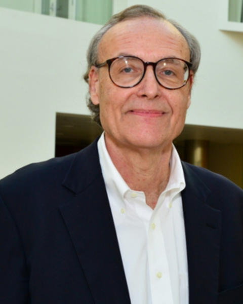
John D. E. Gabrieli is a Professor at the Massachusetts Institute of Technology (MIT) in the Department of Brain and Cognitive Sciences, holding the Grover Hermann Professorship in Health Sciences and Technology and Cognitive Neuroscience. He serves as Director of the Athinoula A. Martinos Imaging Center at the McGovern Institute for Brain Research and leads the MIT Integrated Learning Initiative. His research spans cognitive neuroscience, focusing on memory, thought, and emotion, with clinical applications in dyslexia, autism, and psychiatric disorders. Director, Athinoula A. Martinos Imaging Center (2016–present) Grover Hermann Professor, MIT (2005–present) Faculty, Harvard Graduate School of Education (2006–present) Visiting Professor, Rush Medical College (1990–present) Gabrieli’s work combines brain imaging with behavioral studies to explore learning mechanisms in children, early intervention for reading difficulties, and neuroimaging applications in psychiatric diagnosis. Recent studies examine altered brain activity in dyslexia , neurodevelopmental patterns in autism , and predictive modeling for mental health . His 2024 dataset on adolescent anxiety/depression in the Human Connectome Project highlights longitudinal neuroimaging approaches. Scientific accolades include election to the American Academy of Arts & Sciences (2016), multiple teaching awards at MIT and Stanford, and the Alice H. Garside Lifetime Achievement Award (2017). He contributes to editorial boards of journals like Psychological Science and Biological Psychiatry . His lab emphasizes educational neuroscience , investigating how socioeconomic factors, white matter plasticity, and mindfulness interventions impact learning outcomes. Collaborations span Massachusetts General Hospital, McLean Hospital, and the Harvard Graduate School of Education, integrating clinical and educational applications of cognitive neuroscience.
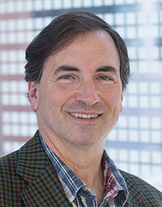
Peter J. Basser is a leading research scientist at the National Institutes of Health (NIH), specifically within the Eunice Kennedy Shriver National Institute of Child Health and Human Development (NICHD), where he heads the Section on Quantitative Imaging and Tissue Sciences (SQITS). His work bridges physics, engineering, and neuroscience to develop non-invasive MRI methods for probing tissue microstructure, particularly in the brain. His educational background is not explicitly mentioned, but his scientific achievements reflect deep training in biophysics and medical imaging. He earned his Ph.D. and has built a career at NIH as a principal inventor of key neuroimaging technologies. Basser's research focuses on quantitative imaging and tissue sciences , especially using diffusion MRI to study brain structure and function. He pioneered Diffusion Tensor MRI (DTI) , Streamline Tractography , and advanced methods like MAP MRI , CHARMED , and AxCaliber , enabling in vivo measurement of axon diameters and microstructural features previously accessible only through histology. His work aims to translate these tools into clinical use for diagnosing developmental disorders, trauma, and neurodegeneration. The 15 most recent articles reflect a consistent focus on developing novel MRI biomarkers, particularly through diffusion and relaxometry methods. They explore water exchange, restriction, glymphatic clearance, latency connectomes, and cortical microstructure, demonstrating a trajectory toward in vivo MRI histology and precision imaging for pediatric and neurological applications. Scientific Awards: National Academy of Engineering (NAE), Inducted 2020 National Academy of Inventors (NAI) Fellow, 2024 Eduard Rhein Technology Award, 2021 ISMRM Gold Medal, 2008 ISMRM Lauterbur Lecturer, 2020 American Society of Neuroradiology Honorary Member, 2019 Victor M. Haughton Award, 2017 ISMRM Fellow, 2010 AIMBE Fellow Best Paper Award, Frontiers in Physics, 2023 Basser leads a dynamic research group that mentors postdoctoral fellows and trainees, many of whom have received prestigious awards. His lab has secured significant grants from the NIH BRAIN Initiative, NICHD, USUHS, and the Bill & Melinda Gates Foundation. The SQITS lab develops open-source software tools like TORTOISE , dmritool , and HI-SPEED , which are widely used in the neuroimaging community. The lab collaborates with institutions such as Uniformed Services University and participates in major initiatives like the Human Placenta Project and the Human Connectome Project. Basser’s vision is to transform clinical MRI scanners into quantitative scientific instruments for precision medicine and large-scale brain mapping. Labs and Teams: Section on Quantitative Imaging and Tissue Sciences (SQITS), NICHD, NIH Neuropathology-Neuroradiology Integration Core (with USUHS) Advanced Translational Neuroimaging Research & Development Core Diffusion – Data Processing Center (DPC)
Susan A. Gauthier is Professor of Neuroscience at the Brain and Mind Research Institute, Professor of Neurology in the Department of Neurology, and Professor of Neurology in Radiology at Weill Cornell Medical College. She serves as Director of Clinical Research at the Judith Jaffe Multiple Sclerosis Center, where she leads a translational research program focused on uncovering the biological mechanisms of multiple sclerosis using advanced quantitative neuroimaging. Dr. Gauthier completed her undergraduate studies at the State University of New York at Buffalo, earned her D.O. from the Philadelphia College of Osteopathic Medicine, and obtained her MPH from Harvard School of Public Health. After completing neurology residency and chief residency at Boston University Medical Center, she was awarded a Clinical Trial Training Fellowship from the National Multiple Sclerosis Society at Brigham and Women's Hospital. Dr. Gauthier's research focuses on developing imaging biomarkers to track inflammation, demyelination, and clinical disability in multiple sclerosis. Her work spans quantitative susceptibility mapping (QSM), TSPO-PET imaging, brain connectivity analysis, and artificial intelligence applications in neuroimaging. She has made significant contributions to understanding chronic active lesions, paramagnetic rim lesions, and their relationship to disability progression in MS. Analysis of Dr. Gauthier's recent publications (2022-2025) reveals a strong focus on quantitative neuroimaging biomarkers for multiple sclerosis. Her work increasingly integrates advanced computational methods including artificial intelligence and machine learning to analyze structural and functional connectivity. A key theme across her recent work is the validation of imaging techniques against pathological findings, particularly regarding chronic active lesions and iron deposition. She has also contributed to several consensus statements that aim to translate imaging research into clinical practice for MS patients. Sylvia Lawry Fellowship in Clinical Trials, National Multiple Sclerosis Society (2002) Dr. Gauthier has served as Principal Investigator on multiple NIH and National Multiple Sclerosis Society grants, including studies on multi-scale imaging assessment of cognitive impairment in MS, quantification of innate immune activity within chronic lesions, and establishing the clinical relevance of chronic active MS lesions. She has built the MS Center's research infrastructure at Weill Cornell and forged strong collaborations with the Department of Radiology. As a dedicated mentor, she has trained numerous students, residents, fellows, and junior faculty who have gone on to academic and clinical leadership positions. Dr. Gauthier leads a research team within the Judith Jaffe Multiple Sclerosis Center that collaborates closely with the Department of Radiology at Weill Cornell. She serves on the Steering and Executive Committees of the North American Imaging in Multiple Sclerosis (NAIMS) Cooperative, which brings together experts from multiple institutions to advance imaging research in MS. Her team utilizes advanced MRI techniques including 7T MRI and PET imaging to study MS pathophysiology.
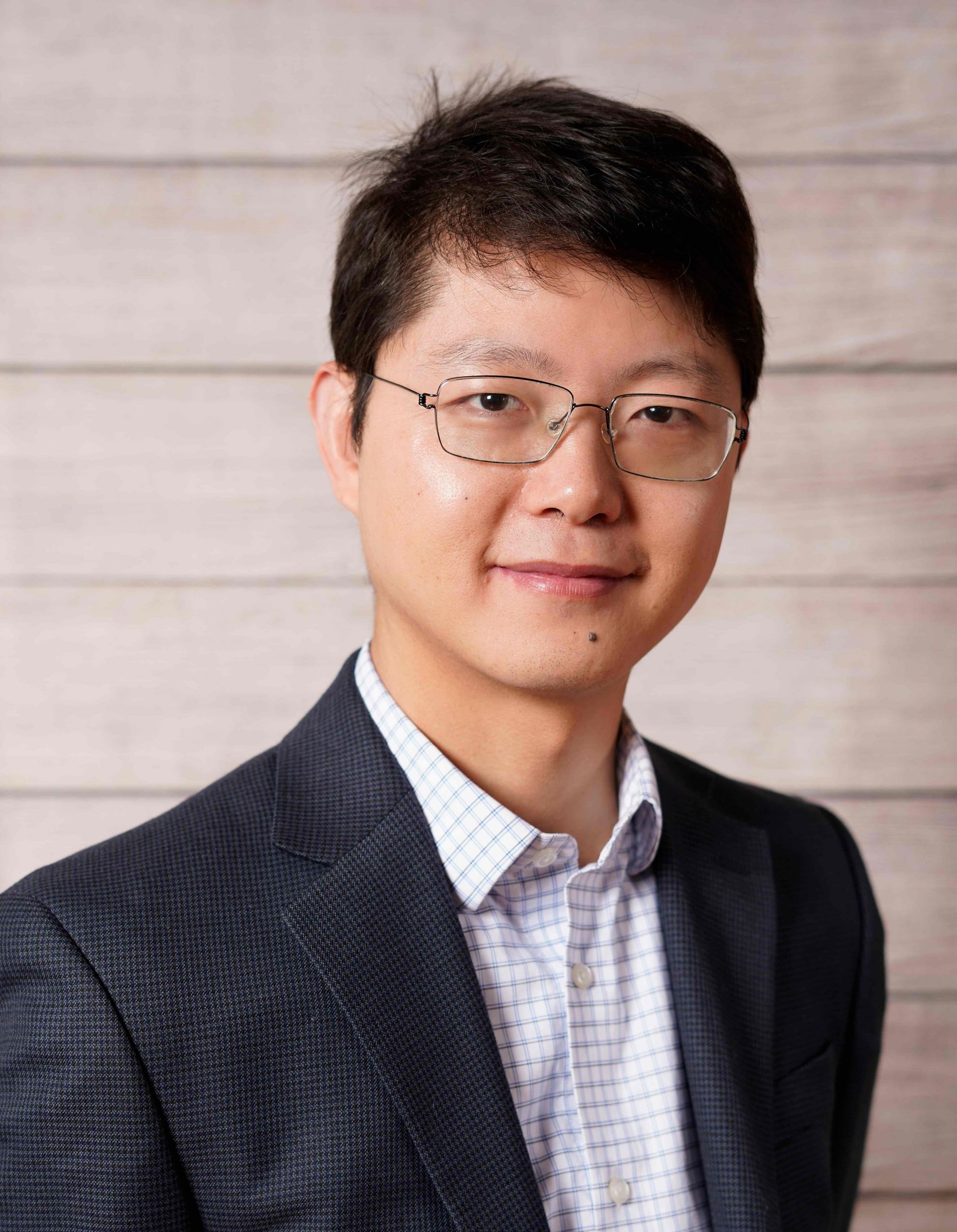
Zhengwu Zhang is an Associate Professor in the Department of Statistics and Operations Research at the University of North Carolina at Chapel Hill. His research focuses on developing statistical and machine learning methods for analyzing high-dimensional neuroimaging data, particularly structural and functional brain connectomics. He leads the UNC Education Program of Intelligence and Connectomics (EPIC), an interdisciplinary initiative training students in brain network analysis. His work addresses challenges in large-scale neuroimaging datasets, including computational efficiency and reproducibility. Zhang completed his Ph.D. in Statistics at Florida State University under Anuj Srivastava. His funding includes NIH grants for CRCNS, structural connectome analysis, and personalized cognitive training. He serves as an Associate Editor for the Journal of the American Statistical Association (Reproducibility). Key contributions include tools like the Surface-Based Connectivity Integration (SBCI) GitHub repository for brain network analysis pipelines. His awards include the 2022 UNC Junior Faculty Development Award and the Oak Ridge Powe Award. Teaching roles include courses on data science, machine learning, and statistical consulting. His research spans brain network dynamics, genetic contributions to connectome structure, and applications of deep learning in neuroscience.
Albert Cardona is a Research Leader at the Medical Research Council Laboratory of Molecular Biology since 2008. His work focuses on connectomics, particularly mapping the wiring diagram of the Drosophila larval brain to understand how neuronal circuits generate behavior. Research Focus: Connectomics, Synaptic Mapping, Neural Circuit Dynamics Model Organism: Drosophila (fruit fly) Techniques: Electron Microscopy, Genetic Manipulation, Computational Modeling His recent publications highlight advancements in whole-brain connectomic mapping, comparative synaptic connectivity analysis, and quantitative neuroanatomy. These works emphasize integrating structural data with functional studies to explain behavioral complexity. Group Members: Shaurya Agrawal Elizabeth Barsotti Jiaqi Chen Marc Corrales Daniel Franco Barranco Peter Hague Ana Jesus Correia Da Silva Shi Yan Lee Laura Lungu Samia Mohinta Scott Wilson Yijie Yin Jiale Zhai
Chao Li is a Principal Research Fellow at the University of Cambridge's Faculty of Mathematics, specializing in AI-driven healthcare solutions. His research focuses on precision medicine through image-based AI and multi-omics integration, with applications in neurological disease modeling, surgical oncology, and clinical AI safety. He leads the Centre for Mathematical Imaging in Healthcare , advancing translational AI for personalized medicine. Research Themes: Image-based AI for precision mental health AI in surgical and interventional oncology Multi-omics approaches for disease characterization Evaluation of AI innovations for clinical translation Publications highlight advancements in multimodal fusion for diagnostics, histology-molecular marker integration, and neuroimaging analytics for mental health. His work bridges computational mathematics with clinical challenges, emphasizing real-world healthcare impact. Labs/Teams: Active member of the Centre for Mathematical Imaging in Healthcare , collaborating across departments to develop AI tools for clinical deployment.
Richard F Betzel is an Associate Professor in Neuroscience at University of Minnesota and affiliated with Indiana University and Northwestern University through collaborative research grants. As a leading network neuroscientist, he develops edge-centric approaches for analyzing brain network organization and dynamics. Current affiliations: University of Minnesota (primary), Indiana University (collaboration), Northwestern University (collaboration) Active research grants from: National Science Foundation (NSF), NIH National Institute on Aging, NIH National Institute of Neurological Disorders & Stroke His research focuses on: Understanding brain network reconfiguration during aging and cognitive tasks Developing computational tools for analyzing connectome architecture Exploring relationships between structural and functional connectivity Investigating network mechanisms in neurodegenerative disorders like Parkinson's Advancing mindfulness meditation research through network neuroscience Recent publications emphasize: Edge-centric network analysis methods Connectome organization across species Dynamic network approaches to social cognition Functional MRI analysis of co-fluctuations Network controllability and modular architecture Applications to both healthy aging and pathological conditions His work contributes to UN Sustainable Development Goals including healthy aging (SDG 3) and scientific knowledge advancement. Current projects include NCS-FO for edge-centric brain mapping (NSF-funded), social cognitive aging research (NIH), and TMS therapy network analysis (NIH).
Dr. Sandra Diaz Pier is a Scientific Lead at the Jülich Supercomputing Centre (JSC) within the Jülich Research Centre , Germany. Specializing in computational neuroscience , high performance computing (HPC) , and machine learning , she bridges neuroscience and advanced computational methods through her research. Education: B.Sc. in Electronic Systems Engineering, Mexico M.Sc. in Computer Science (focus: machine learning, quantum computing), Mexico Second M.Sc. in Electrical Engineering, Ontario, Canada Ph.D. in Computer Science, Germany (2021) Her research focuses on modeling and simulating brain dynamics and plasticity at multiple scales, leveraging HPC to accelerate large-scale neural network simulations. She actively contributes to EU projects like the Human Brain Project (HBP) , Virtual Brain Cloud , and EBRAINS 2.0 , emphasizing infrastructure development and educational training. Her work includes open-source tools such as the NEST simulator , The Virtual Brain , and L2L , enabling efficient parameter exploration and multiscale co-simulation frameworks. The 15 most recent publications highlight her interdisciplinary approach, spanning topics from quantum computing in biomolecular simulations to neural plasticity algorithms and cloud-based brain modeling . These articles reflect her expertise in integrating machine learning , multi-scale simulation , and HPC infrastructure for neuroscience challenges, including seizure propagation, Parkinson’s disease progression, and swarm intelligence in spiking networks. She leads technical coordination in projects like EBRAINS and serves as a task leader in the HBP infrastructure work package , while also organizing workshops and hackathons for open-source tools. Her role involves supporting domain scientists through methodological research and workflow optimization for brain simulations.
Associate Professor Kai-Hsiang Chuang is a Principal Research Fellow at the School of Biomedical Sciences within the Faculty of Health, Medicine and Behavioural Sciences at the University of Queensland. He is also affiliated with the Queensland Brain Institute and the Centre for Advanced Imaging. His research focuses on understanding brain networks, developing advanced imaging techniques, and translating these findings to improve diagnosis and intervention for neurological disorders. Dr. Chuang received his Ph.D. in electrical and biomedical engineering from the National Taiwan University, Taiwan, in 2001. His doctoral research focused on improving the detection of brain activity using functional magnetic resonance imaging (fMRI). Ph.D. in Electrical and Biomedical Engineering, National Taiwan University (2001) Dr. Chuang's research spans multiple areas of brain imaging and neuroscience. His primary focus is on functional brain mapping , where he develops in vivo imaging techniques including functional MRI and multimodal integration with optogenetics, calcium imaging, and electrophysiology. He applies these techniques in both humans and animal models to improve understanding and intervention of brain function, disease processes, and treatment effects. Another key area is brain networks in learning, memory, and dementia . His work explores how brain network wiring and activity underpin cognition and behavior, with particular focus on understanding the causal relationship between brain network activity and memory formation. He develops techniques to modulate behavior by manipulating brain network activity. More recently, Dr. Chuang has expanded into brain waste clearance research, studying the brain's fluid drainage system that clears waste and toxic molecules like amyloid plaques. His lab is developing imaging techniques to track this system's function and understand its regulatory mechanisms, which could provide new treatment targets for dementia. Analysis of Dr. Chuang's recent publications reveals a strong focus on advancing functional MRI techniques for brain network analysis, particularly in rodent models. His work consistently bridges basic neuroscience with clinical applications, especially in understanding memory formation and dementia. A notable trend is the development of multimodal approaches that combine fMRI with optogenetics, calcium imaging, and electrophysiology to establish causal relationships in brain networks. His research increasingly addresses the translation of preclinical findings to human applications, with growing emphasis on Alzheimer's disease mechanisms and potential interventions. Dr. Chuang serves on the editorial boards of multiple prestigious journals including Frontiers in Neuroscience: Brain Imaging Methods , Imaging Neuroscience , and Scientific Reports , reflecting his standing in the field. Editorial Board Member, Frontiers in Neuroscience: Brain Imaging Methods Editorial Board Member, Imaging Neuroscience Editorial Board Member, Scientific Reports Dr. Chuang is actively involved in research supervision, currently serving as Principal Advisor for one PhD student working on "Developing imaging and neuro-technologies for decoding memory formation" and Associate Advisor for two other PhD projects. He has successfully completed supervision of three PhD students on topics related to resting-state networks, memory consolidation, and functional MRI. ARC Discovery Projects (2024-2028): "Decoding the brain network of memory formation" ARC Training Centre for Innovation in Biomedical Imaging Technology (2017-2024) NHMRC-NIH BRAIN Initiative Collaborative Research Grants (2016-2023) Universities Australia - Germany Joint Research Co-operation Scheme (2017-2018) Mater Medical Research Institute Limited grant for mindfulness-based cognitive therapy research (2017-2020) Dr. Chuang leads the Functional and Molecular Neuroimaging Group at the Queensland Brain Institute. His laboratory focuses on understanding the functional connectome of the brain and developing functional and molecular imaging techniques to study brain connectivity associated with behavior. The group has developed various MRI techniques to track neuronal connections, map large-scale brain synchrony, and quantify cerebral blood flow and metabolism in vivo. His research team collaborates extensively with other experts at UQ and internationally, including collaborations with Associate Professor Darryl Eyles, Professor Jürgen Götz, Professor Tianzi Jiang, Dr. Fatima Nasrallah, Professor Linda J. Richards, Professor Pankaj Sah, Professor Elizabeth Coulson, Dr. Patricio Opazo, Professor Feng Liu, and Professor Markus Barth.
Uri Hasson is a Professor at the Princeton Neuroscience Institute, Princeton University, focusing on the neural basis of brain-to-brain human communication, natural language processing, and language acquisition in real-world contexts. His lab challenges traditional experimental models by using deep learning to decode cognition during natural stimuli. Research Highlights: Investigates neural alignment in speaker-listener interactions. Develops computational tools to model language processing in everyday conversations. Explores shared principles between biological and artificial neural networks. Recent Article Trends: 2025–2023 publications emphasize deep language models matching neural responses, event segmentation via Bayesian surprise, and multibrain convergence during storytelling. Key keywords: Neuroscience, Natural Language Processing, Deep Learning, Cognitive Modeling . Awards: NIH Pioneer Award Lab & Collaborations: The Hasson Lab integrates neuroimaging (ECoG, fMRI) with large-scale datasets and AI to study real-life cognition. Collaborations span psychology, computational neuroscience, and machine learning.
Anne-Lise Giraud is a Professor and Group Leader at the Faculty of Medicine, University of Geneva, where she directs a research laboratory focused on the neural mechanisms underlying language and vocal communication. Her work bridges experimental neurophysiology, computational neuroscience, and clinical applications for language disorders. Her research interests encompass: Neural bases of speech and language processing Audio-visual integration in communication Brain-Computer Interfaces for speech communication Neural mechanisms of language disorders (dyslexia, autism, aphasia) Computational modeling of speech processing Comparative studies of vocal communication across species Dr. Giraud's recent publications demonstrate a sophisticated integration of multiple neuroimaging techniques (EEG, fMRI, MEG) with computational approaches to understand both typical and atypical speech processing. A notable trend in her work is the emphasis on neural oscillations as fundamental mechanisms for speech perception, with applications ranging from understanding autism spectrum disorders to developing brain-computer interfaces for communication-impaired individuals. Her research increasingly explores cross-species communication mechanisms, as evidenced by her recent work on dog-human vocal interactions. Her laboratory at Campus Biotech includes multiple graduate students, postdocs, and research assistants working collaboratively on various aspects of language neuroscience.
Dr. Emmanuel Stamatakis is a Senior Research Associate at the University of Cambridge, affiliated with the School of Clinical Medicine and the Division of Anaesthesia. He maintains research ties with the Centre for Speech, Language, and the Brain (CSLB) while focusing on neuroimaging, consciousness studies, and traumatic brain injury (TBI) mechanisms. Institution: University of Cambridge Primary Affiliation: School of Clinical Medicine, Division of Anaesthesia Research Affiliation: Centre for Speech, Language, and the Brain (CSLB) Academic Rank: Research Fellow His research spans neuroscience, neuroimaging, and anaesthesia, with particular emphasis on: Consciousness dynamics under pharmacological and pathological conditions Functional and structural brain connectivity in TBI and delirium Neural mechanisms of anaesthetic agents Evolutionary and comparative neuroscience of brain states Long-term outcomes after neurological trauma Neurotechnology applications in clinical settings Recent publications reveal trends in: Quantifying consciousness through functional gradients and harmonic decomposition Linking anaesthesia-induced neural changes to clinical outcomes Exploring genetic and inflammatory factors in TBI recovery Multi-modal imaging approaches to neurological disorders Translational studies bridging animal models and human conditions Methodological advancements in brain network analysis
Alexandros Poulopoulos, PhD, serves as Associate Professor in the Department of Pharmacology & Physiology at the University of Maryland School of Medicine. His research integrates synthetic biology with developmental neuroscience to pioneer molecular therapeutics for neurogenetic disorders through advanced CRISPR-based genome editing technologies. Education: BSc in Biology, University of Athens, Greece (2003) PhD in Neuroscience, University of Göttingen, Germany (2008) Postdoctoral Fellow, Max Planck Institute for Experimental Medicine (2009) Postdoctoral Fellow (EMBO fellow), Massachusetts General Hospital (2012) Postdoctoral Fellow (HFSP fellow) and Research Associate, Harvard University (2016) Dr. Poulopoulos' research focuses on cortical development, synaptogenesis, and neurogenetic disease mechanisms. His lab develops precision CRISPR agents like Cas9-RC for in vivo somatic genome editing, targeting conditions including epilepsy, autism, schizophrenia, and neurodegeneration. Key investigations explore mTOR signaling pathways, cell adhesion molecules (particularly Neuroligin), and CRISPR delivery systems using in utero electroporation. His work bridges fundamental synaptic biology with therapeutic applications for brain disorders. Analysis of recent publications (2023-2025) reveals three dominant research trajectories: 1) Advancement of prime editing technologies for modeling rare epilepsies (particularly GRIN2A-related disorders), 2) Elucidation of synaptic organization mechanisms through phosphorylation-dependent neuroligin localization and axon guidance principles, and 3) Development of novel delivery platforms including focused ultrasound-mediated blood-brain barrier penetration and nanoparticle systems. These efforts demonstrate a clear progression from basic synaptic biology toward clinically translatable genome editing therapies. Scientific Awards: NIH TARGETED Challenge, phase II winner (2025) Society for Neuroscience Greater Baltimore Chapter President (2024) GPILS Teacher of the Year Award, University of Maryland (2020) NIH Director's New Innovator Award (2019) Harvard Distinction in Teaching Award (2015) Human Frontier Science Program Fellowship (2012) EMBO Fellowship (2010) Max Planck Society Otto Hahn Medal (2009) Dr. Poulopoulos leads the Poulopoulos Lab (poulab.org), which operates within the University of Maryland's Center for Innovative Medicine. His team comprises postdoctoral fellows, graduate students, and research technicians focused on CRISPR agent development and neurogenetic disease modeling. Current funding includes NIH New Innovator Award support for precision genome editing platforms and recent success in the NIH TARGETED Challenge for rare epilepsy therapeutics. He actively mentors PhD candidates through the Graduate Program in Life Sciences (GPILS) and serves as course director for advanced neuroscience modules. The lab employs cutting-edge approaches including single-cell transcriptomics of neuronal compartments, in utero prime editing, and light-sheet imaging of developing cerebellar circuits. Collaborations with clinical neurologists at UMMC and industry partners accelerate translation of their CRISPR-Cas9-RC system toward correcting genomic lesions in neurodevelopmental disorders, with particular emphasis on patient-specific epilepsy models.
Ben Fulcher is an Associate Professor and Senior Lecturer in the School of Physics at The University of Sydney, leading the Dynamics and Neural Systems Group. His research focuses on applying statistical and physics-based methods to study complex systems, with a strong emphasis on neuroscience. He investigates brain structure and function dynamics, particularly exploring the interplay between genetics, neural connectivity, and information processing mechanisms. Fulcher has pioneered tools like hctsa (highly comparative time-series analysis) for analyzing large-scale neuroimaging and physiological data. Research Interests Complex systems analysis Neural dynamics and connectomics Feature-based time-series analysis Gene-brain connectivity relationships Imaging transcriptomics Recent Work Trends Fulcher's recent articles emphasize time-series analysis applications in neuroscience, including studies on anesthesia effects, brain connectivity, and criticality in neural systems. His work bridges computational methods (e.g., generative models, matrix-product states) with biological insights, often involving interdisciplinary collaborations. Key themes include understanding how brain geometry influences dynamics and translating genetic data into connectomic predictions. Grants & Projects 2024: ARC Future Fellowship for sleep research using complex systems methods 2023: Discovery Project on network neuroscience and AI integration Advising & Labs Fulcher supervises research students and directs the Dynamics and Neural Systems Group, which develops open-source tools like the theft R package for time-series analysis. His lab’s work has been featured in media outlets discussing brain structure-function relationships and comparative neurobiology.
Dr. Daniel A. Abrams is a Clinical Associate Professor in the Department of Psychiatry and Behavioral Sciences at Stanford University. His research focuses on the neurobiological basis of social communication impairments in children with autism spectrum disorders (ASD), particularly how atypical reward attribution to social stimuli like voices and faces impacts development. He employs techniques such as functional MRI, psychophysics, and cognitive assessments to identify neural markers of social and memory deficits in ASD. Dr. Abrams received his Ph.D. in Auditory Cognitive Neuroscience from Northwestern University (2008) and a B.F.A. from the University of Arizona (1994). His work has been funded by NIH, NARSAD, and the National Organization for Hearing Research Foundation. He leads the Speech and Social Neuroscience Lab at Stanford Cognitive and Systems Neuroscience Laboratory. Scientific awards include the K01 Research Scientist Development Award (NIH/NIMH, 2014-2017), NARSAD grant (2019-2021), and a 2017 CHRI Pilot Early Career Award. His recent publications highlight neural circuitry in voice processing, memory impairments linked to hyperconnectivity, and efficacy of clinic-home-school collaborative interventions for autistic adolescents. Key research trends: Autism neurodevelopment, reward/salience circuit dysfunction, social communication biomarkers, voice prosody decoding, and memory plasticity. Dr. Abrams’ full description below details his academic trajectory, grants, student collaborations, and clinical trial leadership.