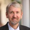
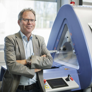
Craig H. Meyer is a Professor in Biomedical Engineering and Radiology & Medical Imaging at the University of Virginia. He holds a Ph.D. from Stanford University and leads the Rapid MRI Research Group, focusing on developing advanced MRI techniques for cardiovascular disease, neural disorders, and pediatrics. His work integrates physics, signal processing, and machine learning to improve MRI acquisition and processing speed. Education: Ph.D. in Biomedical Engineering, Stanford University. Research Interests: Medical and Molecular Imaging, Signal and Image Processing, Biomedical Data Sciences, Biomechanics, and Cardiovascular Engineering. His innovations include fast spiral imaging, conjugate phase reconstruction, and machine learning-enhanced MRI denoising. Awards: Notably includes the Dean’s Award for Excellence in Team Science (2014), Fellowships from NAI (2021), AIMBE (2015), and ISMRM (2013). He also authored two landmark MRI papers recognized as pivotal in the field. Teaching: Courses include BME 6310 (Computation and Modeling in Biomedical Engineering) and BME 8782 (Magnetic Resonance Imaging). He emphasizes translational research, with applications in clinical MRI advancements and collaborative interdisciplinary projects. Labs/Groups: Rapid MRI Research Group focuses on cutting-edge MRI technologies, including real-time cardiac imaging and artifact reduction through deep learning.
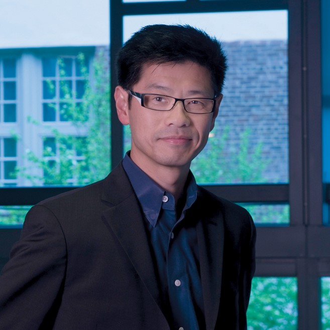
Chi Liu is a Professor of Radiology & Biomedical Imaging at Yale School of Medicine . He serves as Associate Director of Biomedical Imaging Technology at the Yale Biomedical Imaging Institute and Director for Research Faculty Affairs in the Radiology & Biomedical Imaging department. Education : PhD from Johns Hopkins University (2008) Postdoctoral Training : University of Washington (2010) Certification : American Board of Science in Nuclear Medicine (Nuclear Medicine Physics and Instrumentation) His research focuses on quantitative cardiac and oncological PET/CT and SPECT/CT imaging , emphasizing deep learning algorithms , reconstruction algorithms , data correction , and dynamic imaging . Key clinical applications include early detection of chemotherapy-induced cardiotoxicity , multimodality imaging of heart failure , and motion variability elimination in therapy response assessment . The 15 most recent publications reveal a strong emphasis on deep learning techniques for low-dose imaging , motion correction , and cross-tracer generalizability in PET/SPECT systems. These works span applications in cardiac imaging , neuroscience , oncology , and theranostics . Scientific Award : Bruce Hasegawa Young Investigator Medical Imaging Science Award (2012) Contact: chi.liu@yale.edu | ORCID 0000-0002-7007-1037
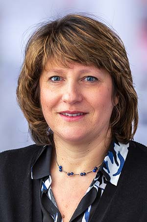
Ruth Etzioni is an Affiliate Professor in the Biostatistics Program and Public Health Sciences Division at the Fred Hutchinson Cancer Center. She leads the Etzioni Lab, focusing on cancer screening, early detection, and overdiagnosis analysis. Her work integrates statistical modeling, epidemiology, and clinical research to address critical questions in prostate and breast cancer control. Dr. Etzioni holds the Rosalie & Harold Rea Brown Endowed Chair and has received a $7.4M NIH Outstanding Investigator Award. Education: PhD in Statistics (Carnegie Mellon University, 1990), MS in Statistics (Carnegie Mellon, 1987), BS in Mathematics (University of Cape Town, South Africa). Research interests emphasize biomarkers, clinical trials, and epidemiological methods. She leads the Biostatistics Core for the Pacific Northwest Prostate Cancer SPORE and participates in the Cancer Intervention and Surveillance Modeling Network (CISNET). Her lab develops models to evaluate screening policies, quantify overdiagnosis, and inform healthcare disparities reduction strategies. Key achievements include groundbreaking work on prostate cancer screening's harm-benefit tradeoffs and contributions to multi-cancer early detection (MCED) frameworks. Recent studies address racial disparities in prostate cancer outcomes, metastasis trends, and the clinical utility of novel diagnostics like PSMA PET imaging. Awards include the Brown Endowed Chair (2020), NCI OIA Award (2023), and recognition for advancing cancer data science. Her lab collaborates with institutions globally and mentors students in biostatistics and translational data science.
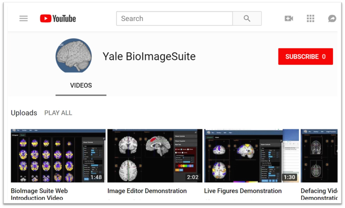
Xenophon Papademetris is a Professor of Biomedical Informatics & Data Science and Radiology & Biomedical Imaging at Yale School of Medicine. He serves as Associate Director of Biomedical Imaging Data Sciences at Yale Biomedical Imaging Institute and directs the Medical Software and Medical Artificial Intelligence Certificate Program. PhD in Electrical and Information Sciences from Yale University (2000) BA from Cambridge University (1994) Postdoctoral Fellowship at Yale University (2002) His research focuses on medical image analysis, machine learning, and biomedical software development. He has developed tools like BioImage Suite Web and contributed to standards committees at the Association for the Advancement of Medical Instrumentation (AAMI). His work spans modalities including MRI, CT, PET, and optical imaging. Recent publications emphasize neuroimaging analysis, explainable AI in healthcare, and multimodal data integration across species. He leads NIH-funded research under the BRAIN Initiative (R24 MH114805) and has authored a textbook on Medical Software published by Cambridge University Press. IEEE Senior Member Yale Brown-Coxe Postdoctoral Fellowship Harding Bliss Prize for Excellence in Engineering He directs the BioImage Suite Project, creating web-based image analysis tools using JavaScript and WebAssembly. His teaching includes both academic courses and a Coursera program on Medical Software with over 14,000 enrollments.
Steven Meikle is a Professor of Medical Imaging Physics and Head of the Imaging Physics Laboratory at the Brain and Mind Centre, University of Sydney. He also serves as Deputy Director (Preclinical) of Sydney Imaging and Deputy Director of the National Imaging Facility's Sydney node. His expertise spans advanced imaging technologies, with a focus on PET/SPECT instrumentation and molecular imaging. He holds a B.App.Sc.(Hons) from the University of Technology Sydney and a PhD from the University of New South Wales. Research focuses include developing novel PET systems like Open-field PET (for freely moving rodents) and Total Body PET, which enhance imaging sensitivity and enable real-time behavioral studies alongside brain function analysis. Collaborations include Tsinghua University (China) and UC Davis (USA). He leads projects on motion correction, quantitative imaging, and AI-driven analysis. Key achievements include over 180 peer-reviewed publications, editorial roles in Physics in Medicine and Biology , and leadership in professional societies. Awards include IEEE Senior Membership and Australian Institute of Physics Fellowship. Current student projects explore Total Body PET applications, motion correction, and radiopharmaceutical evaluation. Teaching roles include medical physics courses in diagnostic radiography and medical physics programs. He advises on imaging ethics, facility implementation, and translational research bridging basic science and clinical applications.
John F Valliant is a Professor in the Department of Chemistry and Chemical Biology at McMaster University. His research focuses on radiopharmaceutical chemistry, molecular imaging, and targeted therapies, particularly in cancer diagnostics and therapeutics. He co-founded Fusion Pharmaceuticals, a company acquired by AstraZeneca for $2.4 billion (US), which specializes in targeted alpha therapy for cancer treatment. His work includes developing radiolabeled imaging agents for prostate cancer (e.g., [18F]DCFPyL), targeted alpha therapies (e.g., FPI-1434), and bioorthogonal chemistry strategies for pretargeted imaging. Valliant has contributed to advancements in photoacoustic imaging, SPECT/PET modalities, and theranostic agents for conditions like bacterial infections and bone diseases. Key projects include the Phase II clinical trial using 18F-DCFPyL PET-MRI for oligometastatic prostate cancer and studies on individualized dosimetry in Lu-177 therapies. He has pioneered platforms for radiolabeled antibody-recruiting molecules and fluorous-phase chemistry for high-specific-activity probes. Valliant’s research bridges disciplines, integrating chemistry, biology, and clinical applications to address unmet medical needs in oncology and diagnostic imaging.
Andrew Sutherland is Professor of Organic Chemistry at the University of Glasgow , School of Chemistry. His research integrates synthetic organic chemistry with molecular imaging, focusing on PET/SPECT tracer development, fluorescent amino acid probes, and transition-metal catalysis for rapid scaffold assembly. Education: Not explicitly listed in the provided text. Research Interests: Molecular imaging of neurological diseases and cancer using PET/SPECT tracers Development of fluorescent amino acid probes for cell imaging Transition-metal-catalyzed transformations (Fe, Cu, Pd, Ni) One-pot multi-reaction processes for drug-like scaffolds and natural products Publication Trends: His 2024–2025 work emphasizes fluorogenic amino acids, radiohalogenation for PET imaging, and iron/copper-catalyzed C–H functionalization. Earlier work includes natural product synthesis and mechanistic studies on halodeboronation. Scientific Awards: None listed in the provided text. Grants & Collaborations: Extensive collaborations with imaging scientists (e.g., Sally Pimlott, Adriana Tavares) and synthetic chemists, evidenced by multi-author papers and joint PET ligand development. Research Group: Leads the Sutherland group , housed in the Joseph Black Building (C5-06), with active projects in chemical biology and organic synthesis.
Trond Vidar Hansen is a Professor at the Department of Pharmacy, University of Oslo , and leads the LIPCHEM research group . He collaborates with institutions including the University of Bergen and Vestlandets Innovasjonsselskap through the VITADEL project, which recently received NOK 5,000,000 in verification support from the Research Council of Norway. His research focuses on the synthesis and biological evaluation of specialized pro-resolving lipid mediators derived from omega-3 fatty acids, with applications in inflammation resolution, neuroinflammation, and drug development. University : University of Oslo Department : Department of Pharmacy Research Group : LIPCHEM Collaborations : University of Bergen, Vestlandets Innovasjonsselskap Research Interests : H Hansen's work centers on the organic synthesis of bioactive lipid derivatives, particularly pro-resolving mediators from omega-3 polyunsaturated fatty acids. His team investigates their roles in inflammatory disease models , neuroinflammation , and PPAR receptor activation , aiming to develop therapeutic agents for conditions like chronic pain, diabetes, and neurodegenerative disorders. The research integrates stereoselective chemistry , biochemical profiling , and pharmacological evaluation to validate these mediators' clinical potential. Recent Awards : 2025: NOK 2,000,000 verification support from Research Council of Norway 2025: Co-leader of NOK 5,000,000 VITADEL project Publications : His articles (2015–2024) reveal a focus on stereoselective synthesis of resolvins, protectins, and maresins, with applications in anti-inflammatory and neuroprotective therapies . Key subfields include omega-3 metabolite profiling , PPAR agonist design , and biosynthetic pathway elucidation , often utilizing human cell models and mouse disease models . Collaborative projects emphasize commercialization of academic research and translational medicine .
James C. Gee is a Professor of Radiologic Science in Radiology at the University of Pennsylvania's Perelman School of Medicine. He serves as Director of the Penn Image Computing and Science Laboratory and Co-Director of the Translational Biomedical Imaging Center , with affiliations in Bioengineering and Applied Mathematics graduate groups. His research focuses on biomedical image analysis, specialization in segmentation, registration, and morphometry applied to neurodegenerative diseases and multi-organ systems. Education : B.S. in Computer Science/Electrical Engineering (University of Washington, 1987), Ph.D. in Computer and Information Science (University of Pennsylvania, 1996) Research : Quantitative medical imaging methods, brain connectomics, neurodegeneration mapping, and translational imaging technologies Publications : 15+ recent works on AI-driven image analysis for Alzheimer's disease, cardiac amyloidosis, and radiomics applications Leadership : Directs MSE-DS Online Degree Program, co-chairs Radiology DCOAP Committee, and founded RISE (Radiology Initiative to Support Inclusive Excellence) His laboratory develops advanced computational tools like ITK-SNAP for biomedical imaging, with applications in both in vivo clinical imaging and ex vivo histology . The work spans cross-disciplinary collaborations in computer science, neuroscience, and clinical medicine.
Privatdozent Dr. Andreas Faust is a leading researcher at the European Institute for Molecular Imaging (EIMI) at the University of Münster, where he heads the Chemical Targeting Lab. His work focuses on developing innovative imaging agents for medical diagnostics, particularly in radiopharmaceutical chemistry and molecular imaging. He maintains strong affiliations with the Department of Nuclear Medicine at the University Hospital Münster and participates in the "Cells in Motion" excellence cluster, contributing to cutting-edge research at the intersection of chemistry, medicine, and imaging technology. Dr. Faust completed his chemistry studies at the University of Münster, earning his Diploma in 1999, followed by his doctoral degree (Dr. rer. nat.) in 2003 with research on artificial caffeine receptors. His academic journey continued with positions at the Department of Organic Chemistry and the Department of Nuclear Medicine before becoming head of the chemistry group at EIMI in 2011. Dr. Faust's research centers on organic and medicinal chemistry with specialization in radiopharmaceutical chemistry . His team develops novel tracers for diagnostic molecular imaging using positron emission tomography (PET), single-photon emission computed tomography (SPECT), optical imaging, and photoacoustic imaging. A significant portion of his work focuses on creating specific ligands for the alarmins S100A8/S100A9 and bacteria-specific tracers based on complex carbohydrates or siderophores. His research has important applications in inflammation imaging, infection diagnostics, and cancer theranostics, with emphasis on improving metabolic stability and target specificity of imaging agents. His publication record demonstrates consistent contributions to molecular imaging, with recent work emphasizing bacteria-specific PET tracers, inflammation imaging targeting S100 proteins, and novel optical imaging probes. The research shows a clear trajectory toward developing clinically applicable imaging agents with improved specificity and metabolic stability, particularly in the areas of infection diagnostics and inflammation monitoring. 2017: Best Poster Award at Symposium "Molecular Imaging Agents in Medicine," Groningen 2009: Young Investigator Award at Deutscher Röntgenkongress, Berlin 2005: Best Scientific Poster Award at 4th Annual Meeting of the Society of Molecular Imaging, Köln Dr. Faust leads multiple significant research projects, including as Coordinator of a project on immune cell distribution imaging (2019-2024) and as Principal Investigator for CRC-project A03 "Targeting of S100A8/A9 for imaging of inflammatory disorders" and research on vascular graft infections (both 2021-2024). His Chemical Targeting Lab comprises a multidisciplinary team working at the intersection of chemistry, microbiology, and medical imaging, securing substantial funding from the Innovative Medicines Initiative and DFG Collaborative Research Centre. The Chemical Targeting Lab maintains state-of-the-art facilities for chemical synthesis, radiochemistry, and biological testing. The lab collaborates extensively with microbiologists, clinicians, and imaging specialists to translate basic research into clinical applications. Current research directions include optimizing bacterial imaging probes for clinical diagnostics and developing new inflammation-specific tracers for early disease detection, with particular focus on S100A9-targeted imaging and siderophore-based bacterial detection systems.
Associate Professor Andre Kyme is an academic staff member in the School of Biomedical Engineering at The University of Sydney. His research focuses on developing enabling technologies for biomedical imaging, including motion compensation in MRI/PET, robotic platforms for image-guided therapy, and cross-disciplinary applications like plant salt uptake analysis using PET. He collaborates with institutions globally and advises students on projects like lameness detection in horses and AI-based motion correction. Research Interests: Kyme's work spans motion correction in medical imaging modalities, medical robotics integration with imaging systems, and innovative applications of imaging technologies in non-traditional fields. His team emphasizes leveraging advancements in computer vision, machine learning, and instrumentation to improve imaging performance and accessibility. Recent Projects: Current research includes MRI-compatible robotic platforms for therapy applications, AI-driven lameness detection in horses, and pediatric neuroimaging improvements. He leads the BREEZE initiative to enhance MRI accessibility for children with cerebral palsy through eye-gaze communication technology. Publications: His work spans 20+ years with over 50 peer-reviewed publications in journals like Physics in Medicine and Biology and IEEE Transactions. Key areas include PET/SPECT/CT motion correction algorithms, robotic systems for medical imaging, and novel imaging applications in plant science. Teaching: Kyme instructs core biomedical engineering courses including thesis supervision and capstone projects at both undergraduate and postgraduate levels. Labs/Teams: Active in the Brain and Mind Centre and Biomedical Imaging, Visualisation and Information Technologies groups at Sydney. Collaborates with industry partners like TeleMedVet and academic institutions including University of California Davis and Chinese University of Hong Kong.
Professor Jerome Liang is a distinguished faculty member at Stony Brook University's Renaissance School of Medicine, holding professorships in Radiology, Biomedical Engineering, Electrical and Computer Engineering, and Computer Science. He serves as Co-Director of Radiology Research and has established himself as a leading expert in medical imaging reconstruction techniques. Dr. Liang's educational background includes a Ph.D. in Physics from City University of New York, postdoctoral training at Duke University, and fellowship at Albert Einstein College of Medicine. His undergraduate degree in Modern Physics was obtained from Lanzhou University in China. His primary research interests focus on advanced medical imaging techniques, particularly low-dose computed tomography image reconstruction, quantitative SPECT reconstruction, high-resolution PET imaging, tissue segmentation from multi-spectral images, computer-aided diagnosis systems, and virtual colonoscopy development. His work bridges engineering principles with clinical applications to improve diagnostic imaging capabilities while reducing radiation exposure. Analysis of his recent publications reveals a strong focus on machine learning applications in medical imaging, particularly in polyp classification, dual-energy CT spectral analysis, and virtual endoscopy. His research consistently aims to enhance diagnostic accuracy while optimizing radiation dose and improving visualization techniques for various medical conditions. 1981 China-US Physics Examination and Application Program (CUSPEA) Winner (Top 25 among 250,000 candidates) 1990 NIH First Investigator Award 1996 American Heart Association Established Investigator Award 1996 Radiological Society of North America Certificate of Merit Award 2002 SUNY Chancellor's Entrepreneur Award 2007 IEEE Society Fellow 2011-2013 SBU, BNL and CSHL Certificates of Excellence in Research and Invention 2013 Stony Brook School of Medicine Award for Excellence in Translational Research Dr. Liang has secured significant research funding including NIH/NCI R01 grants for "Advanced Virtual Colonoscopy for Early Cancer Screening" and "Radiogenomics of Colorectal Polyps." He currently leads active protocols including IRB 93995-MODCR005 focused on integrating virtual and optical colonoscopies with pathological analysis. His laboratory (IRIS - Imaging Research and Informatics) continues to advance medical imaging technology while mentoring the next generation of researchers in this critical field.
Prof. Freek J. Beekman is a Full Professor and head of the Biomedical Imaging section within the Department of Radiation Science & Technology at Delft University of Technology (TU Delft), Faculty of Applied Sciences. He is a leading figure in biomedical imaging, with extensive contributions to nuclear imaging technologies, including SPECT, PET, and CT. His research spans detector development, image reconstruction algorithms, hybrid photonic imaging, and the application of artificial intelligence in medical imaging. Research Interests: His work focuses on advancing imaging modalities through innovations in hardware (e.g., multi-pinhole collimators) and software (e.g., deep learning for attenuation correction). He has pioneered ultra-high-resolution imaging systems, particularly for preclinical and clinical SPECT, and has developed integrated platforms like U-SPECT-BioFluo. His recent research explores glymphatic delivery of nanoparticles, infection imaging, and AI-driven reconstruction techniques, reflecting a strong translational focus. Publication Trends: His most recent publications (2021–2023) emphasize deep learning in SPECT, multi-isotope imaging, high-resolution ex vivo systems, and applications in neuroimaging and oncology. The articles demonstrate a consistent focus on improving image quality, resolution, and clinical utility through physics-informed and AI-enhanced methods. Scientific Awards: NWO Physics Valorization Prize Innovation of the Year Award by the World Molecular Imaging Society (2015, 2018) Edward Hoffman Memorial Award (2017) Bruce Hasegawa Memorial Award (2021) FOM Valorization Award (2013) TU Delft Entrepreneurial Award (2010) Advising and Grants: While specific student names are not listed, his leadership in large collaborative projects and supervision of numerous publications suggests active mentoring. He has secured significant funding through national and international grants, evidenced by his invention of over 20 patent families and successful technology transfer. His founding and leadership of MILabs BV (sold to Rigaku) highlights his impact on commercialization and industry-academia collaboration. Labs and Teams: He leads the Biomedical Imaging research group at TU Delft, which develops cutting-edge imaging systems such as VECTor (SPECT-PET) and EXIRAD-HE. His teams have produced technologies used globally in academic and pharmaceutical research, contributing to tracer development and therapeutic innovation.
Elliot A. Asare, MD, MS, CMQ, FACS serves as Assistant Professor in the Section of Surgical Oncology within the Division of General Surgery at the University of Utah. He specializes in cutaneous malignancies and sarcomas of the retroperitoneum and soft tissues, practicing clinically at both Huntsman Cancer Institute and Intermountain Healthcare Center in Murray. His educational trajectory includes: Phi Beta Kappa graduate with B.S. in Biology and Chemistry (double major) from Randolph-Macon College M.D. from Howard University College of Medicine (Alpha Omega Alpha inductee) General Surgery residency at Medical College of Wisconsin Affiliated Hospitals M.S. in Health Services and Outcomes Research from Northwestern University Complex General Surgical Oncology fellowship at University of Texas MD Anderson Cancer Center Dr. Asare's research program centers on cancer staging methodology, data quality enhancement, prognostication frameworks, and outcomes research with emphasis on health disparities. His work integrates large-scale database analyses (SEER, National Cancer Data Base) to refine AJCC staging systems while addressing inequities in melanoma and sarcoma care across elderly, minority, and rural populations. Recent investigations explore sentinel lymph node biopsy efficacy in aging patients and viral infection impacts on melanoma mortality. His 2023-2025 publications reveal three dominant themes: (1) AJCC Version 9 implementation across multiple cancer types (anal, appendiceal, neuroendocrine), (2) persistent disparities in skin cancer outcomes linked to demographic and geographic factors, and (3) telehealth applications for expanding surgical oncology access. These studies consistently leverage national datasets to validate staging systems while identifying structural barriers to equitable care. Professional recognition includes: Phi Beta Kappa honor society membership Alpha Omega Alpha Medical Honor Society induction Fellowship in the American College of Surgeons (FACS) Dr. Asare actively contributes to surgical oncology education through The SCORE Portal as item writer for soft tissue neoplasm assessments and via publications on medical education frameworks. His institutional affiliations with Huntsman Cancer Institute and Intermountain Healthcare provide platforms for multidisciplinary sarcoma and melanoma management, where he integrates evidence-based staging protocols with patient-centered surgical approaches as evidenced by consistently high patient satisfaction scores (4.9/5 from 166 reviews).
Dag Linnarsson is a Professor of Physiology at Karolinska Institutet, Department of Physiology and Pharmacology, where he has held the position since 1986 and transitioned to Senior Professor in 2009. His research primarily focuses on environmental physiology, with an emphasis on the human body's response to extreme conditions such as microgravity, hypergravity, and spaceflight. He leads the Environmental Physiology Research Group and the Lars Karlsson team, investigating topics like pulmonary function in astronauts, musculoskeletal countermeasures during space missions, and the effects of artificial gravity. His work spans studies on bed rest as a spaceflight analog, lung diffusing capacity, and nitric oxide dynamics under varying environmental pressures. Recent research includes assessing pulmonary nitric oxide levels in astronauts during long-term space missions and exploring the physiological impacts of lunar dust exposure. Linnarsson has also contributed to standardizing bed rest protocols for spaceflight research and evaluating centrifugation as a countermeasure for muscle and bone loss. He maintains affiliations with Karolinska Institutet’s C3 Fysiologi och farmakologi team and collaborates on interdisciplinary projects addressing space medicine challenges. While no formal student listings or awards are explicitly mentioned, his extensive publication record highlights contributions to respiratory physiology, cardiovascular adaptation, and musculoskeletal health in extreme environments.