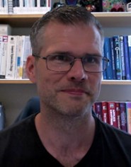
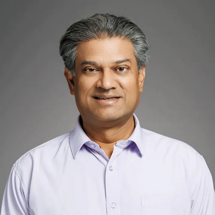
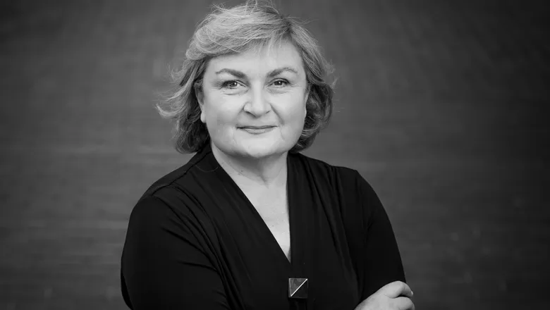
Tommy Löfstedt is an Associate Professor at Umeå University , affiliated with the Department of Computing Science and the Department of Mathematics and Mathematical Statistics. His research focuses on machine learning , computer vision , and medical image analysis , with applications in life sciences, radiation therapy, and biomedical imaging. He leads multiple research projects, including AI-driven delineation in radiation therapy, quantitative MRI for radiotherapy, and machine learning for plant nutrient uptake. Current research emphasizes structured regularization methods to improve model interpretability and robustness. Key applications include medical image segmentation , Alzheimer's classification , and uncertainty estimation in MRI . Recent publications highlight his work on morphological regularization , adversarial attack mitigation , and multi-task learning in medical imaging contexts. His projects span 2022–2026 with funding for pediatric oncology automation and gynecological cancer staging. Affiliated with both computing and mathematical departments, he bridges algorithm development with applied mathematical frameworks in medical and life science domains.
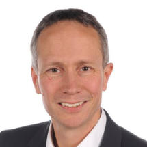
Cornelius Faber is a University Professor in the Department of Radiology at the University of Münster, Germany, where he leads the Experimental Nuclear Magnetic Resonance research group. His work focuses on developing and implementing novel MRI techniques that extend the boundaries of magnetic resonance imaging in terms of spatial and temporal resolution, sensitivity, and specificity for physiological, structural, and molecular changes. He actively participates in the "Cells in Motion" interdisciplinary research initiative at the university. Professor Faber's research spans multiple critical areas in medical imaging and biomedical science. His primary expertise lies in MRI cell tracking , enabling visualization of cellular dynamics in vivo. He has made significant contributions to infection imaging , developing methods to detect and characterize microbial infections using MRI. His work on MR methodology development has advanced quantitative imaging techniques, while his research on multimodal integration in MR and MRI contrast mechanisms has provided deeper insights into molecular and cellular processes. His research bridges physics, engineering, and biomedical applications, with particular relevance to inflammation, cancer, neurological disorders, and cardiovascular disease. Analysis of Professor Faber's extensive publication record reveals a clear evolution from fundamental MRI technique development toward increasingly sophisticated applications in disease models. His recent work demonstrates a strong trend toward multimodal imaging approaches that combine MRI with complementary techniques such as mass spectrometry, optical imaging, and PET. This integration creates comprehensive diagnostic platforms that provide both anatomical and molecular information. A notable pattern is the focus on cellular dynamics, particularly immune cell behavior in inflammatory conditions and tumor microenvironments, with applications spanning neuroscience, oncology, and cardiology. Professor Faber leads a multidisciplinary research team of approximately 15 members, including scientists, doctoral students, technicians, and medical students. His laboratory is deeply integrated with the University of Münster's research infrastructure, particularly the Multiscale Imaging Centre. The group's work contributes significantly to advancing preclinical MRI methodologies while maintaining strong clinical relevance, with numerous publications in high-impact journals across medical imaging, neuroscience, and biomedical engineering disciplines.
Bjoern Menze is a Professor and Rudolf Mößbauer Tenure Track Chair at the Technical University of Munich (TUM), leading the Image-based Biomedical Modeling Group within the Munich School of Bioengineering. His research focuses on medical image computing, tumor growth modeling, and computational physiology, with applications in clinical neuroimaging and personalized radiotherapy design. He holds a Ph.D. in Computer Science from Heidelberg University and has held positions at ETH Zurich, INRIA Sophia Antipolis, MIT, and Harvard Medical School. His academic journey includes a postdoc at MIT’s CSAIL and Harvard Medical School, followed by roles at ETH Zurich and INRIA. His work bridges biomedical imaging with machine learning, emphasizing model-driven analysis of physiological processes. He has been a visiting professor at Maastricht University and contributes to initiatives like the Center for Translational Cancer Research at TUM. Key research areas include tumor growth modeling, quantitative imaging biomarkers, and integrating mathematical models with clinical data. His awards include the MICCAI Young Scientist Award (2014), Leopoldina Fellowship (2009), and DFG Research Fellowship (2008). He advises on medical AI, leads interdisciplinary projects, and publishes extensively in top journals like Nature Neuroscience and IEEE Transactions on Medical Imaging. His lab’s work spans applications such as glioblastoma radiotherapy optimization, whole-body bone lesion detection, and neural connectivity imaging. Collaborations include institutions like Harvard, MIT, and ETH Zurich. He emphasizes translating computational methods into clinical practice for personalized healthcare solutions.
Jed Elison is the Irving B. Harris Professor of Child Development and Distinguished McKnight University Professor at the University of Minnesota’s Institute of Child Development. His research focuses on developmental social neuroscience, structural brain development, and early autism detection. BA in Psychology and English (2005), University of Utah PhD in Psychology (2011), University of North Carolina-Chapel Hill Postdoc in Social Neuroscience (2013), California Institute of Technology Elison’s work examines how attentional orienting drives early cognitive and social development using eye tracking and neuroimaging (MRI, DWI). Key areas include autism , emerging psychopathology , and white matter microstructure . Recent studies model longitudinal trajectories in ASD and explore social-emotional competence. His 2025 articles address infant brain imaging datasets, gesture-vocabulary relationships in autism, and adaptive functioning in corpus callosum agenesis. Collaborative work spans Developmental Science , Pediatrics , and Autism Research . Irving B. Harris Professor of Child Development Distinguished McKnight University Professor Elison advises PhD students in the Cognition and Neurodevelopmental Studies (CNS) Lab, collaborating with Dr. Megan Swanson. The CNS Lab investigates infant brain-behavior associations, particularly in high-risk populations like those with corpus callosum agenesis or congenital CMV . Techniques include MRI , EEG , and behavioral assessments.
Hannah Spitzer is a Research Group Leader at the Institute for Stroke and Dementia Research (ISD) at Ludwig Maximilian University of Munich and an associated Research Group Leader at Helmholtz Munich's Computational Health Center. She leads the Spitzer Lab, focusing on computational analysis of multimodal brain datasets to advance understanding of neurovascular and neurodegenerative diseases. Her educational background includes: PhD in Computer Science from Heinrich-Heine University Düsseldorf and Research Center Jülich (2015-2020) Master's in Computer Science from RWTH Aachen (2013-2015) Bachelor's in Computer Science from RWTH Aachen (2009-2013) Dr. Spitzer's research integrates computational biology and machine learning to decode brain complexity, with emphasis on spatial omics analysis , interpretable image representation learning , and cross-modal data integration . Her group develops tools like squidpy and campa for spatial omics while applying graph neural networks to epilepsy lesion detection through the international MELD project, prioritizing biological interpretability in AI models. Recent publications reveal strong trends in leveraging graph neural networks for subtle brain lesion detection and creating computational frameworks for spatial omics integration. Her work consistently bridges advanced machine learning with clinical neuroscience to uncover disease mechanisms in neurodegeneration and vascular disorders. Dr. Spitzer actively mentors students including current PhD candidate Beatrice Guastella and alumni Deniz Fettahoglu (MSc) and Katia Berr (PhD). Her lab operates through major collaborations including the MELD epilepsy consortium and Helmholtz Imaging Project, with funding supporting computational pipeline development for small-vessel disease prediction and multimodal brain atlasing. The Spitzer Lab comprises postdoc Wasim Aftab and PhD student Beatrice Guastella, working on computational pipelines that integrate histology, spatial omics, and neuroimaging data to decode brain disease mechanisms through interpretable AI approaches.
Professor David Carmichael is a distinguished academic at King's College London , affiliated with the School of Biomedical Engineering & Imaging Sciences and the Department of Biomedical Computing . His research focuses on Magnetic Resonance Imaging (MRI) Physics , EEG-fMRI integration , and epilepsy neuroimaging , with a particular emphasis on mapping epileptogenic networks and optimizing MRI safety for concurrent neurophysiological recordings. Education : PhD in Medical Physics (UCL, 2004), MSci in Physics (UCL, 2000). External Role : Honorary Reader at UCL Great Ormond Street Institute of Child Health. His research interests span advanced neuroimaging techniques, including 7T MRI for pediatric epilepsy, quantitative susceptibility mapping to detect cortical abnormalities, simultaneous EEG-fMRI safety protocols, network-guided neuromodulation for treatment optimization. Recent publications highlight his work on ultra-high field MRI in drug-resistant pediatric cohorts, RF-induced heating safety during combined EEG-fMRI, image quality transfer for low-field MRI in developing regions, motion correction strategies in pediatric scans. He leads critical projects such as 7 Tesla Sodium MRI for Epilepsy (MRC-funded) and Minimal Motion MRI Systems (NIHR-funded), while contributing to global epilepsy research through the King’s Epilepsy Research Collective (KERC) .
Owais Khan serves as an Assistant Professor in the Department of Biomedical Engineering at Toronto Metropolitan University, where he leads research in cardiovascular biomechanics to improve heart disease diagnosis and treatment through engineering-driven approaches combining computational simulations, medical imaging, and biomechanics. His research program focuses on three interconnected pillars: developing physics-based computational models for blood flow simulation in patient-specific anatomies; advancing medical imaging techniques like dynamic CT myocardial perfusion and vessel wall MRI for quantitative physiological assessment; and conducting fundamental biomechanics studies to optimize prosthetic valve designs. This work directly addresses critical clinical challenges including heart surgery complications, aneurysm rupture prediction, and vein graft failure in coronary bypass patients. Khan's publication record demonstrates consistent innovation in cardiovascular computational modeling, with recent work emphasizing personalized medicine through physics-informed neural networks, multi-fidelity uncertainty quantification, and integration of CT perfusion imaging for coronary hemodynamics. His research bridges engineering principles with clinical cardiology to enable virtual treatment planning and risk stratification without additional patient risk. His scientific contributions have been recognized with prestigious awards including the American Heart Association Postdoctoral Fellowship, NSERC Postdoctoral Fellowship, Baxter Young Investigator Award, and MITACS Globalink Research Award. As director of the Cardiovascular Imaging and Modeling Biomechanics Lab (CIMBL), Khan maintains active collaborations with clinicians and radiologists at major hospitals, facilitating direct translation of engineering solutions to clinical cardiovascular medicine through a multi-disciplinary approach focused on personalized treatment strategies.
Melissa Hooijmans is an Assistant Professor at the Vrije Universiteit Amsterdam, affiliated with the Faculty of Behavioural and Movement Sciences and the Physiology department. She also holds a position in the AMS - Ageing & Vitality research group. Her research focuses on advancing MRI techniques for studying muscular dystrophies, skeletal muscle biology, and sports-related injuries. Key areas include diffusion tensor imaging, magnetic resonance imaging applications, and muscle fiber analysis. Her work integrates clinical and biomechanical perspectives, with recent studies on Becker muscular dystrophy, hamstring injury recovery, and exercise physiology. She actively contributes to interdisciplinary collaborations and has published extensively in peer-reviewed journals like NMR in Biomedicine and the Journal of Magnetic Resonance Imaging. Dr. Hooijmans teaches advanced courses such as Master Research Projects and Functional Anatomy, emphasizing hands-on research training for graduate students. She leads initiatives in compositional and functional MRI methodologies, aiming to improve diagnostic precision and therapeutic strategies for muscle-related disorders.
D.S. Fahmeed Hyder is a Professor of Biomedical Engineering at Yale University, with additional appointments in Radiology & Biomedical Imaging. He holds a Ph.D. from Yale University and leads the Hyder Lab, which focuses on advancing quantitative and translational imaging technologies using magnetic resonance methods to study brain function and dysfunction at the laminar level. His research integrates multidisciplinary approaches, including molecular imaging, neurophysiology, and material science. Research Interests: Neurovascular and neurometabolic coupling mechanisms Molecular imaging probes for disease diagnostics Functional MRI (fMRI) and metabolic imaging in health and disease Imaging applications for Alzheimer’s, stroke, and cancer Publications: Dr. Hyder’s recent work emphasizes cutting-edge imaging techniques, such as pH-sensitive biosensors, high-resolution fMRI, and multimodal optical imaging. His research highlights include studies on neurovascular dysfunction in Alzheimer’s models, therapeutic interventions for brain injury, and molecular imaging of tumor microenvironments. Awards: Niels Lassen Award (2003), for cerebral blood flow research Early Career Faculty Award (1998), NSF & NIH Pilot Awards from JDRF & Yale-UCL Collaborative (2008, 2013) Labs/Teams: The Hyder Lab collaborates with institutions like UCL and the James S. McDonnell Foundation, advancing translational imaging solutions for clinical applications.
Kelsey Canada is an incoming Assistant Professor in the Department of Psychological and Brain Sciences at the University of Massachusetts Amherst, starting Fall 2025. She is based in Tobin Hall and will lead a research program focused on developmental cognitive neuroscience. Her research centers on understanding how memory and brain development, particularly in the hippocampus, evolve during childhood and adolescence. She examines how socioeconomic factors influence neurodevelopment, using interdisciplinary methods including MRI , EEG , behavioral testing , and structural equation modeling . Her work emphasizes representative sampling and data integration to uncover modifiable risk and protective factors. Dr. Canada's recent publications highlight her leadership in hippocampal subfield research, quality control in neuroimaging, and collaborative data practices. Her work spans developmental trajectories, methodological innovation, and social determinants of brain health. She earned her PhD from the University of Maryland, College Park, and is establishing her lab at UMass Amherst. Prospective students are encouraged to contact her directly. Research Areas: Developmental Science, Neuroimaging, Cognitive Development Affiliation: Department of Psychological and Brain Sciences, UMass Amherst Dr. Canada has been involved in major collaborative initiatives, including the Hippocampal Subfields Group, and her research is supported by recent publications in top journals such as Human Brain Mapping , Hippocampus , and Developmental Cognitive Neuroscience . While no formal grants or advising roles are listed yet, her active publication record and lab formation indicate a growing research program.
Ronald M Peshock is a Professor of Radiology and Internal Medicine at the University of Texas Southwestern Medical Center (UTSW), with a career spanning over four decades since completing medical school at UT Southwestern in 1976. His academic journey includes Internal Medicine training at Parkland Health & Hospital System (internship 1977, residency 1979) followed by a Cardiology fellowship (1982). Dr. Peshock's research focuses on cardiac imaging, particularly using MRI and CT technologies to assess cardiovascular risk and disease. His work heavily involves the Dallas Heart Study, examining left ventricular hypertrophy, atherosclerosis, and cardiovascular risk factors across diverse populations. In recent years, he has become a prominent researcher in artificial intelligence applications for radiology, with numerous publications on AI implementation in clinical workflows, diagnostic accuracy improvement, and integration of AI into radiology training programs. His publication record shows consistent productivity from 1979 through 2025, with 218 total publications according to available records. Recent work demonstrates particular expertise in cardiac CT, pulmonary embolism imaging, and developing quantitative imaging biomarkers for cardiovascular disease. His Scopus profile indicates an h-index of 67 with 14,914 citations, reflecting significant scholarly impact in his fields. Dr. Peshock maintains active clinical and research affiliations with Parkland Health & Hospital System and serves as a key researcher in the Dallas Heart Study. His work examining ethnic differences in cardiovascular disease, left ventricular hypertrophy patterns, and aortic stiffness has contributed significantly to cardiovascular imaging literature.
Despina Kontos, PhD is the Herbert and Florence Irving Professor of Radiological Sciences at Columbia University Irving Medical Center (CUIMC), with appointments in the Department of Radiology and the Herbert Irving Comprehensive Cancer Center. She serves as the Chief Research Information Officer for CUIMC, Vice Chair of Artificial Intelligence and Data Science Research in the Department of Radiology, and Director of Biomarker Imaging at NewYork-Presbyterian Hospital. Additionally, she holds appointments in the Departments of Biomedical Informatics and Biomedical Engineering. Dr. Kontos received her educational training from prestigious institutions: BS in Engineering from the University of Patras, Greece MSc and PhD in Computer and Information Sciences from Temple University Postdoctoral training in Radiology at the University of Pennsylvania Certificates in Biostatistics and Epidemiology from UPenn, Cancer Biology from Harvard, and AI for Decision Making from Wharton As a computer scientist with expertise in artificial intelligence and machine learning, Dr. Kontos focuses on developing computational methodologies to leverage imaging as quantitative biomarkers for personalized disease prediction, particularly in cancer. Her research program investigates how imaging data can be mined to extract sophisticated phenotypic signatures with diagnostic, prognostic, and predictive value. While her primary focus has been on breast cancer, her lab also pursues related research in lung cancers, evaluating the integration of CT radiomic features with liquid biopsy data to characterize tumor heterogeneity. Dr. Kontos founded and directs Columbia University's Center for Innovation in Imaging Biomarkers and Integrated Diagnostics (CIMBID), a multidisciplinary center dedicated to developing and integrating quantitative imaging and non-imaging biomarkers for personalized disease prediction. Through CIMBID, she has built a vibrant scientific ecosystem that brings together expertise across Columbia's campuses, linking basic science, engineering, clinical medicine, public health, and health services research. Analysis of Dr. Kontos's publication record reveals a strong focus on applying AI and machine learning to biomedical imaging, particularly for cancer risk prediction and personalized treatment. Her work demonstrates a progression from foundational methodological development to clinical translation, with increasing emphasis on multi-modal biomarker integration. Recent publications show expansion into new disease areas including Alzheimer's disease prediction, while maintaining her strong focus on breast and lung cancer applications. Dr. Kontos has received significant recognition for her contributions to the field: Academy for Radiology and Biomedical Imaging Research Distinguished Investigator Award (2020) Eastern Cooperative Oncology Group - American College of Radiology Imaging Network ECOG-ACRIN Young Investigator Award of Distinction for Translational Research (2014) Dr. Kontos has been highly successful in securing research funding, with numerous grants from federal agencies including the National Institutes of Health (NIH) and the Department of Defense (DOD), as well as private foundations such as the American Cancer Society (ACS) and the Radiological Society of North America (RSNA). Her leadership extends to mentoring students and postdoctoral researchers through her roles at CIMBID and the Department of Radiology. As the founding director of CIMBID, Dr. Kontos leads a multidisciplinary team that includes the Computational Imaging Biomarker Group (CBIG), the Laboratory of AI and Biomedical Science (LABS), and several other affiliated research labs. The center leverages Columbia's institutional strengths in engineering, data science, and clinical medicine to advance personalized healthcare through AI and imaging technologies.
Professor Kate Tchanturia is a leading academic at King's College London , affiliated with the Institute of Psychiatry, Psychology & Neuroscience and serving as a Professor in the Department of Psychological Medicine . She is a Consultant Clinical Psychologist at the South London and Maudsley NHS Foundation Trust and holds visiting professor roles at Ilia State University (Georgia) and Tbilisi State Medical University. Her work bridges clinical practice, research, and education in eating disorders and autism. Doctor of Clinical Psychology, Tbilisi State University (1998) PhD in Experimental Psychology, Tbilisi Academy of Sciences (1989) MSc in Psychology, Tbilisi State University (1982) Her research focuses on neuropsychological, cognitive, and emotional aspects of eating disorders , particularly in women and autistic individuals. She explores treatment innovations, cultural adaptations, and recovery trajectories through translational studies. Recent projects include the PEACE Pathway for autism-inclusive care and SOOTH-ED with sensory technology. Her 15+ recent articles highlight collaborations in autism and eating disorder comorbidity , cognitive remediation therapy , and neuroimaging . Key awards include the National Award of Georgia , President of the Eating Disorders Research Society , and Academia Europaea membership . She mentors PhD students and supervises clinical trainees.
Ann Ragin, PhD, is a Research Professor in the Department of Radiology at the Feinberg School of Medicine, Northwestern University. Her work focuses on quantitative MRI and brain network analysis to investigate aging effects and viral infections including HIV and COVID-19. She maintains active affiliations with the Institute for Public Health and Medicine (IPHAM), Northwestern University Clinical and Translational Sciences Institute (NUCATS), Northwestern University Institute of Neuroscience (NUIN), Robert J. Havey, MD Institute for Global Health, and Simpson Querrey Institute for Epigenetics. Her academic credentials include a PhD from Northwestern University (1987) and postgraduate training at the University of Chicago (1989), where she completed a postdoctoral fellowship in Quantitative Methods. Dr. Ragin's research program integrates advanced imaging techniques with clinical neuroscience: Development of quantitative MRI methodologies for in vivo brain measurement Brain network analysis for early neural injury detection 4D flow imaging applications in cerebral blood flow assessment Investigation of neuroinflammation in HIV/AIDS and aging populations Structural-functional neuroimaging correlations in viral infections Her recent publications reveal a dual research trajectory: computational neuroscience innovations (tensor-based graph networks for brain analysis) and clinical applications in cardiopulmonary imaging (pulmonary hypertension biomarkers). This work bridges machine learning, neurology, and cardiology through collaborative NIH-funded studies with the AIDS Clinical Trials Group. Professional leadership includes continuous membership in the AIDS Clinical Trials Group (2013-present), Conference on Retroviruses and Opportunistic Infections, and International Society for Magnetic Resonance in Medicine (2002-present), with prior service on the Chicago Society for Neuroscience Council (2003-2005). While specific grant details remain undisclosed per institutional policy, her ACTG membership and multi-institute affiliations indicate substantial involvement in federally funded research. No advisee information appears in public profiles, though her 99 publications suggest extensive mentorship within collaborative projects. Current initiatives focus on multimodal brain network analysis for HIV-related neurocognitive disorders and quantitative MRI biomarkers for pulmonary hypertension, leveraging Northwestern's advanced imaging infrastructure across multiple research consortia.
Dr. Xi Chen is an Assistant Professor in the Department of Integrative Neuroscience at Stony Brook University. He holds a Ph.D. from the University of Texas at Dallas (2019). His research focuses on cognitive aging, Alzheimer’s disease (AD) biomarkers, and the interplay between brain structure/function and cognitive decline. Dr. Chen employs multi-modal neuroimaging techniques (e.g., MRI, PET) to investigate neural mechanisms underlying age-related cognitive changes and AD progression. His work emphasizes early detection of AD pathology and resilience factors in aging populations. Education: Ph.D., University of Texas at Dallas, 2019 Research Interests: Dr. Chen explores individual differences in cognitive aging, AD biomarkers, and successful aging through multi-modal approaches. Key topics include amyloid and tau pathology’s impact on memory and brain function, socioeconomic disparities in cognitive health, and the role of prior knowledge in memory retention. His lab uses advanced imaging techniques to identify early biomarkers for interventions targeting neurodegenerative diseases. Publications Trends: Recent studies highlight the lab’s focus on tau pathology’s role in cognitive decline, functional MRI correlates of memory in aging populations, and the predictive value of biomarkers like plasma p-tau217 for AD progression. These works underscore the lab’s commitment to bridging basic neuroscience and clinical applications for early AD detection. Lab & Collaborations: Dr. Chen leads the Cognitive Health and Neurodegeneration Lab, which integrates quantitative modeling, neuroimaging, and clinical data to advance understanding of aging and AD. Collaborative efforts include studies on metacognition, cortical thickness, and tauopathy’s effects on cognition.