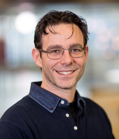
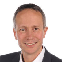
Massimo Mischi is a Full Professor at the Faculty of Electrical Engineering of the Eindhoven University of Technology (TU/e) and chairs the Signal Processing Systems (SPS) Division , the largest division at TU/e with over 250 researchers. He founded the Biomedical Diagnostics (BM/d) Lab in 2012, which now includes 180 researchers and clinical/industrial advisors, focusing on biomedical signal processing for diagnostics and monitoring.
Craig H. Meyer is a Professor in Biomedical Engineering and Radiology & Medical Imaging at the University of Virginia. He holds a Ph.D. from Stanford University and leads the Rapid MRI Research Group, focusing on developing advanced MRI techniques for cardiovascular disease, neural disorders, and pediatrics. His work integrates physics, signal processing, and machine learning to improve MRI acquisition and processing speed. Education: Ph.D. in Biomedical Engineering, Stanford University. Research Interests: Medical and Molecular Imaging, Signal and Image Processing, Biomedical Data Sciences, Biomechanics, and Cardiovascular Engineering. His innovations include fast spiral imaging, conjugate phase reconstruction, and machine learning-enhanced MRI denoising. Awards: Notably includes the Dean’s Award for Excellence in Team Science (2014), Fellowships from NAI (2021), AIMBE (2015), and ISMRM (2013). He also authored two landmark MRI papers recognized as pivotal in the field. Teaching: Courses include BME 6310 (Computation and Modeling in Biomedical Engineering) and BME 8782 (Magnetic Resonance Imaging). He emphasizes translational research, with applications in clinical MRI advancements and collaborative interdisciplinary projects. Labs/Groups: Rapid MRI Research Group focuses on cutting-edge MRI technologies, including real-time cardiac imaging and artifact reduction through deep learning.
Professor Guy Williams is a leading academic at the University of Cambridge with a focus on imaging science and clinical neurosciences, affiliated with Downing College and the Wolfson Brain Imaging Centre . Holding a PhD in Physics from his initial Natural Sciences degree, he specializes in nuclear magnetic resonance (NMR) and MRI techniques for brain imaging. Education: BA, PhD in Physics His research centers on non-invasive imaging of brain structure and function, particularly in traumatic brain injury (TBI) and dementia. His work involves developing novel MRI pulse sequences and advanced data analysis algorithms, including AI-based diagnostic tools. He leads studies on white matter integrity post-trauma, longitudinal dementia assessment, and applications of MRI in disorders of consciousness and addiction. Recent publications highlight collaborations in traumatic brain injury outcomes, AI-guided dementia prediction, and neuroimaging of post-COVID cognitive deficits. His team's work on ultra-high field laminar fMRI and distortion correction methods has advanced clinical neuroscience applications. Key techniques include diffusion tensor imaging (DTI), 7 Tesla MRI, and positron emission tomography (PET/MR). His research spans from basic NMR physics to clinical translation, with a strong emphasis on multi-site studies and real-world diagnostic implementation.
Dr. Merry Mani is an Associate Professor in Radiology and Imaging Sciences and Biomedical Engineering, specializing in biomedical imaging and signal processing. Her work focuses on advancing MRI-based imaging technologies to study neurological disorders such as Alzheimer's, Autism, and Epilepsy. She holds a Ph.D. in Electrical and Computer Engineering from the University of Rochester (2014) and completed a postdoctoral fellowship at the University of Iowa School of Medicine (2018). Her research combines biophysical modeling with machine learning to explore brain microstructures. Key achievements include the NNARSAD Young Investigator Grant and NIH-funded projects like 'Fast Multi-dimensional Diffusion MRI with Sparse Sampling'. Her lab develops cutting-edge reconstruction methods like qModeL and MUSSELS, prioritizing high spatio-temporal resolution imaging. Major contributions span diffusion MRI acquisition, model-based deep learning, and clinical applications in neurodegenerative diseases. Notable grants include NIH R01EB031169 for Alzheimer’s neurodegeneration studies and projects on rTMS for depression. Her work bridges imaging innovation with clinical impact, aiming to improve diagnosis and treatment through advanced imaging biomarkers.
Dr. Peter Fokker is a Researcher at Utrecht University's Faculty of Geosciences, specifically within the Department of Earth Sciences and the Experimental Rock Deformation/HPT group. He is affiliated with the Research Programme in Earth Sciences Utrecht (DES/IVAU) and has been actively publishing in geomechanics, subsidence modeling, and induced seismicity for over three decades. His work primarily focuses on the application of geomechanical principles to understand and model subsurface processes related to resource extraction and geothermal energy. Dr. Fokker's research interests span several interconnected domains in geomechanics and subsurface engineering. His primary focus is on experimental rock deformation , studying how rocks behave under various stress conditions. He has made significant contributions to subsidence modeling , particularly in the context of gas field depletion in the Netherlands. His work on induced seismicity has helped understand the relationship between subsurface operations and seismic events. Additional interests include geothermal energy systems , reservoir engineering , and the application of data assimilation techniques to improve subsurface characterization. His research often bridges theoretical models with practical applications in energy resource management. An analysis of Dr. Fokker's recent publications (2020-2025) reveals a strong focus on practical applications of geomechanics to real-world challenges. His work increasingly integrates InSAR technology and data assimilation methods to monitor and model subsidence processes. There's a clear emphasis on geothermal energy applications , reflecting growing interest in sustainable energy solutions. His research also demonstrates a sophisticated approach to modeling complex reservoir behaviors across multiple scales, from laboratory experiments to field-scale operations. The interdisciplinary nature of his work is evident in collaborations spanning geology, engineering, and environmental science. Dr. Fokker has supervised multiple research projects and students throughout his career, as indicated by the "Supervised Work (4)" reference in his profile. His research has been supported by various grants focused on subsidence modeling, geomechanics of energy resources, and induced seismicity. He has been involved in significant collaborative efforts, including the Dutch National Scientific Research Program on Land Subsidence. Dr. Fokker is part of the Experimental Rock Deformation/HPT group at Utrecht University, which conducts laboratory experiments and develops theoretical models to understand rock behavior under various conditions. His work contributes to the broader research ecosystem focused on sustainable resource management and understanding subsurface processes, with particular relevance to the Dutch context of gas extraction and land subsidence.
Jonas Bylander is a Professor at Chalmers University of Technology in the Department of Microtechnology and Nanoscience, specifically within the Quantum Technology division. He leads a research group focused on developing quantum computers using superconducting circuits.
Bertan Bakkaloglu is the On Semiconductor Professor in the School of Electrical, Computer and Energy Engineering at Arizona State University (ASU), where he has been since 2004. Prior to ASU, he worked at Texas Instruments focusing on analog, mixed-signal, and RF SoC development for communication transceivers. His research expertise spans RF and mixed-signal IC design, wireless/wireline communication systems, and broadband communication systems. Education: Ph.D. in Electrical Engineering, Oregon State University (1995) M.S.C. in Electrical Engineering, University of Houston (1992) Research Interests: RF and mixed-signal integrated circuits Power management ICs (including LDO regulators and DC-DC converters) High-efficiency power delivery systems Radiation-hardened electronics for space applications MEMS-based sensor systems Biomedical circuits for implantable devices Grants & Collaborations: Over 40+ funded research projects with institutions like NASA/JPL, BAE Systems, and NSF, focusing on power electronics, space systems, and biomedical applications Key projects include radiation-hardened converters, self-calibrating DACs, and implantable medical device circuits Industry partnerships with Texas Instruments, Space Micro, and FLIR Professional Activities: Technical committee member for IEEE Radio Frequency Integrated Circuits Conference Founding chair of IEEE Solid-State Circuits Society Phoenix Chapter
Eduardo Mercado III is a Professor in the Department of Psychology at the University at Buffalo, College of Arts and Sciences. His research focuses on bioacoustics, cognitive psychology, and marine ecology, particularly the vocal behavior of humpback whales and its implications for understanding human impact on marine ecosystems. He is also known for his work in perceptual learning, autism spectrum disorder, and comparative cognition. Scientific Awards Guggenheim Fellowship Harvard Radcliffe Institute Fellowship Research Trends His recent publications emphasize bioacoustic analysis of humpback whale songs, including their spectral entropy, cyclical variations, and adaptive adjustments to anthropogenic noise. Additional work explores perceptual learning mechanisms in autism, neural network modeling for acoustic classification, and cognitive processes in canines and rodents. Projects Mercado’s “Singers as Sentinels” project combines acoustic analysis of humpback whale songs with public awareness initiatives about ocean noise pollution. The project will produce a book, Why Whales Sing and Dolphins Don’t , and a web-based interface for public engagement.
Dr. Gary Glover is a Professor of Radiology (Radiological Sciences Lab) at Stanford University , with courtesy appointments in Psychology and Electrical Engineering. His work focuses on the physics and mathematics of MRI, particularly rapid scanning methods using spiral k-space trajectories for functional brain imaging and multimodal neuroimaging (fMRI/EEG/fPET/fNIRS) combined with neuromodulation techniques like TMS and transcranial ultrasound. Academic Appointments: Radiology, Psychology, Electrical Engineering Professional Affiliations: Bio-X, Stanford Cancer Institute, Wu Tsai Neurosciences Institute Research Interests include: Development of blood oxygen level-dependent (BOLD) and viscoelastic contrast in MRI Functional MR Elastography for brain activation mapping Optimization of MR-ARFI for transcranial ultrasound guidance Automated spinal cord segmentation (EPISeg) using machine learning Scientific Awards : National Academy of Engineering (2013) Gold Medal, ISMRM (2000) Steinmetz Award, General Electric (1985) Lauterbur Lecture, ISMRM (2018) Recent Publications analyze: Fast fMRI sampling and spurious signal correction Dissociated patterns in default mode network anti-correlations Neural correlates of collaborative behavior in triadic fMRI Salience network contributions to depression pathophysiology
Guido Pintacuda is a CNRS Research Director and Head of the Lyon High-Field NMR Center (CRMN) at École Normale Supérieure de Lyon since 2019. His work centers on advancing solid-state NMR methodologies with ultra-fast magic-angle spinning (MAS) to achieve atomic-level resolution in complex biomolecular and materials systems that are intractable to conventional techniques. Educational background: Undergraduate studies (1992-1997) and PhD in Sciences (1998-2002) at Scuola Normale Superiore in Pisa, Italy; postdoctoral research at Karolinska Institutet (2001-2004) and Australian National University (2004). Research interests focus on pushing NMR frontiers through high-field instrumentation and fast MAS (up to 160 kHz), with dual objectives: (i) biomolecular structure determination for membrane proteins, amyloid fibrils, and viral assemblies; (ii) solid-state NMR of paramagnetic materials like battery cathodes and catalysts. His innovations include proton detection in fully protonated proteins and DNP-enhanced sensitivity. Recent publications (2021-2024) show heavy emphasis on proton-detected NMR under fast MAS for structural biology, alongside growing work in paramagnetic materials. Key trends include method development for μs–ms dynamics, miniature rotor protocols for membrane proteins, and collaborations with Bruker for 150+ kHz probe technology. Scientific awards: ERC Consolidator Grant (P-MEM-MAS, 2015-2021) Sackler Prize (2017) ISMAR Fellow (2020) Mentoring and grants: Principal investigator for major projects including ERC (2.5 M€), ANR CTRbyNMR (384 k€), and EU PANACEA (5 M€, co-coordinator). Actively mentors PhD student Clément Ollier and postdocs (Z. Sun, S. Medina-Gomez) at ENS Lyon and international schools. Labs and teams: Directs CRMN (UMR 5082 CNRS/ENS Lyon/UCBL), a world-class NMR facility with unique high-field equipment. Leads a research group developing 150+ kHz MAS probes in partnership with Bruker Biospin and maintains strong ties to the University of Delaware (T. Polenova) and European networks.
Koenraad Muylaert is a Full Professor at the Faculty of Science, KU Leuven, and head of the Biology department at KU Leuven Kulak. His research focuses on microalgae ecology and phytoplankton physiology , with applications in eutrophication studies , wastewater treatment , and biofuel production . Based in Kortrijk, Belgium, he works with international teams in Ecuador, Qatar, and Belgium. Current projects on mountain lake eutrophication and urban aquatic systems Specializes in nano-material flocculation and omega-3 fatty acid production from microalgae Research Trends from his recent articles show emphasis on: Microalgae harvesting innovations (cellulose nanocrystals, PDMAEMA polymers) Comparative processing techniques (DAF vs sedimentation, drying methods) Biotechnological applications in flavor chemistry and microbiome interactions Laboratory operates at KU Leuven's Kortrijk campus, with strong collaborations in environmental engineering and food science . His work bridges fundamental ecological research with industrial biotechnology for sustainable solutions.
Dr. Neal Bangerter is a Visiting Professor in the Department of Bioengineering at Imperial College London's Faculty of Engineering. He specializes in medical imaging (MRI), artificial intelligence, machine learning, and signal processing. Dr. Bangerter holds adjunct appointments at INSEAD, the University of Utah, and Brigham Young University. His research focuses on ultra-high field MRI, AI applications in healthcare, and data-driven bioscience technologies. He leads the London Collaborative Ultra-High Field Scanner (LOCUS) project and advises companies on AI and innovation strategies. Education: B.S. in Physics (UC Berkeley), M.S. and Ph.D. in Electrical Engineering (Stanford University). Career highlights include roles at McKinsey & Company, Microsoft, and Reactrix, as well as founding BYU's Medical Imaging Research Center. He has pioneered cross-faculty initiatives like the Crocker Innovation Fellowship Program. Research interests include novel MRI pulse sequences, AI in medical imaging, and large-scale health data analysis. His work spans collaborations with Stanford, Oxford, Cambridge, and Siemens Healthcare. He teaches executive education at INSEAD, focusing on bridging technical concepts with business strategies. Key awards include the David Evans Chair at Brigham Young University. His contributions to the UK Biobank Neuroimaging study and development of MRI techniques like RAFO-4 highlight his impact on advancing imaging technologies and AI applications in healthcare.
Douglas C. Noll is the Ann and Robert H. Lurie Professor of Biomedical Engineering and Professor of Radiology at the University of Michigan. He holds key roles as Co-Director of the Functional MRI Laboratory, Co-Lead of the NeuroImaging Core at the Michigan Alzheimer’s Disease Research Center, and collaborator at the Michigan Institute for Imaging Technology and Translation (MIITT). His affiliations include the Michigan Neuroscience Institute, Center for Computational Medicine and Bioinformatics, and Michigan Concussion Center. His research focuses on advancing MRI and fMRI technologies to study brain function and neurological disorders. Key projects include rapid image acquisition, artifact elimination, physiological modeling, and MRI-guided therapies like histotripsy. Recent work emphasizes pre-clinical MRI-guided focused ultrasound systems and collaborations with neuroscientists to map brain organization in health and disease. Notable contributions include developing the Oscillating Steady State Imaging (OSSI) technique, the TOPPE framework for MRI sequence prototyping, and tools like FieldMapNet MRI for off-resonance correction. His lab addresses challenges in high-resolution fMRI, real-time motion compensation, and translational imaging for clinical applications. Current efforts span improving MRI hardware-software integration, advancing non-invasive brain therapies, and applying machine learning to enhance image reconstruction and artifact correction. Collaborations bridge engineering, neuroscience, and clinical medicine to tackle complex neurological conditions like Alzheimer’s and brain tumors.
Yonghyun Ha is an Associate Research Scientist in the Department of Radiology & Biomedical Imaging at Yale School of Medicine. His research focuses on advancing magnetic resonance imaging (MRI) technologies, particularly in low-field MRI systems, RF pulse design, and imaging hardware innovation. He collaborates with experts like Duy Phan, Haifan Lin, and Nikhil Malvankar, contributing to projects such as RF pulse distortion compensation and novel RF coil development. Research Interests: His work spans low-field MRI systems , RF engineering , imaging algorithm optimization , and hardware design . He explores applications like point-of-care imaging and cost-effective MRI solutions. Articles Trends: Recent publications address gradient-free imaging, field-cycling magnets, and deep learning for data compression. His work bridges engineering and clinical needs, emphasizing practical MRI advancements. Advising & Grants: No formal advisees are listed, but his collaborations suggest involvement in interdisciplinary research teams. No specific grants are mentioned, but his projects imply funding through institutional or NIH channels. Labs/Teams: Active in Yale’s Radiology & Biomedical Imaging department, contributing to MRI technology development and translational research initiatives.
Cornelius Faber is a University Professor in the Department of Radiology at the University of Münster, Germany, where he leads the Experimental Nuclear Magnetic Resonance research group. His work focuses on developing and implementing novel MRI techniques that extend the boundaries of magnetic resonance imaging in terms of spatial and temporal resolution, sensitivity, and specificity for physiological, structural, and molecular changes. He actively participates in the "Cells in Motion" interdisciplinary research initiative at the university. Professor Faber's research spans multiple critical areas in medical imaging and biomedical science. His primary expertise lies in MRI cell tracking , enabling visualization of cellular dynamics in vivo. He has made significant contributions to infection imaging , developing methods to detect and characterize microbial infections using MRI. His work on MR methodology development has advanced quantitative imaging techniques, while his research on multimodal integration in MR and MRI contrast mechanisms has provided deeper insights into molecular and cellular processes. His research bridges physics, engineering, and biomedical applications, with particular relevance to inflammation, cancer, neurological disorders, and cardiovascular disease. Analysis of Professor Faber's extensive publication record reveals a clear evolution from fundamental MRI technique development toward increasingly sophisticated applications in disease models. His recent work demonstrates a strong trend toward multimodal imaging approaches that combine MRI with complementary techniques such as mass spectrometry, optical imaging, and PET. This integration creates comprehensive diagnostic platforms that provide both anatomical and molecular information. A notable pattern is the focus on cellular dynamics, particularly immune cell behavior in inflammatory conditions and tumor microenvironments, with applications spanning neuroscience, oncology, and cardiology. Professor Faber leads a multidisciplinary research team of approximately 15 members, including scientists, doctoral students, technicians, and medical students. His laboratory is deeply integrated with the University of Münster's research infrastructure, particularly the Multiscale Imaging Centre. The group's work contributes significantly to advancing preclinical MRI methodologies while maintaining strong clinical relevance, with numerous publications in high-impact journals across medical imaging, neuroscience, and biomedical engineering disciplines.