Madhavi Ganapathiraju is an Associate Professor in the Department of Biomedical Informatics at the University of Pittsburgh, affiliated with the School of Computing and Information. She holds additional roles in the Intelligent Systems Program and Integrated Systems Biology Program. Her research focuses on systems biology, particularly protein-protein interaction prediction, and applies machine learning to translational bioinformatics and genomics of diseases. Education includes a PhD from Carnegie Mellon University (Language Technologies Institute) and master's/bachelor's degrees from Indian institutions. Her work bridges computer science and medicine, with notable contributions in schizophrenia interactome studies, congenital heart disease genetics, and drug repurposing via computational methods. Awards include NIH BRAINS Award (2011) and grants from Stanley Medical Research Institute. She leads projects like the MHAIN Interactome and Mesothelioma interactome studies. Active in funding, she has served as PI/co-PI on multiple NIH and foundation grants totaling over $5M. Labs/Teams: Leads the Computational Biomedical Informatics Lab, collaborates with geneticists and clinicians on translational projects. Current focus includes cilia biology in heart defects, AI-driven drug discovery, and interactome-based precision medicine strategies.
Holger Stark is a Professor in Molecular Electron Cryomicroscopy at the University of Göttingen and Director at the Max-Planck-Institute for Multidisciplinary Sciences. His research focuses on 3D structural determination of macromolecular complexes using cryo-EM, including spliceosomes, ribosomes, and metabolic enzymes. He leads the Structural Dynamics department, developing novel methodologies for complex stabilization and computational image analysis. Education includes a Dr. rer. nat. in Biochemistry from Freie Universität Berlin (1996). Key career milestones include postdoctoral research at Imperial College London, junior and BioFuture group leadership roles at Max-Planck-Institute, and directorship since 2015. His work bridges biochemistry, biophysics, and computational biology, achieving world-record cryo-EM resolutions. Research interests emphasize structural mechanisms of mRNA processing and metabolic pathways. Collaborative projects include spliceosome dynamics and fatty acid synthesis. His lab also develops imaging and computational tools, such as the COW software package.
Dr. Marcus Bischoff is a Lecturer in the School of Biology at the University of St Andrews. His research focuses on understanding the mechanisms governing morphogenesis—the process by which cells coordinate behaviors to shape tissues and organs. Using Drosophila abdominal epidermis as a model, he combines live 4D microscopy with genetic tools to study signal regulation, cytoskeletal dynamics, and mechanical forces in development. Key interests include how cells interpret signals to execute migration, constriction, and other behaviors, with implications for wound repair and cancer. His research has been funded by grants from The Wellcome Trust and BBSRC, including projects investigating morphogen transport, cytoskeletal coordination, and biofilm inhibition. He advises PhD students Aimee Bebbington and Carey Li. Publications highlight contributions to fields like exosome-mediated Hedgehog transport, caspase activation dynamics, and anti-biofilm cyclic peptide design. Collaborations span institutions globally, emphasizing interdisciplinary approaches to developmental biology challenges.
Giovanna Barba-Spaeth is a Group Leader in the Structural Virology Unit at the Institut Pasteur in Paris, where she leads the FlavImmunity research group. Her work focuses on the structural and functional dynamics of enveloped viruses, particularly flaviviruses (dengue, Zika, yellow fever, West Nile) and SARS-CoV-2. Her research interests center on understanding the conformational changes in viral envelope proteins during virus entry, fusion, and egress, and how these processes are modulated by the cellular environment and neutralizing antibodies. She employs cutting-edge techniques such as cryo-electron microscopy (cryo-EM and cryo-ET), single-particle fluorescence microscopy, and functional assays under BSL-3 conditions. The 15 most recent publications highlight a strong focus on flavivirus immunology, antibody-dependent enhancement (ADE), vaccine responses, and structural mechanisms of viral entry and neutralization. Key themes include cross-reactivity between dengue and Zika, the impact of prior flavivirus immunity on yellow fever vaccination, structural insights into envelope protein dynamics, and the development of tools like CoronaFISH for visualizing SARS-CoV-2 RNA. Human type I IFN deficiency does not impair B cell response to SARS-CoV-2 mRNA vaccination. Analysis of mRNA vaccination-elicited RBD-specific memory B cells reveals strong but incomplete immune escape of the SARS-CoV-2 Omicron variant. Prior flavivirus immunity skews the yellow fever vaccine response to cross-reactive antibodies with potential to enhance dengue virus infection. She mentors several researchers, including PhD student Sina Sommer and MSc student Jolyn Ooster, and collaborates with laboratories at LMU Munich. Her group is internationally recognized for its contributions to structural virology and flavivirus immunology, with publications in top-tier journals such as Nature, Nature Immunology, and Nature Communications.
Bjørn Torger Stokke is a Professor in the Department of Physics at the Norwegian University of Science and Technology (NTNU) . His research focuses on biophysics at the intersection of life sciences and engineering, with emphasis on the physical properties of biological macromolecules and their assemblies, such as hydrogels. Key areas include biopolymer interactions, polyelectrolyte complexes, and responsive hydrogels as biosensors. His work employs advanced techniques like atomic force microscopy (AFM), total internal reflection fluorescence microscopy (TIRFM), and dynamic force spectroscopy. Collaborations with NTNU NanoLab enable microfluidic chip fabrication for hydrogel research. Notable projects include studying amyloid structures, Toll-like receptors, and enzymatic mode-of-action through combined experimental and computational approaches. Publications span topics like hydrogel characterization, cancer cell mechanics, and plant cell wall analysis. His research bridges material science with biomedicine, addressing applications in diagnostics and therapeutics. Collaborations span national and international institutions, emphasizing interdisciplinarity.
Adalberto Claudio Quiros serves as a Lecturer in Artificial Intelligence within the School of Cancer Sciences at the University of Glasgow, where he bridges advanced machine learning techniques with oncological research to transform cancer diagnostics and treatment. His work focuses on developing AI-driven solutions for analyzing histopathological data to improve cancer outcome prediction and therapeutic decision-making across multiple malignancies. His research program centers on self-supervised learning applied to digital pathology, with core interests including: Development of deep learning frameworks for histomorphological pattern discovery Predictive modeling of treatment response and cancer progression Unsupervised representation learning from unlabeled whole-slide images Integration of AI with clinical oncology for precision medicine applications Quantitative analysis of tumor microarchitecture and spatial heterogeneity Analysis of his publication trajectory reveals a concentrated effort in leveraging self-supervised learning to decode histopathological features predictive of clinical outcomes. His recent work demonstrates consistent innovation in applying these techniques across diverse cancers—including cutaneous squamous cell carcinoma, mesothelioma, osteosarcoma, and lung adenocarcinoma—with particular emphasis on identifying morphological biomarkers for recurrence, metastasis, and treatment resistance. This research program shows increasing clinical translatability through collaborations with oncology and pathology departments. No scientific awards are documented in available materials. Regarding academic mentoring, no student names or advising activities are mentioned in the provided information. Similarly, details about research grants, laboratory infrastructure, or collaborative research teams are not specified in the source content.
King-Wai Yau is a Professor of Neuroscience at the Solomon H. Snyder Department of Neuroscience, Johns Hopkins University School of Medicine. His research focuses on sensory transduction processes in vision and olfaction, particularly phototransduction in rods, cones, and intrinsically photosensitive retinal ganglion cells (ipRGCs), as well as olfactory transduction mechanisms. Yau’s work explores how light and odorant signals are converted into electrical signals, with emphasis on cyclic nucleotide pathways, calcium feedback mechanisms, and signal amplification. He investigates topics such as light adaptation, melanopsin signaling, and the structural-functional relationships of photoreceptors in mammals and teleosts. Yau’s lab has pioneered studies on ipRGCs, revealing their dual roles in conveying rod/cone signals and intrinsic light detection. Key discoveries include the role of melanopsin in non-image-forming vision (e.g., circadian rhythms, pupillary reflex), the molecular basis of phototransduction in these cells, and comparisons with traditional rod/cone pathways. His work also addresses olfactory receptor neuron transduction, including odorant-receptor specificity and cAMP-mediated signaling. Yau has advised multiple graduate students and postdoctoral fellows, including Yaqing YE, Zuying CHAI, and others. His lab integrates electrophysiology, molecular genetics, and imaging techniques to study retinal physiology and sensory biology. Current projects include understanding phototransduction kinetics, light-induced signaling cascades, and the evolutionary adaptations of sensory systems. His research has been published in top-tier journals such as Nature , Science , and Neuron , with over 15 highlighted publications since 2002. Yau serves on the faculty of the Neuroscience Training Program and Biochemistry, Cellular and Molecular Biology Graduate Program.
Prof. Dr. Nikolai Klymiuk is an Associate Professor of Cardiovascular Translation in Large Animal Models at the Technical University of Munich (TUM), Department of Cardiology. He leads the TUM School of Medicine and Health's research on genetically modified large animal models for biomedical applications, focusing on monogenic hereditary diseases and xenotransplantation. His work emphasizes translational potential, advanced intervention techniques, and comprehensive model characterization. Education: Biochemistry studies at University of Vienna, followed by research at Ludwig Maximilian University of Munich. Director, Institute of Farm Animal Genetics (2018–present). Associate Professor at TUM since 2019. Research highlights include: Development of pig models for Duchenne muscular dystrophy and Usher syndrome. Pioneering cardiac xenotransplantation studies achieving 3-month survival in baboons. Advances in gene editing (e.g., CRISPR/Cas9) and somatic cell therapy. Key achievements include the 2017 European Society of Toxicologic Pathology Award and the 2014 Walther Baier Prize. His lab collaborates extensively on preclinical trials for heart, kidney, and lung xenotransplantation. Current projects focus on optimizing xenograft compatibility, reducing immune rejection, and refining gene therapy delivery in porcine models.
Dr Sarah Dean is a Senior Lecturer in Healthcare/Biomedical Science at the University of the West of England (UWE Bristol), affiliated with the HAS - Applied Sciences department within the School of Applied Science. She specializes in Cellular Pathology and Cancer Biology, focusing on undergraduate and postgraduate education. Her roles include Placement Coordinator for the School of Applied Science and Module Leader for courses such as Applied Scientific Practice, Tissue and Tumour Science, and Professional Practice in Applied Science. Her research centers on biomarkers in breast cancer, particularly Triple Negative Breast Cancer (TNBC), with emphasis on metabolic pathways, prognosis, and cellular mechanisms. She employs both in vivo and in vitro models to investigate biomarkers like BCAT isoforms, GRP78, and IGFBP-3. Collaborations include work with Dr. Claire Perks at the University of Bristol and membership in the NHS Research Ethics Committee. Publications highlight her contributions to understanding tumorigenesis, chemoresistance, and molecular pathways in breast cancer. Key areas of exploration include EMT induction under hypoxia, PI3K/Akt signaling modulation, and the role of PTEN in TNBC progression. While no scientific awards are listed, her work bridges clinical and laboratory research, emphasizing translational outcomes. Teaching responsibilities span cellular pathology, histology, and professional practice, with a focus on hands-on placement modules. Her research extends to acute myeloid leukemia drug resistance and low-abundance protein visualization techniques, demonstrating versatility in oncology research.
Jun Hee H. Lee is a Professor of Molecular and Integrative Physiology and Research Professor at the Institute of Gerontology within the University of Michigan's Medical School. He serves as Associate Director of Academic Programs in Molecular and Integrative Physiology. His research focuses on stress, aging, and metabolism, particularly exploring stress adaptation mechanisms that enhance cellular resilience and longevity. Lee's work integrates molecular biology, genomics, and technology development to understand age-related diseases and metabolic disorders. Research affiliations include the Center for Computational Medicine and Bioinformatics, Caswell Diabetes Institute, Rogel Cancer Center, and others. Key areas of investigation include Sestrin signaling, autophagy dysfunction in diseases like NAFLD, and single-cell spatial transcriptomics. Recent breakthroughs include the Seq-Scope technology for spatial gene expression visualization, which earned him the 2024 Hanseong Science Award in Life Sciences. Key Technologies: Spatial transcriptomics, single-cell analysis Clinical Focus: Metabolic disorders, cancer, cardiovascular health Interdisciplinary Collaborations: Bioinformatics, computational medicine, systems biology Lee's lab actively develops tools to study cellular stress responses at unprecedented resolution, with applications in drug discovery and precision medicine. His mentorship supports students and researchers in translating molecular insights into clinical solutions.
Ralph E. Kleiner is an Associate Professor of Chemistry at Princeton University, leading the Kleiner Lab. His research focuses on understanding RNA function and regulation through chemical biology approaches, specifically investigating RNA-binding proteins (RBPs), RNA modifications, and developing chemical tools for studying RNA synthesis, turnover, and structure. Key areas include post-transcriptional regulation, epitranscriptomics, and therapeutic targeting of RBP interactions. Education and affiliations are not explicitly detailed in the provided text, but his lab is located in the Frick Laboratory. Research highlights include developing tools like metabolic labeling and RNA editing platforms to study RNA-protein interactions, as well as characterizing RNA-modifying enzymes. Research Interests: The lab explores the role of RBPs in RNA lifecycle processes, the functional significance of RNA modifications (e.g., 5-formylcytosine, m6A), and the development of chemical methods for RNA imaging and structural probing. Recent advancements include SNIPER-seq for RNA structure analysis and methods for studying ribonucleoprotein granules. Awards and Honors: 2023: ICBS Young Chemical Biologist Award 2023: Kavli Fellow 2019: NSF CAREER Award 2019: Sloan Research Fellowship 2017: Sidney Kimmel Foundation Scholar Award Lab and Collaborations: The Kleiner Lab collaborates across disciplines, integrating chemistry and biology to address fundamental and translational questions. Recent projects include preprints on DUS enzymes and RNA structure probing, and mentoring graduate students such as Tyler Schwarz and Crystal (PhD graduates). The lab also emphasizes outreach and training through its website and educational initiatives.
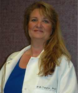
Patricia R. Taylor, PhD is an Associate Professor in the Department of Ophthalmology and Visual Sciences at Case Western Reserve University's School of Medicine, with a secondary appointment as Research Health Scientist at the Cleveland VA Medical Center. Her research focuses on immunological mechanisms underlying diabetic retinopathy, particularly the role of IL-17A cytokine signaling. Education: BSc Microbiology (University of Akron, 2005), MSc Biology (University of Akron, 2007), PhD Biomedical Sciences (Kent State University/NEOMED, 2010), Postdoc in Ocular Immunology at Case Western Reserve University (2014). Research Interests: Investigates IL-17A's role in retinal oxidative stress, inflammation, and vascular damage. Current projects include NEI R01-funded studies targeting TRAF signaling proteins and VA Merit-funded clinical trials assessing IL-17A's role in anti-VEGF resistance. Grant Funding: Active NEI R01 and VA Merit awards. Research emphasizes translational approaches to identify novel therapeutics for retinal pathogenesis. Key Labs/Teams: Collaborates with Cleveland VA Medical Center and the Institute of Pathology. Her work bridges immunology and ophthalmology to address diabetic retinopathy mechanisms.
Jakub Nedbal is a Researcher at King’s College London’s Department of Physics within the Faculty of Natural, Mathematical & Engineering Sciences. He holds a Master’s in Physics from Masaryk University (Czech Republic) and a Master’s in Immunology from King’s College London, alongside a PhD focused on imaging techniques in B cell biology. His research specializes in developing advanced fluorescence-based imaging technologies, particularly leveraging SPAD arrays and time-correlated single-photon counting (TCSPC) techniques. Notable contributions include creating the first fluorescence lifetime flow cytometer using TCSPC and co-developing a fast TCSPC microscope with parallelized SPAD array detectors. Current work focuses on spatially resolved TCSPC detectors for thick-sample imaging and low-phototoxicity techniques. Outside academia, Jakub served as Product Development Lead for a startup commercializing TCSPC array cameras until 2018. His innovations have been applied to art conservation via fluorescence lifetime imaging (FLIM) for varnish analysis and have advanced microalgae cultivation bioreactor designs. His research interests span fluorescence lifetime imaging, microscope development, SPAD arrays, and photosynthesis studies. Recent work highlights include a novel dual-excitation fluorometer for photosynthesis research and a SPAD array camera for lightsheet FLIM. His techniques address challenges in live-cell imaging phototoxicity and high-throughput FRET screening.
Michael Held is an Associate Professor in the Department of Chemistry and Biochemistry at Ohio University's College of Arts and Sciences, where he leads an active research program focused on plant cell wall biochemistry. His work integrates molecular, biochemical, and biophysical approaches to understand the regulation and assembly of plant cell walls, with a particular emphasis on extensin glycoproteins and post-transcriptional regulatory mechanisms. His educational background includes a Ph.D. and B.S. from Ohio University, followed by postdoctoral training at Michigan State University and Purdue University, establishing a strong foundation in plant molecular biology and biochemistry. Held's research centers on two major areas: (1) the self-assembly of plant cell wall polymers, particularly the role of extensins as structural scaffolds, and (2) the post-transcriptional regulation of cell wall biosynthesis via small RNAs derived from cellulose synthase antisense transcripts. His lab employs advanced techniques such as small RNA next-generation sequencing (sRNA-NGS), bioinformatics, and biophysical assays to uncover novel regulatory networks in plant development. The recent publications highlight a strong trend in plant glycobiology, with a focus on arabinogalactan-proteins, glycosyltransferases, and gene co-expression networks. His development of PlantNexus, a database for barley and sorghum, reflects his commitment to open science and interdisciplinary collaboration in plant genomics. Specific protein interactions between rice members of the GT43 and GT47 families to form various central cores of putative xylan synthase complexes (2024) Knockout of eight hydroxyproline-O-galactosyltransferases cause multiple vegetative and reproductive growth defects (2023) PlantNexus: A Gene Co-expression Network Database and Visualization Tool for Barley and Sorghum (2022) Functional characterization of hydroxyproline-galactosyltransferases for Arabidopsis arabinogalactan-proteins synthesis (2021) Extensins: Self-assembly, crosslinking, and the role of peroxidases (2021) Dr. Held actively mentors students and collaborates with researchers across institutions, contributing to projects involving CRISPR-Cas9 gene editing, mass spectrometry for glycan detection, and the functional characterization of glycosyltransferases. His lab has received support for research in plant wall biosynthesis, though specific grants are not detailed in the text. He is a key contributor to the PlantNexus initiative, which provides valuable tools for plant biologists studying barley and sorghum. The Held Lab is located at the Biochemistry Research Facility, 350 W. State St., Athens Campus, and maintains a research focus on uncovering the molecular mechanisms governing plant cell wall integrity, development, and function. The lab combines classical biochemical methods with modern genomics and bioinformatics to address fundamental questions in plant biology.
David Linden is a Professor of Neuroscience at the Solomon H. Snyder Department of Neuroscience, Johns Hopkins University School of Medicine. His research focuses on neuronal plasticity, particularly structural and functional changes in the adult mammalian brain in response to injury, learning, hormonal cycles, and pharmacological interventions. He employs in vivo two-photon microscopy to visualize dynamic changes in neural circuits, with a special emphasis on serotonin and norepinephrine axon regeneration. His research interests include cellular and molecular neuroscience , neural circuits , synaptic plasticity , and the neurobiology of disease . He investigates how experience shapes brain structure and function, including how estrogen affects spine dynamics, how brain injury triggers axonal regrowth, and the molecular basis of long-term synaptic depression. His work bridges molecular mechanisms with behavioral outcomes. The most recent publications reveal a strong trend toward understanding functional axon regeneration in the central nervous system, particularly in catecholaminergic systems. His lab has demonstrated that serotonin and norepinephrine axons can regrow across injury sites and restore function—a rare phenomenon in the adult brain. Other studies explore hormonal modulation of plasticity, receptor dynamics in learning, and cerebellar circuit stability. These works span in vivo imaging , molecular neuroscience , and behavioral neurobiology . David Linden is affiliated with the Cellular and Molecular Medicine and Neuroscience Training Program graduate programs at Johns Hopkins. While no formal list of students is provided, his publications include numerous trainees and collaborators, indicating active mentorship. There is no mention of external grants, but his consistent publication record in top journals suggests sustained funding. His lab closed in November 2024, marking the end of active research operations. He has not received any explicitly mentioned scientific awards in the provided text. However, his body of work has significantly contributed to the understanding of neuroplasticity and regeneration. David Linden led the Linden Lab, which specialized in in vivo imaging of neural plasticity and axon regeneration . The lab utilized advanced microscopy to track structural changes in real time, particularly in cortical and cerebellar circuits. Research teams included neuroscientists, molecular biologists, and imaging specialists. The lab closure in 2024 indicates the conclusion of this research program.