Dr. Dragomir Milovanovic is a Group Leader at the German Center for Neurodegenerative Diseases (DZNE) , affiliated with Charité CrossOver Berlin. His research focuses on understanding how intrinsically disordered regions (IDRs) in presynaptic proteins regulate synaptic vesicle (SV) clustering and cytosol solubility in the crowded nerve terminal environment. Key Themes: Phase separation, biomolecular condensates, neuronal metabolism, and implications for neurodegenerative diseases like Parkinson's and Alzheimer's. The lab investigates how IDRs balance solubility and spatial organization in nerve terminals, using techniques such as confocal imaging and single-molecule tracking . Their work has shown that synapsin 1 forms liquid-like condensates that sequester lipid vesicles, maintaining SV mobility while enabling clustering. Notable findings include the discovery of electric potential at condensate interfaces and the development of the Dipping Contacts model for studying condensate-organelle interactions. These studies provide insights into normal synaptic function and pathological protein aggregation in neurodegenerative diseases.
Michael A. Silver is an Associate Professor in the School of Optometry and Vision Science at UC Berkeley, with affiliations in the Helen Wills Neuroscience Institute and the Department of Psychology. His research focuses on cognitive neuroscience, particularly the neurophysiological and neurochemical substrates of visual perception, attention, and learning. He leads the Silver Lab, which investigates topics such as visual attention mechanisms, perceptual learning, cholinergic pharmacology, and the effects of psychedelics on brain function. Silver holds a Ph.D. from UCSF and has been recognized with grants from the NIH, NSF, and private foundations. Education: B.S. Biological Sciences and Chemistry (Carnegie Mellon, 1991); Ph.D. Neuroscience (UCSF, 1999). Awards include the Howard Hughes Medical Institute Predoctoral Fellowship and Hellman Family Faculty Fund. He mentors graduate students and postdoctoral researchers in vision science and neuroscience. Research interests span visual neuroscience, including the impact of psychedelics (e.g., psilocybin) on neural substrates of perception and cognition. His lab collaborates with institutions like Stanford University and UC Irvine to explore therapeutic applications of psychedelics and age-related auditory decline. Key projects include cholinergic modulation of attention and perceptual learning, and functional subdivisions of the lateral geniculate nucleus. Recent articles highlight studies on attentional modulation of visual crowding, effects of psychedelics on predictive coding, and GABA levels in amblyopia. Silver teaches courses on visual cognitive neuroscience and supervises over 20 graduate students and postdocs. His grants total over $5M, supporting work on nicotine therapy for auditory decline and psychedelic science.
Josie Clowney is an Associate Professor in the Department of Molecular, Cellular, and Developmental Biology at the University of Michigan, where she has held faculty position since 2017. Her research investigates the genomic algorithms that construct neural circuits during development, using Drosophila as a model to study how chemosensory systems drive both instinctual behaviors and learning. She teaches Bio 172 and an upper-level seminar on cellular diversity and scientific writing, and mentors graduate students through MCDB, CMB, NGP, and BIOINF PhD programs. Her educational background includes: Ph.D. in Biomedical Sciences (2012) from the University of California, San Francisco B.S. in Cellular and Molecular Biology (2005) from the University of Michigan, where she conducted research with Cunming Duan Clowney's research centers on understanding how definitive neuronal parameters are encoded in genomic information and translated into cellular architectures. Her lab hypothesizes that developmental algorithms for learning circuits versus instinctual circuits differ fundamentally in their genomic requirements, with chemosensory circuits serving as key models. Using fruit flies for their tractable brain organization, her work bridges computational principles and biological implementation to uncover universal brain organization rules. Analysis of her 15 most recent publications (2016-2025) reveals consistent focus on Drosophila mushroom body development, neural sexual differentiation, and spatial constraints in circuit formation. Key themes include non-deterministic mechanisms diversifying cell surface expression, chromatin dynamics in circadian regulation, and how input density tunes sensory responses. Her work integrates genomics, neuroanatomy, and behavior to model how compact genomic information generates complex neural architectures. No scientific awards were mentioned in the provided text. Dr. Clowney advises graduate students through multiple PhD programs at the University of Michigan, though specific student names and grant details are not provided in the source material. Her teaching includes foundational undergraduate coursework and advanced seminars emphasizing scientific writing. The active publication record spanning 2016-2025 indicates sustained research funding supporting her lab's investigations into neural circuit development. The Clowney Lab, housed in the Biological Sciences Building (4218 BSB), employs Drosophila genetics and neuroanatomical techniques to dissect developmental algorithms of brain wiring. Current projects explore how spatial constraints structure learning circuits, mechanisms of neural sexual differentiation, and the genomic encoding of circuit diversity. The lab collaborates within Michigan's neuroscience community through the Program in Biology and participates in interdisciplinary initiatives studying brain evolution and function.
Peter Jonas is the Magdalena Walz Professor for Life Sciences at the Institute of Science and Technology Austria (ISTA) since 2022, and has been a Professor at ISTA since 2010. Previously, he served as Professor of Physiology and Department Head at the University of Freiburg, Germany (1995–2010) and Associate Professor at the Technical University of Munich (1994–1995). Current Role: Group Leader, Cellular Neuroscience Key Techniques: Nanophysiology, Presynaptic Patch-Clamp, Two-Photon Ca2+ Imaging, Optogenetics, Functional Anatomy His research focuses on synaptic signaling mechanisms in the hippocampus, particularly how glutamatergic and GABAergic synapses contribute to network functions. He investigates: Biophysical signaling and plasticity at mossy fiber synapses Role of synaptic properties in higher network functions Calcium channel-vesicle coupling dynamics Modeling of synaptic and network-level phenomena The group employs in vitro and in vivo approaches, combining experimental and computational methods to decode brain function. Recent work highlights the structural and functional analysis of 'giant' cortical presynaptic terminals. Scientific Awards include: Magdalena Walz Professor for Life Sciences (2022) Peter Seeburg Integrative Neuroscience Prize (2021) EMBO Membership (2019) FWF Wittgenstein Award (2016) DFG Gottfried Wilhelm Leibniz Award (2006) His students and postdocs include PhD candidates Silvia Jamrichova, Peipeng Lin, Rebecca Morse Mora, and Priyansha Verma, alongside postdocs Katharina Lichter, Andrea Navas Olive, and Jake Watson. The lab also collaborates with technical staff and scientific computing experts.
Barbara Karten is a Professor and Department Head of the Department of Biochemistry and Molecular Biology at Dalhousie University’s Faculty of Medicine. She holds a PhD from Karl-Franzens University Graz. Her research focuses on cholesterol metabolism in the brain, particularly in neurodegenerative diseases like Niemann-Pick Type C (NPC) disease. Key areas include mitochondrial cholesterol import, synaptic dysfunction in NPC1 deficiency, and lipid trafficking mechanisms. Education: PhD, Karl-Franzens University Graz Research Interests: Cholesterol homeostasis in neurons and glial cells Role of mitochondrial cholesterol in neurodegeneration Mechanisms of NPC1 protein in endosomal-lysosomal pathways Impact of cholesterol imbalances on synaptic function Her lab investigates how cholesterol distribution defects contribute to neuronal energy deficits and synaptic failure in NPC disease. Awards: None explicitly listed in the text. Advising & Grants: Current students include Jena Barter (MSc), Stanley Ibeh (PhD), and Emma Williams (Honours). Funding sources are not detailed here, but research aligns with NIH/CIHR priorities in neurodegenerative disease. Labs/Teams: Her lab is part of Dalhousie’s Department of Biochemistry and Molecular Biology, collaborating with groups in the Faculty of Medicine. A lab website is available for further details.
Kyle E Miller serves as an Associate Professor at Michigan State University with cross-program appointments in the Integrative Biology Faculty, Neuroscience Program, and Genetics & Genome Sciences Program. His laboratory operates from room 337 of the Natural Science Building, where he leads research on fundamental mechanisms of neuronal development. Dr. Miller's research program centers on elucidating the biophysical and molecular mechanisms underlying axon growth. His laboratory employs an integrative methodology combining time-lapse microscopy in living Drosophila embryos and cultured neurons, mathematical modeling of cytoskeletal dynamics, targeted gene disruption, and biophysical force measurements. This multidisciplinary approach specifically investigates organelle biogenesis, transport kinetics, and cytoskeletal element degradation during neurite outgrowth, with translational aims toward improving treatments for traumatic brain injury, spinal cord injury, stroke, and chronic neurodegenerative conditions. Analysis of his 15 most recent publications (2017-2025) reveals three dominant research trajectories: (1) evolutionary hypotheses connecting cytokinesis to neuronal development, (2) active fluid modeling of cytoskeletal mechanics in axonal elongation, and (3) molecular motor regulation of organelle transport (particularly mitochondria and lysosomes). These works consistently bridge neuroscience, cell biology, and biophysics through innovative combinations of in vivo imaging and computational approaches. No scientific awards are documented in the available materials. Dr. Miller maintains an active research laboratory conducting both basic and translational neuroscience investigations, though specific grant funding details and student mentorship records are not provided in the source documentation. The Miller Lab utilizes Drosophila embryonic models and primary neuronal cultures to dissect the mechanical and molecular drivers of axonal elongation, with particular emphasis on microtubule dynamics, molecular motor function, and the evolutionary origins of neuronal structures.
James J. Cheetham is an Associate Professor in the Department of Biology at Carleton University within the Faculty of Science. He holds a B.Sc. and Ph.D. from McMaster University and serves as an Undergraduate Co-op Advisor. His research bridges membrane biochemistry, computational biology, and biophysics. Education: B.Sc. (McMaster University) Ph.D. (McMaster University) Research Interests: Protein-lipid interactions in synaptic vesicle regulation Computational modeling of presynaptic nerve terminals Biophysical techniques for membrane analysis Application of machine learning to protein interaction prediction Research Trends: His publications span molecular neuroscience, computational modeling, and bioinformatics. Recent work focuses on antimicrobial mechanisms and fungal membrane disruption, while earlier studies emphasize synaptic protein dynamics and algorithm design for biological simulations. Advising & Techniques: He employs liposomes, atomic force microscopy, and fluorescence spectroscopy alongside bioinformatics tools like PIPE. Collaborations include neuroscience (e.g., synapsin studies) and computational science (e.g., DEVS models).
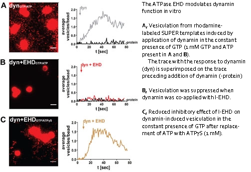
Lennart Brodin is a Professor at the Department of Neuroscience, Karolinska Institutet. He leads the 'Presynaptic mechanisms – Lennart Brodin group,' focusing on molecular mechanisms controlling synaptic vesicle cycling and their links to neurodegenerative diseases. His research integrates cellular and molecular neuroscience to study synaptic vesicle trafficking, including roles of EHD ATPases, epsin proteins, and actin cytoskeleton dynamics. Key areas include APP processing in Alzheimer’s pathology and presynaptic mechanisms regulating neurotransmitter release. Brodin’s work spans experimental models from lamprey giant synapses to mammalian systems. Research Interests: Presynaptic vesicle trafficking and recycling Mechanisms of neurodegeneration, particularly Alzheimer’s disease Role of EHD proteins in dynamin regulation Endophilin and synapsin functions in synaptic vesicle organization Actin cytoskeleton interactions with synaptic machinery Recent Article Trends: Brodin’s recent work emphasizes protein liquid phase components in nerve terminals, synaptic vesicle reserve pool organization, and molecular pathways regulating synaptic vesicle dynamics under high activity conditions. His 2023 studies highlight BAR domain proteins and actin cytoskeleton roles in vesicle pool stability. Grants & Labs: Leads the Brodin group, part of the Department of Neuroscience. Research is supported by grants focused on synaptic mechanisms in health and disease. Collaborations include studies on α-synuclein’s role in synaptic vesicle phases and retromer’s synaptic functions.
Professor Peter Jonas is a leading neuroscientist at IST Austria, specializing in synaptic transmission mechanisms and their role in neural circuits. He holds the Magdalena Walz Professorship for Life Sciences and leads the Jonas Group, focusing on nanophysiological studies of synapses using advanced techniques like optogenetics and flash-and-freeze electron microscopy. His research bridges biophysical mechanisms with network functions in the hippocampus and cortex. Education & Career: MD, University of Giessen (1987) Professorships at University of Freiburg (1995–2010), Technical University of Munich (1994–1995), and currently at IST Austria since 2010 Research Interests: Synaptic signaling and plasticity at glutamatergic/GABAergic synapses Role of presynaptic calcium channels and vesicle dynamics Neural coding in hippocampal circuits Mechanisms of gamma oscillations via inhibitory interneurons Awards & Honors: ERC Advanced Grants (2010, 2016), Gottfried Wilhelm Leibniz Award (2006) Member of EMBO, Leopoldina Academy, and Academia Europaea Lab & Team: Jonas Group at IST Austria includes postdocs, PhD students, and technical staff Current projects: Mossy fiber synapse plasticity, synaptic ultrastructure, and in vivo network modeling
Xuelin Lou, PhD, is a Professor in the Department of Cell Biology, Neurobiology and Anatomy at the Medical College of Wisconsin (MCW). He serves as Director of the Advanced Cell Imaging Core, leading cutting-edge imaging infrastructure. His research bridges neuroscience, cell biology, and biophysics, focusing on synaptic communication mechanisms and their dysfunction in neurodegenerative diseases (Alzheimer's/Parkinson's) and metabolic disorders like diabetes. Key research areas include: Presynaptic vesicle trafficking and exocytosis-endocytosis coupling Insulin granule dynamics in pancreatic beta cells Super-resolution microscopy applications (PALM, STORM, STED) Phosphoinositide signaling in membrane trafficking Methodologies employed: Single-molecule localization microscopy Live-cell imaging (TIRF/Confocal) Mouse genetics and disease models Optogenetics Electrophysiology (patch-clamp, capacitance measurements) Recent publications emphasize dynamin protein roles in insulin secretion, synaptic plasticity, and phosphoinositide distribution at the nanoscale. His work highlights interdisciplinary approaches to uncover mechanisms underlying synaptic dysfunction and metabolic diseases. Current initiatives include developing novel imaging tools and recruiting postdoctoral fellows with expertise in live-cell imaging, optogenetics, or molecular biology.
Timothy Ryan is a Professor of Biochemistry at Weill Cornell Medicine, leading a lab focused on understanding the molecular and biophysical mechanisms underlying synaptic function. His work integrates quantitative optical tools to study single synapse biology, emphasizing nerve terminal bioenergetics and metabolic vulnerabilities in neurodegenerative diseases. He earned a Physics degree from McGill University, followed by a PhD in Physics at Cornell University under Watt Webb. After postdoctoral training in Stanford's Molecular and Cellular Physiology department, he launched his independent career at Weill Cornell. Major affiliations include the HHMI Janelia Research Campus and the Kavli Neural Systems Institute. Key research themes include synaptic ATP dynamics, metabolic control of neurotransmission, and the role of mitochondria in synaptic health. His lab's innovations include fluorescent sensors like iATPSnFR2 for real-time ATP monitoring. Over 30 students and postdocs have trained under him, many securing academic and industry roles globally. Notable awards include the McKnight Technological Innovation in Neuroscience Award (twice), NINDS Javitz Award, and election to the National Academy of Sciences (2024) and American Academy of Arts and Sciences (2024). His work has been featured in Nature, Neuron, and Science, with recent focus on synaptic metabolic deficits in Parkinson's disease and mitochondrial roles in synaptic function.
Rod Murphey serves as a Professor in the Department of Biological Sciences at Florida Atlantic University's Jupiter campus, where his research centers on molecular mechanisms of synapse formation, plasticity, and competition within the central nervous system. His work integrates advanced methodologies including laser neuron ablation, genetic circuit manipulation, and electrophysiological testing to investigate fundamental neural processes. His educational foundation includes: Ph.D. in Biological Sciences from the University of Oregon (1970) Dr. Murphey's laboratory employs Drosophila melanogaster as a primary model system to examine critical neurodevelopmental processes. Key investigations focus on axon guidance molecules (particularly netrin and frazzled) functioning as synaptogenic factors, synaptic competition dynamics in neural circuit assembly, and regulatory mechanisms involving ubiquitination and autophagy. His research extends to collaborative studies on oxidative stress-induced degeneration of DOPA neurons with implications for Parkinson's disease pathogenesis, demonstrating translational relevance to neurodegenerative disorders. Analysis of his publication record (2009-2017) reveals consistent emphasis on genetic and molecular dissection of synaptic development and degeneration in Drosophila . Recurring themes include protein-specific functions (Highwire, PHR proteins, netrin) in axon termination, neural pruning, and circuit formation across genetics, neuroscience, and cell biology disciplines. This body of work establishes foundational insights into neural connectivity mechanisms with direct relevance to neurodegenerative disease processes. Dr. Murphey directs an active research laboratory at FAU's Jupiter campus utilizing confocal microscopy, molecular genetics, and cellular neurophysiology approaches. His work operates within a collaborative framework involving multiple FAU faculty investigating oxidative stress pathways in nervous system pathology, reflecting integrated institutional research efforts in neuroscience.
Moritz Armbruster is a Research Assistant Professor in the Department of Neuroscience at the Tufts University School of Medicine. He holds a PhD from Rockefeller University (2012) and a BS from Cooper Union (2006). His research focuses on astrocyte-neuron interactions, particularly how astrocytic functions such as glutamate clearance and potassium buffering modulate neuronal activity. Key techniques include astrocyte/neuronal electrophysiology and advanced live imaging of glutamate, calcium, and voltage sensors. His work has revealed novel synaptic specificity in astrocytic glutamate clearance and its rapid kinetics. Notable contributions include studies on traumatic brain injury’s impact on cortical connectivity, Kir4.1 channel roles in respiratory control, and interneuron maturation in epilepsy models. Armbruster has been funded by grants such as the Epilepsy Foundation of America (2014-2015) and maintains professional affiliations with the American Epilepsy Society and Society for Neuroscience. Research Themes: Astrocyte physiology, glutamate dynamics, synaptic modulation, traumatic brain injury, epilepsy mechanisms Key Methods: In vivo electrophysiology, laser-scanning imaging, molecular genetics, optogenetics Impact: Over 60 scholarly articles with significant contributions to understanding glial-neuronal communication
Prof. Dr. Silvio O. Rizzoli is a full Professor and Director of the Department of Neuro- and Sensory Physiology at the University Medical Center Göttingen, Faculty of Medicine. He leads a pioneering research group in synaptic function and super-resolution microscopy, with a focus on STED imaging and quantitative synaptology. His laboratory is part of multiple interdisciplinary programs including IMPRS in Molecular Biology, Neurosciences, and Cellular and Molecular Physiology of the Brain. BSc in Biochemistry, University of Bucharest (1996–2000) PhD in Physiology, University of Colorado, Denver (2000–2004) Postdoctoral Fellow, Max Planck Institute for Biophysical Chemistry (2004–2007) Group Leader, European Neuroscience Institute Göttingen (2007–2012) Professor (W3), University Medical Center Göttingen (2012–2014) Director, Department of Neuro- and Sensory Physiology (2014–present) His research focuses on overcoming the diffraction limit in optical microscopy to study synaptic vesicle recycling, protein domain patterning, and the molecular architecture of synapses. Using STED, electron microscopy, and NanoSIMS, his lab investigates the functional organization of presynaptic compartments, developing concepts like stoichiometric biology to link molecular distribution with function. His group has demonstrated the role of a small pool of 'perfect' vesicles in neurotransmission and their buffering function in vivo. The recent publications reveal a strong emphasis on super-resolution imaging, synaptic proteomics, and vesicle dynamics. The work integrates advanced microscopy with biochemical and biophysical approaches to analyze protein clusters, vesicle trafficking, and nanoscale organization in neurons. Key themes include the development of nanobody-based imaging (Imageomics), expansion microscopy (ONE Microscopy), and ultra-resolution techniques (ULTRARESOLUTION). Prof. Rizzoli leads major funded projects including: Imageomics (Horizon 2020 FET-OPEN) ULTRARESOLUTION (ERC Synergy Grant) The Neuron Nanomap NanoSIMS 50L He is the Director of the Göttingen EXM Forum (GEF25), promoting accessibility of super-resolution microscopy. His lab collaborates widely across engineering, physics, and life sciences to develop next-generation imaging tools. Though no formal students are listed, his leadership in training environments like IMPRS indicates active mentorship. The lab’s work has broad implications for neuroscience, diagnostics, and biomedical imaging.
Pascal Kaeser is a Professor of Neurobiology at Harvard Medical School, leading the Kaeser Lab in the Department of Neurobiology. His research focuses on molecular mechanisms of neurotransmitter and neuromodulator release sites, with emphasis on synaptic plasticity and dopamine release. Dr. Kaeser's research centers on: Architecture and plasticity of neurotransmitter release sites Molecular composition and function of active zones Mechanisms of dopamine and neuromodulator release Implications for neuro-psychiatric disorders His lab employs molecular and biochemical methods, conditional gene targeting in mice, super-resolution and electron microscopy, and electrophysiological and optogenetic analyses to dissect synaptic mechanisms. Analysis of recent publications (2023-2025) reveals dominant themes in dopamine release dynamics, active zone protein organization, and voltage-gated calcium channel targeting. Key trends include the role of specific proteins in synaptic vesicle priming, molecular basis of neuromodulation, and links between synaptic gene sets and disorders like schizophrenia. The Kaeser Lab operates from the Armenise Building, Room 310, at Harvard Medical School in Boston, MA.