Stefan Kluge is a Professor at the Department of Intensive Care within the University Medical Center Hamburg-Eppendorf . With over 150 publications in 2025-2024 alone, his work focuses on critical care medicine, sepsis, ARDS, and cardiogenic shock. His recent research explores ARDS impact on liver cirrhosis outcomes Hyperoxia effects in traumatic brain injury Biomarkers for cardiogenic shock prognosis Novel retrograde limb perfusion techniques during ECPR Dual-energy CT for body composition analysis He contributes extensively to German national guidelines for nosocomial pneumonia and participates in multi-center critical care trials. Stefan Kluge's collaborations span neuro-intensive care , cardiovascular research , and infection/immunity domains. His work integrates machine learning applications and medical informatics to improve ICU decision-making.
Klaus Püschel is a Professor at the Institute of Forensic Medicine, University Medical Center Hamburg-Eppendorf (UKE). His research focuses on Forensic Pediatrics Skeletal Regeneration in Orthopedics Postmortem Analysis of Trauma Osteoporotic Bone Pathology Medico-Legal Death Classification Research Trends: His publications emphasize forensic case studies, particularly in pediatric deaths from traffic accidents, drowning, and historical exhumations. He investigates cement leakage in spinal procedures and allograft integration in revision surgeries. Key Publications: Recent works include analyses of 3-year-old drowning cases , 60-year exhumation studies , and osteoporotic vertebral augmentation complications .
Gerhard Gründer serves as Professor and Head of the Department of Molecular Neuroimaging at the Central Institute of Mental Health (ZI Mannheim), part of Heidelberg University's Medical Faculty Mannheim and the Mannheim Comprehensive Neuroscience Campus (MCNC). Established in 2018, his department pioneers neurobiological research on mental disorders using integrated PET/fMRI imaging on a Siemens Biograph mMR scanner, alongside translational psychopharmacology and therapeutic drug monitoring initiatives. His research spans neuroimaging of mental disorders, psychopharmacology of antipsychotics in schizophrenia, and translational neuroscience of dopamine/opioid systems in addiction. Key methodologies include simultaneous PET-MR imaging, early-phase clinical trials with a dedicated six-bed research unit, and pharmacogenetic approaches to personalize treatment. The department functions as a central hub for neurochemical studies across the ZI Mannheim research ecosystem. Analysis of his publication record reveals consistent focus on schizophrenia therapeutics, antipsychotic mechanisms, and neurobiological substrates of reward processing. Recent work (2019-2023) emphasizes cognitive effects of antipsychotics, dopamine adaptations in alcoholism, and social reward neurocircuitry, demonstrating strong integration of clinical trials with advanced neuroimaging biomarkers. The department operates cutting-edge infrastructure including the Siemens Biograph mMR PET/MR scanner (operational since 2019) at the Center for Innovative Psychiatry and Psychotherapy Research (ZIPP), alongside a six-bed unit for experimental psychopharmacology. It maintains active collaborations with international research centers and pharmaceutical industry partners for multi-center trials of novel therapeutics.
Prof. Dr. Manuel Stetter is a Professor of Practical Theology at the University of Rostock . He studied Protestant Theology at Tübingen and Leipzig universities, earning his doctorate in 2016 and habilitation in 2022. His career includes roles as a Research Assistant (2010-2015), Regional Church Assistant (2018-2022), and Teaching Assignments at the State University of Music and Performing Arts, Stuttgart. Education: Protestant Theology (Tübingen, Leipzig, 2004-2010) Doctorate (Tübingen, 2016) Habilitation (Tübingen, 2022) Key Research Areas: Funeral practices and the social presence of the dead Homiletics (sermon analysis and rhetorical theology) Digital religious communication (YouTube confessional practices) Ritual theory and postcolonial theological frameworks Publications & Projects: The Constitution of the Dead (monograph, 2024) Co-editor of Predigtanalysen (2025) and rerum religionum series DFG-funded YouTube confessional practices project 2025 conferences on postmortem recognition and ethnography of religion Awards: Doctoral Prize, University of Tübingen (2017)
Dr. Alfred Anwander is a Senior Researcher in the Department of Neuropsychology at the Max Planck Institute for Human Cognitive and Brain Sciences in Leipzig, Germany. His work focuses on diffusion MRI as a biomarker for brain development and plasticity, particularly in language-related pathways and neuroplasticity mechanisms during second language acquisition. He has held research positions at the institute since 2000, contributing to projects such as the DFG-funded "Human microstructural connectomics" and "Dynamic Connectome" initiatives. Education 1991–1996: Diploma in Electrical Engineering from the University of Karlsruhe 1995–2000: MSc (DEA) and PhD in Signal and Image Processing from INSA Lyon (France), with a thesis on model-based color image segmentation. Research Interests Anwander’s research integrates advanced neuroimaging techniques to study structural connectivity in the human brain. Key areas include: Diffusion MRI applications in developmental neuroscience and neuroplasticity Language-related white matter pathways Evolutionary comparisons of brain connectivity in primates Methodological advancements in high-resolution MRI and tractography Grants & Projects DFG project "Human microstructural connectomics: Computational modelling and validation with histology and CLARITY (MiCo-MRI)" DFG project "The dynamic connectome underlying language in the brain (DynaCon)" Evolution of Brain Connectivity (EBC) initiative Labs & Teams He collaborates within the institute’s Cortical Networks and Language Research groups, focusing on interdisciplinary approaches to connectomics and neuroimaging technology development.
Daniel Kido, MD, is a Professor and Vice Chair in the Department of Radiology at the School of Medicine. His expertise lies in advanced magnetic resonance imaging techniques, particularly susceptibility-weighted imaging (SWI) and quantitative susceptibility mapping (QSM), with applications in pediatric and adult neurological conditions including stroke, dementia, traumatic brain injury, and neonatal brain disorders. His research interests center on neuroradiology , brain iron metabolism , cerebral microbleeds , hypoxic-ischemic injury , and MR spectroscopy . He has extensively studied the role of SWI in detecting microhemorrhages in dementia and trauma, visualizing deep medullary veins in infants, and assessing cerebral oxygenation using QSM. His work bridges clinical imaging with neuropathological validation, especially in neurodegenerative diseases. The trend in his recent publications shows a strong focus on quantitative neuroimaging biomarkers for predicting outcomes in stroke, dementia, and pediatric trauma. His work increasingly integrates preclinical models (e.g., neonatal piglets) with human studies to validate imaging findings. Collaborations with experts like E. M. Haacke and J. P. Jacobson highlight his role in advancing SWI as a clinical and research tool. No scientific awards were mentioned in the provided text. Dr. Kido has contributed to numerous peer-reviewed articles, book chapters, and conference presentations, often in collaboration with multidisciplinary teams. While no formal advising or grant information is listed, his long-standing research output suggests significant involvement in academic mentorship and funded research. He has played a key role in developing and validating high-resolution MRI techniques for detecting occult vascular lesions and brain injury. He is part of a prominent neuroimaging research group focused on SWI development and clinical translation , working closely with physicists and neurologists to expand the diagnostic utility of MRI in neurological disorders.
Dr. Marc Christian Zimmermann is an Adjunct Professor at the Institute for Mathematics, University of Rostock, affiliated with the Faculty of Mathematics and Natural Sciences. His research bridges mathematics, medical imaging, and artificial intelligence. Recent publications highlight his work in quantitative MRI analysis , AI-driven diagnostic tools , and musculoskeletal radiology , focusing on improving imaging accuracy and developing explainable AI systems for dementia detection. He contributes to international collaborations like the Federated Tumor Segmentation (FeTS) challenge. Key areas include 23Na MRI sodium quantification , age-related cartilage analysis , and language models in radiology . His contact details include an email at marc.zimmermann@uni-rostock.de and an office in Ulmenstraße 69, Rostock.
Sarah Briest is a Research Fellow at the University of Tübingen's Collaborative Research Center 1391 "Other Aesthetics" since 2020. Previously, she served as a research assistant at the Free University of Berlin (2016) and completed her doctoral process at Ruhr University Bonn (2013). Her research focuses on early modern English literature, collaborative authorship, plague literature, and interdisciplinary intersections between literary studies and archaeology. Education : Master's Degree (Ruhr University Bochum), PhD (Ruhr University Bonn) Briest explores authorship metaphors like 'brainchild' across literary history, tracing their evolution from early modern poetics to contemporary debates. Her work spans interdisciplinary analyses of plague pamphlets, emblem books, and modern Shakespearean adaptations, including contributions to digital humanities projects like The Literary Encyclopedia. Recent publications address collaborative writing in Detection Club novels, postmortem agency in early modern texts, and allegorical representations in comics. She teaches courses on early modern literature, text/image relationships, and Shakespearean stagings at institutions including Ruhr University Bochum and the University of Tübingen.
Prof. Dr. habil. Till Sieberth holds a Professorship for Photogrammetry at Jade University and is a current member of the IAPG Institute. He leads the Laboratory for optical 3D metrology and maintains an office in Room G 210 with contact number +49 441 7708 - 3198. His academic work bridges theoretical geoinformatics with practical applications across multiple domains. Dr. Sieberth's research focuses on Photogrammetry , Optical 3D Metrology , and Forensic Imaging , with significant contributions to medical applications and cultural heritage preservation. His work demonstrates a consistent pattern of developing practical, cost-effective solutions for complex measurement challenges. Notable achievements include creating a cost-effective 3D documentation device for forensic medicine and investigating 3D bone reconstruction techniques for surgical applications. His research increasingly integrates traditional photogrammetric methods with deep learning approaches to enhance accuracy and efficiency in 3D measurement tasks. Analysis of Dr. Sieberth's recent publications (2024-2025) reveals a strategic focus on applying 3D metrology to forensic science and medical contexts. His work shows progression from basic measurement techniques to sophisticated applications involving deep learning for organ segmentation in postmortem CT scans and advanced 3D documentation systems. The consistent publication in both specialized workshops (Oldenburg 3D Days) and peer-reviewed journals demonstrates his commitment to both theoretical advancement and practical implementation. Dr. Sieberth actively supervises graduate research and leads multiple significant projects: Co-Head of the "Provenance and Collection Research Digital" project (2024-2028), funded by zukunft.niedersachsen, addressing colonial-era artifacts in German museums Co-Head of the "Model adaptation of generic rotor blades" project (2023-2026), funded by the Federal Ministry for Economic Affairs and Energy for wind turbine assessment Co-Head of the long-term "Geometric monitoring of the Bremer Kogge" project (since 2017) with the German Maritime Museum As director of the Laboratory for optical 3D metrology, Dr. Sieberth oversees research that spans forensic documentation, surgical applications, cultural heritage preservation, and industrial measurement systems. His laboratory serves as an interdisciplinary hub where geoinformatics principles are applied to solve real-world problems across diverse sectors, from crime scene reconstruction to wind turbine maintenance and historical ship preservation.
Oliver Krebs is a Researcher at the Institute of Forensic Medicine within the University Medical Center Hamburg-Eppendorf. His work focuses on forensic pediatric pathology , with case studies involving child deaths from traffic accidents, drowning, and exhumation after decades. He also investigates forensic entomology (blow fly monitoring) and biometric evidence tampering (fingerprint manipulation). Key Research Areas : Forensic Pediatrics, Postmortem Interval Estimation, DNA Analysis of Crime Scene Evidence, Forensic Ethics. Recent Articles highlight his interdisciplinary approach, combining pathology, entomology, and legal medicine. Notable trends include studies on exhumation protocols , forensic identification of infants , and digital tools for medical education . Infrastructure links to UKE's research facilities, including forensic labs and collaborations with institutions like the Vice Dean's Office for Research.
Dr. Lauren Parsons is an Associate Professor of Pathology at the Medical College of Wisconsin and Medical Director of Pathology & Laboratory Medicine at Children’s Wisconsin. She holds leadership roles as Program Director for the Anatomic and Clinical Pathology Residency Program, Director of the Pediatric Pathology Fellowship, and serves as a central pathology reviewer for the Children’s Oncology Group Renal Tumor Committee. Her clinical expertise spans pediatric cytopathology, surgical pathology, placental pathology, and pediatric/perinatal autopsy. She actively contributes to pediatric renal tumor clinical trials and educational initiatives. Dr. Parsons completed her MD at the Medical College of Wisconsin (2009), followed by residency in Anatomic and Clinical Pathology (2010-2015), a Cytopathology Fellowship (2013-2014), and a Pediatric Pathology Fellowship (2015-2016). Her research focuses on pediatric renal tumors, thyroid nodules/cancer, and minimally invasive biopsy techniques. She has co-authored over 12 peer-reviewed articles since 2020, emphasizing advancements in tumor diagnosis, prognosis, and international pathology collaboration. Her recent publications highlight innovations in pediatric renal tumor classification, TI-RADS diagnostic accuracy for thyroid nodules, and global pediatric pathology service delivery. She is a key contributor to multidisciplinary pediatric thyroid programs and pediatric oncology trials, aiming to improve diagnostic precision and treatment outcomes for rare pediatric malignancies.
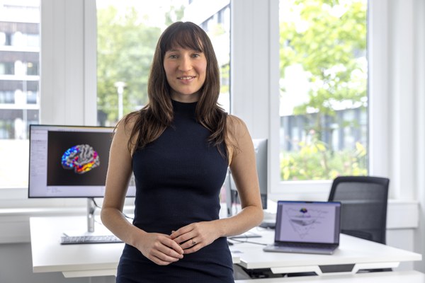
Dr. Casey Paquola leads the Multiscale Neurodevelopment Team at the Institute of Neuroscience and Medicine (INM-7), Research Center Jülich. His research focuses on understanding neurodevelopmental processes through multiscale frameworks that integrate postmortem histology, transcriptomics, and in vivo neuroimaging (e.g., MRI, EEG). The team studies cytoarchitecture maturation, connectivity, and functional dynamics across early life to elucidate mechanisms of neurodevelopmental disorders. Their work emphasizes microstructure as a bridge between postmortem and in vivo data, identifying principles like large-scale cortical gradients. Research methods include microscopy, neuroimaging, and computational modeling to construct brain models. Key areas of study include the default mode network, epilepsy-related brain changes, and the interplay between genetics and brain structure. The team develops tools like BrainSpace and Micapipe for analyzing structural-functional relationships in neuroimaging datasets. Led initiatives: ENIGMA-Toolbox for transdiagnostic analysis, BrainStat for statistical neuroimaging analysis. Focus areas: Cortical development gradients, functional resilience, and translational neuroimaging. Collaborations: Multidisciplinary teams combining computational, clinical, and experimental neuroscience. Future work aims to advance precision medicine through personalized brain mapping and understanding neurodevelopmental disorders' multiscale origins.
Anna Suling is a Research Fellow and Head of the Clinical Studies Working Group at the University of Hamburg's Faculty of Medicine. She is affiliated with the Institute of Medical Biometry and Epidemiology within the Center for Experimental Medicine at the University Medical Center Hamburg-Eppendorf (UKE). Her expertise lies in medical biometry, clinical trial design, and epidemiological analysis. Dr. Suling leads research in cardiovascular medicine, rare diseases, and geriatric mental health, with a focus on improving clinical outcomes through evidence-based methodologies. Key research interests include atrial fibrillation management, stroke rehabilitation, and the application of biostatistics in clinical trials. She has contributed to major studies such as the EAST-AFNET 4 trial on atrial fibrillation and the NETS trial investigating transcranial stimulation post-stroke. Her work bridges statistical rigor with clinical relevance, addressing both common and rare disease challenges. Educations: Dr. rer. medic (Medical Doctorate) from the University of Hamburg Publications span interdisciplinary topics including rare disease family interventions, cardiac biomarkers, and surgical outcomes. Awards include the 2022 Biometrics Certificate from GMDS/IBS-DR, recognizing her methodological contributions. Collaborations with international consortia like EAST-AFNET highlight her role in advancing translational research. Recent work emphasizes pragmatic trial design, such as the CARE-FAM-NET study evaluating mental health interventions for rare disease families. She also explores systemic issues like sex differences in myocardial infarction outcomes and the impact of ERAS protocols in pancreatic surgery. Awards: Zertifikat „Biometrie in der Medizin“ (2022) Her advisory role in clinical studies ensures methodological excellence across diverse projects, from neurorehabilitation to genetic analyses in atrial fibrillation. Active in teaching biostatistics and mentoring early-career researchers, Dr. Suling’s work underscores the synergy between rigorous science and patient-centered care.
Igor Meglinski is a Professor at Aston University, affiliated with the College of Engineering and Physical Sciences and the Department of Mechanical, Biomedical & Design Engineering. He is part of the Engineering for Health initiative and the Aston Research Centre for Health in Ageing. With a PhD, his expertise spans biophotonics, biomedical imaging, and optical spectroscopy. He actively supervises PhD students, with contact details provided for project inquiries. His research focuses on advanced optical techniques such as laser speckle contrast imaging, dynamic light scattering, and neuroimaging applications. Recent work includes non-invasive blood flow assessment and postmortem hemodynamic studies in mouse brains. Collaborations span global institutions, reflecting his commitment to interdisciplinary biomedical optics. Notable contributions include editorial roles and datasets like the 'Noninvasive control of the transport function of fluorescent coloured liposomal nanoparticles.' His research emphasizes translational applications in healthcare diagnostics and aging-related health challenges.
PD Dr. Jakob Matschke is a researcher at the Institute of Neuropathology of the University Medical Center Hamburg-Eppendorf (UKE). With over 187 publications, his work spans critical areas in neuropathology , neuroimmunology , and neurodegenerative diseases . Current Affiliation: Institute of Neuropathology, UKE Research Themes: Brain tumor biology, post-COVID-19 neurological effects, prion diseases, immune-tumor interactions, and neurodegenerative tauopathies His research on malignant gliomas explores somatic mutations in HLA class genes and antigen presentation mechanisms, while recent studies on SARS-CoV-2 examine neuroinflammatory scars and blood-brain barrier dysregulation. Collaborations include teams in clinical immunology (C3i) and the Neuro-Immune Network Hamburg . Key trends in his 15 most recent articles (2022-2025) include: Neuro-oncology: Tumor growth kinetics, glioblastoma survival disparities, spinal cord tumors Neuro-immunology: CD4 T cell responses, MPXV immune evasion, post-COVID-19 microglial activation Neurodegeneration: Prion protein shedding, TDP-43 cross-seeding, anti-IgLON5 disease pathology Prominent methodologies involve epigenomics , spatial immune mapping , and RT-QuIC assays for prion detection. His work often intersects clinical case studies with translational research, particularly in postmortem analyses of infectious and degenerative neurological conditions.