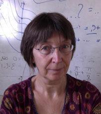
Kaye Morgan is an Associate Professor in the School of Physics and Astronomy at Monash University, specializing in X-ray imaging technologies with applications in medical and respiratory research. She holds an Australian Research Council Future Fellowship and has held prestigious positions including a Hans Fischer Fellowship at Technische Universität München. Her research focuses on advancing X-ray optics methodologies, particularly phase contrast X-ray imaging (PCXI) and dark-field imaging, to enhance resolution, speed, and sensitivity. These techniques are applied to study airway health in cystic fibrosis and other respiratory diseases, using synchrotron facilities like SPring-8 and the Munich Compact Light Source. She has pioneered single-grid imaging and propagation-based dark-field approaches, enabling real-time visualization of lung dynamics and treatment efficacy. Morgan leads multiple high-impact projects funded by ARC and international collaborations, with over 85 publications in journals like Optics Express and Scientific Reports. Her work contributes to UN Sustainable Development Goals related to health and innovation. Key achievements include developing lab-based X-ray sources for clinical translation and quantifying lung microstructure through advanced imaging algorithms.
Dr. Amir Tavakoli Taba is a Senior Lecturer in Medical Imaging Sciences at the University of Sydney, where he co-directs the Medical Image Optimisation and Perception Group (MIOPeG). He specializes in improving medical imaging accuracy, particularly in breast cancer diagnosis, through advancements like phase-contrast tomography and AI integration. His work bridges technological innovations (e.g., low-dose imaging) with clinical practice, emphasizing quality control and radiologist performance analysis. Education: PhD (University of Sydney) MEngSc (University of New South Wales) BSc (University of Tehran) Research Interests: Dr. Taba’s research focuses on phase-contrast computed tomography (PCT), AI-driven diagnostic tools, and clinical workflow optimization. His projects include the world’s first PCT clinical trial (scheduled for 2024 in Melbourne) and collaborations with institutions like ANSTO, Harvard Medical School, and the University of Iowa. He also investigates radiologist expertise development and the role of social networks in medical decision-making. Grants & Awards: NHMRC Synergy Grant (IMPACT: Implementation of X-ray Phase-Contrast Tomography) International recognition, including the SPIE Medical Imaging Award Advising & Labs: Current students: Mohammed ALANAZI (abdominal CT optimization), Jenna ARBID (phase-contrast imaging) Labs: MIOPeG, part of the Sydney Vital and Sydney Catalyst cancer research networks Teaching: Courses in imaging technologies, medical image perception, and clinical capstone projects for diagnostic radiography students.
Dr. Kelly Crossley is a Senior Research Fellow at the Fetal and Neonatal Health Research Group, The Ritchie Centre, Hudson Institute of Medical Research, and an Adjunct Senior Research Fellow in the Department of Obstetrics and Gynaecology at Monash University, within the Faculty of Medicine, Nursing and Health Sciences. She is actively involved in perinatal research and is accepting PhD students. Senior Research Fellow – Hudson Institute of Medical Research Adjunct Senior Research Fellow – Department of Obstetrics and Gynaecology, Monash University Dr. Crossley earned a Bachelor of Science (Honours) from Monash University (1991–1995), focusing on fetal behavioural states regulated by progesterone. Her research centers on perinatal physiology, particularly the respiratory and cardiovascular transition at birth. She investigates mechanisms of brain injury in newborns and develops interventions to improve spontaneous breathing in preterm infants. Her work employs advanced preclinical models in rabbits and sheep and leverages synchrotron imaging from Australian and Japanese facilities. Key areas include neonatal resuscitation, congenital diaphragmatic hernia, transient tachypnoea, and mechanical ventilation. The recent publications highlight a strong trend in preclinical neonatal physiology, focusing on resuscitation techniques, lung development, and circulatory recovery. Her work bridges animal models and clinical translation, especially in oxygenation strategies and cord clamping. Reducing respiratory distress after caesarean delivery Phase-contrast neuroimaging for early detection of brain injury Improving neonatal transition in CDH Reducing pulmonary hypertension in CDH Mechanisms to improve newborn respiratory function via X-ray imaging Dr. Crossley has led multiple research projects as a Primary Chief Investigator and collaborates widely with clinicians and scientists. She has secured active and completed grants from diverse sources, including Congenital Diaphragmatic Hernia Australia. While no awards are listed, her leadership in high-impact, peer-reviewed research underscores her scientific contributions. She is a key member of the Fetal and Neonatal Health Research Group at The Ritchie Centre, Hudson Institute, where she conducts collaborative, multidisciplinary research integrating physiology, imaging, and clinical innovation.
Associate Professor Marcus Kitchen is affiliated with Monash University's School of Physics and Astronomy and the Victorian Heart Institute. He specializes in developing advanced X-ray imaging techniques, including Phase Contrast Imaging, Compton Scatter Imaging, and Ultra-Low Radiation Dose methods. His research focuses on enhancing diagnostic capabilities in lung and brain imaging while minimizing radiation exposure. Collaborations with institutions like the Hudson Institute aim to improve neonatal care, particularly for preterm infants with underdeveloped lungs. Key projects include translating synchrotron-based methodologies for clinical and industrial applications. Research interests span biomedical imaging, material discrimination, and radiation dose reduction. He leads or co-leads over 30 projects, including 'X-ray Scatter Imaging: Vast Information with Minimal Radiation' (2025–2028) and 'IMPACT: IMplementation of x-ray PhAse-Contrast Tomography to transform cancer diagnosis' (2022–2026). His work aligns with UN Sustainable Development Goals focused on health and innovation. Publications emphasize novel imaging modalities and their applications in respiratory and neurological disorders. Despite no explicit awards listed, his contributions to reducing radiation exposure and advancing lung mechanics research are significant. He supervises graduate students and actively seeks PhD candidates. His lab integrates theoretical, computational, and experimental approaches to bridge gaps between fundamental physics and clinical medicine.
Marie-Christine Zdora is a Research Fellow in the School of Physics and Astronomy at Monash University. She holds adjunct roles including Adjunct Scientist at ETH Zürich (2022–2020) and Postdoctoral Researcher at Paul Scherrer Institut (2021–2021). Her academic journey includes a PhD in Physics from University College London (2015–2020), and master’s degrees from TU Munich and the University of Sydney. Her research focuses on advanced X-ray imaging techniques, particularly phase-contrast imaging using near-field speckles, dark-field imaging, and multi-modal approaches. Key areas include medical imaging applications, biological tissue analysis, and synchrotron-based tomography. She leads projects such as Multi-scale, multi-modal X-ray imaging using speckle and collaborates internationally on diagnostic radiology innovations. Zdora has received prestigious awards including the Springer Thesis Award (2020) and the Award of Congressi Stefano Franscini (2016). She serves as an Associate Editor for Optics Express and actively supervises students and mentors interns. Her work contributes to SDGs related to health, innovation, and sustainable development.
Samantha Jane Alloo is a Research Fellow at Monash University's School of Physics and Astronomy , specializing in X-ray imaging techniques. She holds a PhD in Medical Physics (2024) and degrees in Mathematics and Physics from the University of Canterbury. Education PhD in Medical Physics, University of Canterbury (2021–2024) Bachelor of Science with First-Class Honours in Medical Physics, University of Canterbury (2020–2021) Bachelor of Science in Mathematics and Physics, University of Canterbury (2017–2019) Her research focuses on speckle-based X-ray imaging , particularly phase-contrast and dark-field imaging. She has pioneered the multimodal intrinsic speckle-tracking (MIST) algorithm for rapid, high-resolution imaging and currently investigates Fokker-Planck equation applications in paraxial X-ray imaging to expand sample information extraction. Her publications span X-ray imaging , computational techniques , and biomedical applications , with recent work on dark-field tomography and phase-contrast tomography for unknown materials. She has secured international grants and led experiments at the European Synchrotron Radiation Facility. Scientific Awards Best Oral Presentation at Materials@UC Conference (2020) Best PhD Student Oral Presentation at UC-SPCS Symposium (2021) Best Poster Prize at Australian Synchrotron Meeting (2022) Best Student Oral Presentations at NZPEM Conferences (2021, 2022) She actively collaborates with researchers like Andrew Stevenson and Kaye Morgan, aiming to apply her expertise to diverse physics-related fields for future work.
Dr. Thinh Le is a Postdoctoral Research Fellow at the School of Computer Science , University of Technology Sydney , where he works at the Centre for Quantum Software and Information (since 2023). His academic journey includes research fellowships at the Institute for Quantum Optics and Quantum Information Vienna (2020-2023) and Leibniz University Hannover (2019-2020). He earned his PhD in Physics (2015) and BSc in Physics (2011) from the National University of Singapore . Thinh's research focuses on quantum information science , bridging theoretical foundations and experimental implementations. His work addresses critical challenges in quantum computing (error correction, fault tolerance), quantum tomography (device-independent characterization), and quantum software stack development. He investigates quantum randomness generation, Bell nonlocality, and entanglement distillation for quantum networks, with applications in quantum cryptography and secure communication. His recent publications examine advanced topics like trapped-ion lattice surgery (2025), quantum correlation boundaries (2025), and quantum backflow phenomena (2023). Earlier work established foundational limits for real quantum theory (2021) and developed practical entanglement distillation protocols (2018). Scientific awards include the Lise Meitner Postdoctoral Fellowship (2020-2023) and Humboldt Postdoctoral Fellowship (2019-2020). His funded research projects focus on quantum computing error correction and device-independent quantum tomography , aligning with his theoretical expertise guided by experimental applications.
Michelle Croughan is a Research Fellow at the School of Physics and Astronomy, Monash University, where she conducts advanced research in x-ray imaging and biomedical physics. Her work integrates physics and physiology to study lung development and structural changes in disease models. Her research focuses on developing and applying novel x-ray imaging techniques such as directional dark-field and phase-contrast imaging to study microstructural changes in biological tissues. She applies these methods to preclinical models of cystic fibrosis and neonatal lung conditions, particularly in understanding lung aeration and vascular development in newborns with diaphragmatic hernia. Recent publications demonstrate a strong trend in combining computational image processing with synchrotron-based x-ray techniques to achieve high-resolution, quantitative insights into lung physiology and bone microstructure. Her work bridges physics, engineering, and medicine. Effect of prenatal diaphragmatic hernia on pulmonary arterial morphology (2025) Directional dark-field retrieval with single-grid x-ray imaging (2023) Fast implicit diffusive dark-field retrieval (2023) Sustained inflation improves lung aeration (2023) Accurate measures of regional lung air volumes (2022) Michelle Croughan leads research projects funded by the Australian Institute of Nuclear Science and Engineering (AINSE), including an active grant on directional dark-field imaging of small animal bones and teeth in cystic fibrosis models. She previously led an International Synchrotron Access Grant. There is no public information on student supervision or formal advising roles. She is part of a collaborative research network involving experts in medical physics and neonatal physiology, including Dr. Marcus Kitchen and other researchers at Monash University. Her work is conducted using advanced imaging facilities, likely including synchrotron sources, and focuses on translational preclinical applications.
Jelena Schmalz is a Lecturer in Mathematics at the School of Science and Technology, University of New England. She holds an M.Sc. in Mathematics from Moscow State Lomonossov University and a Ph.D. in Physics from UNE. Her background includes extensive teaching experience in first-year mathematics, number theory, and cryptography courses. Dr. Schmalz's research focuses on applied mathematics with applications to X-ray imaging, tomography, sport science, cryptography, and tertiary education. Her doctoral work investigated direct and inverse problems in X-ray phase-contrast imaging. Her publications demonstrate consistent focus on mathematical applications and educational innovation, with recent work exploring mathematical education during COVID-19 and security protocols in banking systems. Earlier research established foundations in X-ray physics and tomography techniques.
Peter Munro serves as an Adjunct Senior Research Fellow in the School of Engineering at The University of Western Australia, specifically within the Department of Electrical, Electronic and Computer Engineering. His research profile indicates active engagement in advanced optical imaging techniques with significant contributions to the field. Dr. Munro's research expertise centers on tomography, particularly optical coherence tomography, phase contrast imaging, X-ray imaging, and image formation processes. His work bridges physics, engineering, and biomedical applications, with special focus on wave optics phenomena and biomedical imaging systems. The research demonstrates sophisticated understanding of numerical aperture, Maxwell's equations, Gaussian distributions, and detector technologies in imaging systems. Analysis of his publication record reveals consistent focus on improving imaging techniques through rigorous simulation and experimental validation. His work spans theoretical modeling of light propagation, development of novel imaging methodologies, and practical applications in biomedical contexts, particularly in understanding how microscopic structures affect image formation. Dr. Munro has secured multiple research grants, demonstrating his ability to lead and contribute to significant research initiatives in optical imaging. His collaborative approach is evident through numerous co-authored publications with researchers across various institutions. His research contributes to UN Sustainable Development Goals related to quality education, industry innovation, and good health and well-being, reflecting the practical applications of his imaging technologies in healthcare and scientific advancement.
Michael Morgan is a Professor in the School of Physics and Astronomy at Monash University. His research focuses on theoretical physics, including topological defects, particle cosmology, geometric phases, and X-ray imaging techniques. He has contributed to advancements in electron microscopy and phase contrast imaging, particularly in nanoscale field mapping and vector tomography. His teaching interests include electrodynamics, scattering theory, and quantum mechanics. Professor Morgan has led or participated in multiple research projects funded by organizations like the Australian Research Council (ARC). Notable projects include the 'Nanoscale field mapping in functional materials' and 'Electron Tomography of Electromagnetic Fields'. He has also been recognized with the Vice-Chancellor's Diversity and Inclusion Award in 2020. His work spans interdisciplinary collaborations, addressing challenges in materials science, medical imaging, and computational physics. Key contributions include developing novel microscopy techniques and applying mathematical models to reconstruct physical phenomena. His research has been published in high-impact journals, with a focus on enhancing precision and resolving power in imaging technologies.
Dr. Konstantin Pavlov is an Adjunct Associate Professor at the University of Canterbury's School of Science and Technology, where he leads the X-ray Physics and Imaging Research Group. His research focuses on novel X-ray imaging techniques using laboratory and synchrotron radiation sources, with significant contributions to phase-contrast imaging, coherent diffractive imaging, and tomographic reconstruction algorithms. Research interests center on advancing X-ray physics methodologies, including: Phase-contrast imaging and tomography Coherent diffractive imaging techniques Statistical dynamical X-ray diffraction theory Hybrid phase-retrieval approaches for complex materials Publications demonstrate consistent innovation in X-ray physics, covering nanolens aberrations, phase retrieval methods, and deterministic coherent imaging. Recent work emphasizes experimental validation of theoretical models in synchrotron environments.
Dr. Erin McGillick is a NHMRC Peter Doherty Early Career Research Fellow at The Ritchie Centre, Hudson Institute of Medical Research, and an Adjunct Research Fellow in the Department of Obstetrics and Gynaecology at Monash University, Faculty of Medicine, Nursing and Health Sciences. Her research focuses on fetal and neonatal physiology, particularly the mechanisms of lung development and the transition from fetal to newborn life in compromised pregnancies. Her educational background includes a PhD in Medical Science from the University of South Australia (awarded 2016) and a Bachelor of Laboratory Medicine with First Class Honours from the same institution (awarded 2012). Her research integrates molecular biology, functional physiology, and advanced imaging techniques such as synchrotron X-ray imaging to understand how conditions like gestational diabetes, maternal overnutrition, and intrauterine growth restriction impair lung development and increase respiratory complications at birth. Dr. McGillick’s recent publications highlight a strong trend in preclinical models (lambs, rabbits, sheep) to study lung aeration, respiratory patterns, and interventions such as sustained inflation at birth. Her work spans neonatal respiratory physiology, cardiorespiratory transition, and the use of ultrasound and phase-contrast imaging for real-time assessment of lung function. These studies aim to generate preclinical evidence for future clinical trials to improve outcomes for premature and compromised infants. Her scientific achievements have been recognized with several prestigious awards: Australian Institute of Policy and Science Victorian Young Tall Poppy Science Award (2018) Australian Society for Medical Research International Research Award (2017) Best Early Career Researcher Oral Presentation, Fetal and Neonatal Workshop of Australia and New Zealand (2017) Fresh Science Finalist (2017) Monash University Bridging Postdoctoral Fellowship (2017) Dr. McGillick has secured competitive research funding and leads projects such as Improving the fetal to neonatal transition in compromised newborns , where she serves as Primary Chief Investigator. She collaborates extensively with physiologists, physicists, and clinicians across institutions. She currently serves as Director of the Australian Society for Medical Research (since November 2019), reflecting her leadership in the scientific community. While no formal students are listed, her role as a principal investigator suggests mentorship of junior researchers. Her lab is embedded within The Ritchie Centre, a leading perinatal research hub, where multidisciplinary teams work on translational neonatal science.
Professor Brian Abbey is a Professor at La Trobe University, holding roles such as Deputy Director of the La Trobe Institute for Molecular Science (2022–present), co-founder of AlleSense (2024–present), and Research Director of the School of Molecular Science (2020–2022). He leads research in coherent optics, nanotechnology, and X-ray science, with a focus on imaging materials at atomic and cellular levels. His work combines optics, synchrotron science, and XFEL technologies for applications in biophysics and medical diagnostics. PhD in Physics from the University of Cambridge (2007) MSci from University College London MRes from Imperial College London MEng from the University of Oxford His research interests span Optics , Nanofabrication , and X-ray Science , with breakthroughs in plasmonic biosensors and colorimetric histology. He has pioneered techniques like plasmon-enhanced phase microscopy and multi-hit serial femtosecond crystallography. Abbey has secured over $38M in external funding since 2014, including ARC and NHMRC grants, and holds five licensed patents. His awards include the Australian Museum Eureka Prize (2022) , Victoria Prize for Physical Sciences (2022) , and Victorian Young Tall Poppy Science Award (2017) . He has supervised 12 PhD graduates and currently mentors six students, emphasizing early-career researcher development. Abbey’s labs, including FARLabs (online education platform), and collaborations with Stanford and Oxford focus on applications in cancer diagnostics and immune system imaging.
Dr Dania Abu Awwad is a Lecturer in Diagnostic Radiography at the University of Sydney , within the Discipline of Medical Imaging Science at the Sydney School of Health Sciences. She holds a PhD (2021) and BAppSc (2018) from the University of Sydney. Her roles combine academic teaching, clinical practice in Sydney and Western Sydney Local Health District hospitals, and private imaging clinics. Education: PhD in Medical Imaging (2021), University of Sydney Bachelor of Applied Science (Diagnostic Radiography) with Honours (2018), University of Sydney Research Interests: Evidence-based practice in radiography Radiation dose optimization Infection prevention and control in CT imaging Breast cancer screening techniques Trauma imaging protocols Her work bridges clinical practice and education, with a focus on improving diagnostic accuracy and patient safety in imaging environments. Publications Trends: Over 15 peer-reviewed articles since 2020, with recent focus on: Infection control practices in CT imaging (multiple 2024 studies) Global radiotherapy accessibility analysis (The Lancet Oncology, 2024) Breast screening efficacy and radiographer training Novel imaging techniques like the modified trauma axial shoulder view Grants & Advising: Recipient of 2022 Lectureship Support Package from the Faculty of Medicine and Health Supervising two HDR students: Jenna (breast phase-contrast imaging) and Frances (IPC guidelines for MRI departments) Labs/Teams: Active in the University of Sydney's Medical Imaging Sciences research group, contributing to both clinical and educational initiatives.