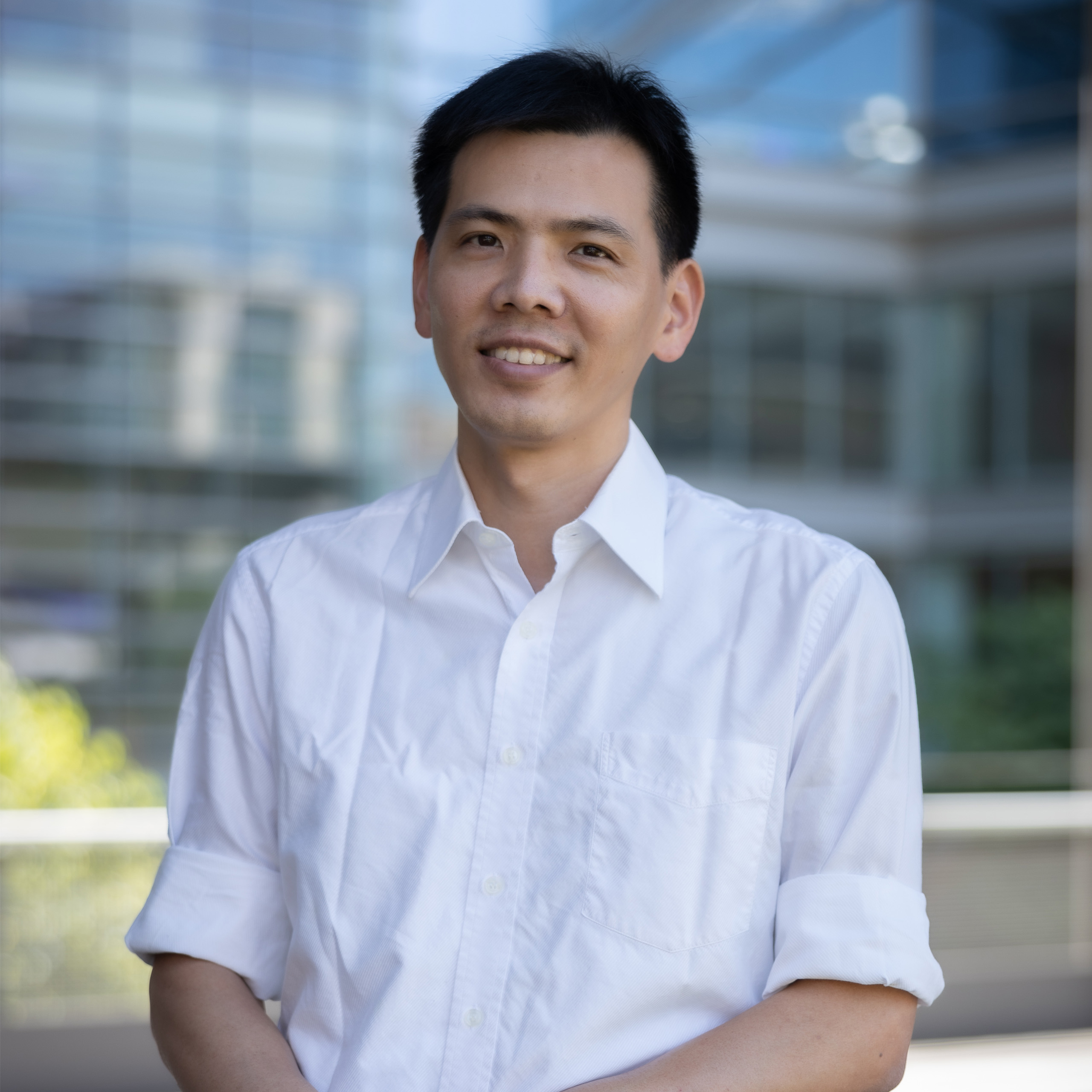
Edward Nieh, Ph.D., is an Assistant Professor of Pharmacology at the University of Virginia, leading the Nieh Lab. His research focuses on systems neuroscience, particularly motivated behaviors such as feeding, social interaction, and novelty-seeking, and their disruption in disorders like drug addiction and eating disorders. The lab employs advanced techniques including optogenetics, virtual reality for rodents, and cellular-resolution imaging, emphasizing cutting-edge neuroscience methodologies. Research interests span biotechnology, computational biology, molecular pharmacology, and neuroscience. The lab investigates how behaviors are encoded by large neuronal populations and studies sex differences in these contexts. Recent work explores neural circuits underlying reward-seeking behaviors, dopamine signaling pathways, and the interplay between homeostasis and motivation. Key technical innovations include the development of open-source microscopy tools like TWINKLE and methodologies to correct calcium imaging artifacts. Collaborations span computational modeling and experimental neurophysiology. Scientific contributions include studies on thalamo-amygdala circuits, dopamine’s role in aversive signaling, and the neural basis of observational learning. The lab’s findings bridge basic neuroscience with translational applications in neuropsychiatric disorders. Edward Nieh’s work is supported by grants focusing on neuroimaging, drug addiction mechanisms, and novel behavioral paradigms. His interdisciplinary approach integrates molecular, circuit, and behavioral levels of analysis to understand complex brain functions.
Rikard Blunck is an Accredited Professor in the Department of Physics at the University of Montreal, affiliated with the Faculty of Arts and Sciences. His research focuses on ion channel biophysics, membrane protein dynamics, and structural biology. He employs advanced techniques like fluorescence spectroscopy, molecular dynamics simulations, and single-molecule imaging to study voltage-gated potassium channels, mechanosensitive channels, and disease-related mutations in ion channels. Key research interests include the structural determinants of ion channel function, mechanisms of channel inactivation, and the impact of genetic variants on channel physiology. His work spans from fundamental biophysics to clinical implications in neurology and cardiology, particularly in channelopathies linked to epilepsy, migraine, and ataxia. Notable contributions include studies on the Shaker Kv channel, TACAN pain sensors, and structural analysis of Kv2.1/Kv6.4 heterotetramers. His methodologies include voltage-clamp fluorometry, single-subunit counting, and computational modeling to probe conformational changes during channel activation and inactivation. Blunck has published extensively, with recent work investigating cooperative C-type inactivation mechanisms, KCNG4 genetic variants in migraine, and stoichiometry determinations of ion channel complexes. His research bridges molecular mechanisms with clinical applications, aiming to identify therapeutic targets for channel-linked disorders.
Nazzareno D'Avanzo is an Accredited Professor in the Department of Physiology at the University of Montreal's Faculty of Medicine. His research focuses on ion channel physiology, particularly exploring the regulation of HCN (hyperpolarization-activated cyclic nucleotide–gated), Kir (inward rectifier potassium), and other ion channels by lipids, endocannabinoids, and pharmacological agents. He investigates molecular mechanisms underlying channel function, including lipid interactions, drug binding, and mutations' effects on channel behavior. His work spans biophysics, neuroscience, and pharmacology, with notable studies on cannabinoid effects on membrane dynamics, norquetiapine's inhibition of HCN channels, and phosphoinositide regulation of potassium channels. Computational modeling complements experimental approaches, as seen in studies using molecular dynamics simulations to predict lipid-channel interactions. Key research themes include channel-drug interactions (e.g., antipsychotic metabolites), lipid regulation of membrane proteins, and the structural basis of channel selectivity. His contributions advance understanding of neurological disorders linked to ion channel dysfunction and inform drug development strategies.
Dr. Wei-Tang Chang is an Assistant Professor in the Department of Radiology at the University of North Carolina School of Medicine. His research focuses on advancing ultrahigh-resolution functional and diffusion MRI techniques, with emphasis on improving spatial and temporal resolution while reducing scan times. Key projects include submillimeter isotropic-resolution fMRI for hippocampal subfield analysis (funded by NIH R21), novel dMRI approaches to overcome resolution limits, and clinical translation of robust imaging methods resistant to motion/noise artifacts. Dr. Chang holds a PhD in Biomedical Engineering from National Taiwan University and completed postdoctoral training at the Martinos Center for Biomedical Imaging (MGH), Massachusetts General Hospital, and Singapore BioImaging Consortium (SBIC). His research innovations include SORDINO fMRI for awake rodent imaging, pPRISM diffusion MRI for submillimeter resolution, and ZTE pulse sequences for ultra-fast acquisitions. Awards include the 2011 OHBM Trainee Award for work on MEG source localization and fMRI temporal resolution breakthroughs. Current work bridges basic neuroimaging science with clinical applications, particularly in neurodegenerative disease biomarker development and rodent disease model studies. Education: PhD in Biomedical Engineering, National Taiwan University Postdoctoral Fellowships: Martinos Center for Biomedical Imaging (MGH) Singapore BioImaging Consortium (SBIC) Key Technologies Developed: ZTE pulse sequences (25 ms temporal resolution fMRI) pPRISM diffusion MRI (navigator-free submillimeter imaging) Draining-vein suppression layer-dependent fMRI Awards & Funding: NIH R21 Grant (2019) for hippocampal subfield fMRI OHBM Trainee Award (2011) Dr. Chang's translational focus involves adapting laboratory innovations for clinical use, with particular interest in Alzheimer's disease biomarkers through hippocampal network analysis and Huntington's disease models using rodent functional connectivity studies. His lab actively develops open-source MRI reconstruction algorithms and collaborates internationally on multi-center neuroimaging projects.
Patricia Boya is a Full Professor in the Department of Medicine at the University of Fribourg, within the Faculty of Mathematics, Natural Sciences and Medicine. She is a leading researcher in autophagy, mitophagy, and lysosomal biology, with a focus on retinal and neurodegenerative diseases. Her research interests center on the molecular mechanisms of autophagy and mitophagy, particularly their roles in retinal health, aging, neurodegeneration, and cell death. She investigates how mitochondrial dynamics, lysosomal function, and autophagic flux contribute to diseases such as retinitis pigmentosa, glaucoma, ALS, and Parkinson’s. Her work combines molecular biology, cell imaging, and preclinical models to uncover therapeutic targets. The analysis of her recent publications reveals a strong focus on mitophagy regulation, neuroinflammation (especially via the cGAS/STING pathway), and the development of neuroprotective compounds. She has contributed to high-impact reviews and guidelines in autophagy research, and her work spans fundamental mechanisms to translational applications. She is actively involved in scientific leadership, including the "Women in Autophagy" initiative to promote gender equity in science and the "DRIVE" international PhD training program in autophagy. Dr. Boya has published over 150 articles in top journals including Nature , Nature Communications , Autophagy , and Cell Death & Disease . Her work is highly collaborative, involving interdisciplinary teams across Europe. She leads a research group focused on cellular recycling mechanisms and their implications for vision and aging, contributing significantly to the understanding of autophagy in health and disease.
José Antonio Del Río Fernández is a distinguished Professor and Group Leader at the Institute for Bioengineering of Catalonia (IBEC) , where he directs the Molecular and Cellular Neurobiotechnology group. His work bridges developmental neurobiology , neurodegeneration , and lab-on-chip technology , focusing on diseases like Alzheimer's and Parkinson's . He has been active in academic research for over 30 years, previously affiliated with institutions such as the University of Barcelona , Frankfurt University , and Imperial College London . Education : PhD , University of Barcelona (1994) Research Interests center on neurodegenerative disease mechanisms , particularly the spreading of tau and α-synuclein in conditions like Alzheimer's and Parkinson's, and axon regeneration after CNS injury. His group pioneers lab-on-chip devices to model neurobiological processes and test regenerative therapies. Collaborations span institutions such as Imperial College , i3A, Zaragoza , and Biodonostia Hospital . Scientific Contributions include over 170 publications, with a H-index of 48 (Web of Science) and 55 (Google Scholar), and significant advancements in understanding prion protein interactions in neurodegeneration. His recent work explores optogenetics for axon regeneration and multitarget compounds for Alzheimer's therapy. He has mentored 24 PhD students , 26 Master's students , and 15 postdoctoral fellows , reflecting a strong commitment to academic training. Scientific Awards : Fundación Francisco Cobos CSIC Award (2001) Generalitat of Catalunya Award of Young Researcher (2003) Research Trends demonstrate a focus on neurodegenerative disease mechanisms , lab-on-chip platforms , and axon regeneration strategies . His collaborations with institutions like Imperial College London and University of Zaragoza highlight interdisciplinary approaches integrating biomaterials , optogenetics , and computational modeling .
John Kubie is an Associate Professor in the Department of Cell Biology at SUNY Downstate Medical Center's School of Medicine. His research career spans several decades, with publications dating from the 1980s to the present day. Dr. Kubie has maintained a consistent focus on understanding the neural mechanisms underlying spatial cognition and navigation. Dr. Kubie's primary research interest centers on hippocampal place cells and their role in spatial navigation and cognitive mapping. His work combines experimental neuroscience with computational modeling to understand how populations of neurons represent space and contribute to navigation behavior. He has extensively studied how place cells—pyramidal neurons that fire only when an animal is in specific locations—form the basis of cognitive maps that help rats navigate their environment. His research has explored how place cells adapt to novel environments, how they're affected by hippocampal lesions, and how their firing patterns relate to spatial memory. His publication record shows a consistent trajectory from early foundational work on place cell properties in the 1990s to more recent investigations into hippocampal remapping, neural network dynamics, and computational models of spatial cognition. Recent publications indicate continued active research in computational neuroscience, including studies on theta-resonant pyramidal neurons and the relationship between grid cells and hippocampal function. Dr. Kubie has maintained a long-standing collaboration with Dr. Robert U. Muller, as evidenced by numerous co-authored publications spanning decades. After Dr. Muller's passing, Dr. Kubie co-authored a memorial piece honoring his colleague's contributions to neuroscience. His laboratory has developed sophisticated techniques for recording from multiple neurons simultaneously and has created behavioral tasks to assess spatial capabilities in rats. The work bridges experimental neuroscience with computational approaches, seeking to develop computer models that can solve navigational problems similar to those solved by the biological hippocampus.
Daan Brinks is an Assistant Professor at Delft University of Technology in the Department of Imaging Physics within the Faculty of Applied Sciences. He leads the Brinks Lab, which operates at the intersection of physics, biochemistry, optics, mathematics, and nanofabrication, focusing on developing novel imaging tools for neuroscience applications. His research spans both fundamental biophysics and practical biomedical applications, with significant collaborations including Erasmus MC. Faculty of Applied Sciences, Delft University of Technology Department of Imaging Physics (ImPhys) Brinks Lab leader Founding member of BIOlab (Biomedical Intervention Optimization lab) Lead of a convergence Health and Technology Consortium Dr. Brinks' academic journey began with an MSc in Molecular Nanophotonics from the University of Twente (2002-2007), followed by a PhD at ICFO Institute Barcelona (2007-2012). He then completed prestigious fellowships at Harvard University as a Rubicon Fellow (2012-2014) and HMMI Fellow (2014-2017) before joining TU Delft as an Assistant Professor in 2017. His research interests center on voltage imaging techniques to monitor neural activity, optogenetics for neural control, nonlinear optical microscopy for enhanced resolution, and AI applications in bioimaging . The lab develops tools to transduce information in neurons into detectable photons, addressing questions from biophysical principles to behavioral consequences and from subcellular compartments to complete organisms. Current projects include Voltage nanoscopy using plasmonic enhancement, Absolute Voltage Imaging through fluorescence lifetime measurements, Multiphoton Voltage Imaging for deep tissue applications, and advanced image analysis with machine learning. The publications reveal a strong focus on developing novel optical tools for neuroscience, particularly genetically encoded voltage indicators and plasmonic enhancement techniques. His work bridges physics, molecular biology, and neuroscience, with applications ranging from fundamental understanding of neural circuits to cancer cell identification. The research shows progression from fundamental physics (early career) to increasingly applied neuroscience and biomedical applications (recent work), with publications in top journals including Nature, Science Advances, and Nature Biomedical Engineering. Rubicon Fellow (2012-2014) HMMI Fellow (2014-2017) Publications in Nature, Science Advances, Nature Biomedical Engineering Media coverage in major outlets including Delta TU Delft and Trouw Dr. Brinks actively mentors students and researchers, with his lab welcoming enthusiastic students, PhD candidates, and postdocs interested in multidisciplinary projects at the junction of optics, molecular biology, and neuroscience. His research has received external funding through fellowships and likely additional grants supporting his lab's operations. The Brinks Lab collaborates extensively with both academic and medical institutions, particularly evident in the cancer cell research with Erasmus MC. The lab maintains strong physical infrastructure including advanced microscopy systems and nanofabrication capabilities, supporting their work in voltage imaging, plasmonics, and single-cell analysis. They have developed several hardware and software interfaces for automated interaction with excitable tissues and model dynamics in hybrid systems, reflecting their interdisciplinary approach to neuroscience questions.
Dr. Sulin Zhang is a Professor in the Department of Engineering Science and Mechanics at Pennsylvania State University , where she investigates the interplay between mechanical forces and materials behavior across materials science , biology , chemistry , and medicine . Her work spans solid-state battery technology , biofilm mechanics , and cellular mechanobiology . 2025 : Stack Pressure Effects in Silicon Anodes 2024 : 3D Neural Probes, Liquid Metal Composites 2023 : Biofilm Nematic Ordering, Plant Cell Wall Mechanics Her research themes focus on stress generation and mechanical regulation in systems ranging from lithium-ion anodes to biofilm morphogenesis . She employs in situ microscopy , agent-based modeling , and machine learning to analyze mechanical-chemical-electrochemical couplings. Key trends include anisotropic material failure , nanoparticle-cell interactions , and self-organizing microbial systems . Recent funding includes a $2.14M NIH grant (2022) for nerve regeneration scaffolding development. While no explicit awards are listed, her work has been highlighted in Nature Physics and PNAS Nexus , indicating high visibility in interdisciplinary research.
Pınar ÖZ is an Associate Professor at Uskudar University, Department of Molecular Biology and Genetics, since 2015. Her research spans neurobiology, computational neuroscience, and neurodevelopmental disorders, with a focus on membrane properties, adult neurogenesis, and orexin system dynamics. Boğaziçi University, Institute of Biomedical Engineering (2012–2014) Max Planck Institute for Dynamics and Self-organization (2011–2012) Research Interests: Computational modeling of neuronal excitability Neurodevelopmental disorders, including autism models Role of orexin in sleep deprivation and sensorimotor gating Neuroprotective agents (e.g., zeatin) in ischemic models Evolutionary bioinformatics of neurochemical pathways Applications of agent-based modeling in neurogenesis Recent Trends: Publications from 2015–2025 emphasize interdisciplinary approaches combining in vitro and in vivo models with computational tools to study hippocampal plasticity, neurodegeneration, and pharmacological interventions. Scientific Awards: Georg Christoph Lichtenberg Scholarship (Lower Saxony Ministry of Science and Culture, Germany, 2008–2009) Projects: Investigates personalized drug monitoring, neuroprotective effects of zeatin, orexin system's role in psychosis models, and computational frameworks for neurogenesis. She also supervises graduate theses on topics like neurodevelopmental inhibition networks and age-dependent simulations.
James Morizio serves as an Adjunct Professor in the Department of Electrical and Computer Engineering at Duke University, leveraging 35+ years of expertise in analog CMOS microelectronics for biomedical applications. His research bridges electrical engineering with neuroscience through disruptive sensor interface technologies for neural recording/stimulation and ultrasonic microfluidics. His educational background includes a Ph.D. in Electrical Engineering from Duke University (1995) M.S. in Electrical Engineering from University of Colorado, Boulder (1984) B.S. in Electrical Engineering from Virginia Polytechnic Institute and State University (1982) Morizio's research focuses on developing high-performance neural interfaces and acoustofluidic systems. Key areas include wireless neural instrumentation for closed-loop electrophysiology, CMOS-based ultrasonic transducers for microfluidic manipulation, and low-power VLSI design for implantable devices. His work emphasizes translating microelectronics innovations into biomedical solutions for neuroscience and diagnostics. His publication portfolio demonstrates consistent contributions to neural engineering and microfluidics, with recent trends showing increased focus on acoustofluidic diagnostics (e.g., AIMDx chip) and osseointegrated neural interfaces for prosthetic control. The research spans fundamental circuit design to translational applications in cancer metabolism and neuroprosthetics. Scientific recognition includes 15-year service award from Triangle BioSystems International/Harvard Bioscience Inc. (2016) Current research is supported by major grants including Neuro-CROWN (ultra-flexible electrode arrays), digital acoustofluidic systems for biomedical automation, and osseointegrated neural interfaces for prosthetic control. His teaching portfolio spans advanced VLSI design and special topics courses, though specific advising roles are not documented. Collaborative work involves interdisciplinary teams in neuroscience and biomedical engineering, particularly through partnerships with Triangle BioSystems International and translational projects using ovine models for neural interface validation.
Leonard Edward White is an Associate Professor in Neurology at Duke University, with additional appointments in Psychology and Neuroscience, Orthopaedic Surgery, and Neurobiology. He serves as Associate Director of the Duke Institute for Brain Sciences and Director of Undergraduate Studies of Neuroscience. His academic career spans over three decades since earning his Ph.D. from Washington University in St. Louis in 1992. Dr. White's research focuses on the structure and function of the mammalian brain, particularly through the development of advanced magnetic resonance methods for interrogating brain structure. His work combines light sheet microscopy with MRI techniques to provide new insights into microscopic brain structure, whole-brain connectivity, and how neural tissue constrains connectivity in animal models. He maintains a sustained interest in how early sensorimotor experience influences neural circuit formation and maturation in the cerebral cortex, as well as the intersection of brain sciences with humanities. His recent publications (2020-2025) reveal a strong emphasis on high-resolution brain imaging techniques, particularly MRI and light sheet microscopy for creating detailed brain atlases. His work spans multiple species (mouse, rat, human) and addresses fundamental questions in neuroanatomy, connectomics, and developmental neuroscience. Notably, he has been developing the Duke Mouse Brain Atlas and exploring the impact of prenatal drug exposure on brain development. Dr. White has secured significant research funding, including the current 'Ultra-high Resolution Structural Connectome Atlases of the Animal Brain' grant (2022-2026) from the University of Pittsburgh, and previously led NIH-funded projects on visual cortex development spanning nearly two decades. He is deeply involved in medical education, serving as Director of Undergraduate Studies of Neuroscience and developing innovative approaches to teaching neuroanatomy. His educational scholarship includes work on integrating art into medical education and revitalizing neuroanatomy teaching methods. He also maintains an active presence in neurohumanities, exploring the intersection of neuroscience with arts and humanities.
Dr. Robert J. Usselman serves as an Assistant Professor of Chemistry in the College of Engineering and Science at Florida Institute of Technology , where he also acts as Deputy Director of Computational Research At Florida Tech (CRAFT) . His research bridges quantum physics and cellular biology, focusing on magnetic resonance, biophotonics, and redox biochemistry. Specializes in quantum biological clocks and ROS production mechanisms Leads a multidisciplinary lab with active undergraduate/graduate students Develops computational infrastructure for AI and quantum research Research interests include: Quantum-Classical Interface in Biological Systems Magnetic Field Effects on Cellular Redox Balance Autofluorescence Lifetime Microscopy ROS Dynamics in Cellular Metabolism Recent publications highlight quantum biological mechanisms in ROS production, magnetic field modulation of cellular processes, and multiphoton imaging techniques. His work integrates theoretical quantum biology with practical applications in bioenergetics and disease models. Dr. Usselman actively mentors students in biotechnology, biomedical engineering, and physics, while advancing collaborations in AI-driven computational research through CRAFT. His lab combines experimental and computational approaches to study redox signaling and quantum sensing in biological systems.
Marianne Fyhn is a Professor at the University of Oslo's Section of Physiology and Cell Biology within the Faculty of Mathematics and Natural Sciences. She leads the Centre for Integrative Neuroplasticity (CINPLA), a strategic research initiative integrating experimental biology with computational physics/mathematics to study brain information processing and plasticity. PhD in Neuroscience (NTNU, 2000-2005) MSc in Physiology (University of Tromsø, 1997-1999) Bachelor in Arctic Biology (University Courses in Svalbard, 1995-1996) BSc in Biology (University of Bergen/Oslo, 1992-1995) Her research focuses on neural plasticity mechanisms in cortical structures, using large-scale neuronal recordings and transcranial two-photon microscopy to study synaptic and population code changes during sensory learning. Key findings include discovering grid cells in mice and demonstrating how experience modifies cortical circuits for long-term memory. Recent publications analyze perineuronal nets' role in memory stabilization, grid cell conformal mapping, and topological population coding in visual cortex. These works reveal intersections between neural circuit dynamics , computational modeling , and neurodegenerative processes . 2008: European Brain and Behaviour Society Award 2007: Eppendorf-Science Prize in Neurobiology 2006: Donald B. Lindsley Prize & I.K. Lykke Award Fyhn serves as course manager for advanced courses including MBV1020 - Physiology and MBV4340 - Advanced Neurobiology . Her lab develops educational tools like Neuronify for neural circuit simulation and open-source platforms for electrophysiological data analysis.
Jefferson W. Kinney is a Professor and Founding Chair of the Department of Brain Health at the Kirk Kerkorian School of Medicine, University of Nevada Las Vegas (UNLV) . He co-directs both the Pam Quirk Brain Health and Biomarker Laboratory and the Translational Mechanisms and Drug Discovery Laboratory , focusing on neurodegenerative diseases. Colorado State University - PhD National Institute of Mental Health - Intramural Research Training Fellowship The Scripps Research Institute - Helen Dorris Fellowship Dr. Kinney's research spans cellular and molecular mechanisms of Alzheimer's disease , with emphasis on: Neuronal-glial interactions in AD pathogenesis Role of GABA B receptors in memory deficits Diabetes-induced AD risk (insulin signaling, hyperglycemia) Neuroinflammatory pathways in AD and traumatic brain injury (TBI) Translational biomarker discovery in clinical samples GPCR signaling in microglial activation His publications reveal a focus on GABAergic dysfunction , metabolic comorbidities , and translational models bridging preclinical and clinical AD research. Key techniques include: Transgenic mouse models (APP/PS1) Cellular/molecular biology (qRT-PCR, Western blot) Behavioral assays (Barnes maze, fear conditioning) Immortalized microglia cultures (BV2 cells) Scientific Honors: Reg Grundy and Joy Chambers-Grundy Chair for Brain Health Helen Dorris Fellowship in Neuropharmacology NIMH Intramural Research Training Fellowship Dr. Kinney leads two interconnected laboratories that synergize preclinical mechanisms ( Translational Mechanisms and Drug Discovery Laboratory ) with clinical biomarker discovery ( Pam Quirk Brain Health and Biomarker Laboratory ).