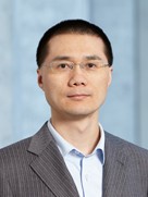
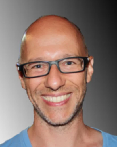
Prof. Dr. Jing Wang is a Full Professor at the Department of Civil, Environmental and Geomatic Engineering at ETH Zürich. His research focuses on air pollution control, nanoparticle transport, and environmental health and safety (EHS) impacts of nanomaterials. He has held roles including Assistant Professor at ETH Zürich (2010–present), Research Assistant Professor at the University of Minnesota (2007–2010), and postdoctoral associate in Particle Technology (2005–2007). Education: Bachelor’s in Engineering (2000) – Tsinghua University, Beijing Master’s in Computer Sciences (2003) – University of Minnesota PhD in Aerospace Engineering (2005) – University of Minnesota Research Interests: Air/water filtration technologies Nanoparticle emission reduction and measurement Multiphase flow mechanics Environmental impacts of nanomaterials Collaborations: Industrial partnerships include 3M, BASF, Boeing, Intel, Samsung, and others in nanoparticle measurement and filtration solutions. Honors: 2011 Smoluchowski Award (Association for Aerosol Research) 2006 ‘Best Dissertation’ Award (University of Minnesota) 2004 Doctoral Dissertation Fellowship Teaching: Courses include Air Pollution Control, Environmental Engineering Seminars, and Excursions for Environmental Engineers. Labs/Teams: Leads the Particle Technology Lab and collaborates with the Institute of Environmental Engineering at ETH Zürich.
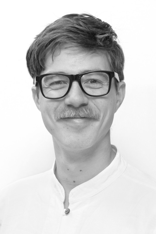
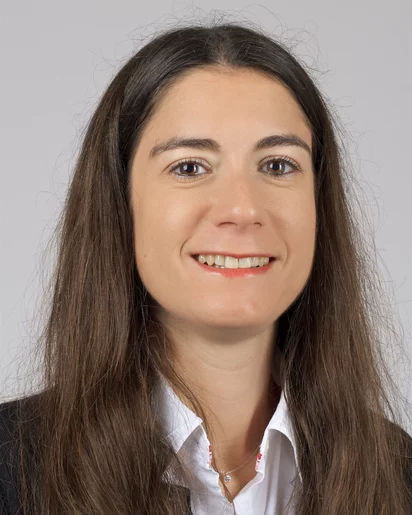
Dr. Cooper Harshbarger is a Lecturer at the Department of Health Sciences and Technology at ETH Zurich , Switzerland. His research bridges biomechanics and acoustofluidics, focusing on spinal surgery and microscale cell manipulation technologies. Email: cooper.harshbarger@hest.ethz.ch Research Interests : Dr. Harshbarger specializes in biomechanical analysis of spinal structures and acoustofluidic device development . His work explores: Biomechanics of the lumbar spine and osteoligamentous complexes Acoustically-driven microfluidic systems for medical diagnostics Cell focusing/trapping technologies using sharp-edge acoustofluidics Scientific Contributions : Recent publications highlight his dual expertise in spinal fusion biomechanics and microscale fluid control , with applications in cancer diagnostics and cell manipulation. Key technologies include BAW-based systems and programmable acoustofluidic chips.
Swiss Federal Institute of Technology in LausanneSwitzerland
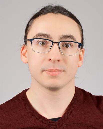
Georg Fantner is an Associate Professor at the Swiss Federal Institute of Technology Lausanne (EPFL) with dual appointments in the School of Engineering (STI) within the Institute of Bioengineering and the School of Life Sciences (SV) for teaching. He directs the Laboratory for Bio- and Nano-Instrumentation (LBNI) and holds leadership roles including President of the Open Science Strategic Committee and the Association des Professeurs de l'EPFL. Research Focus: Bioinstrumentation, Nanotechnology, Scanning Probe Microscopy, and Metrology Teaching: Structural Mechanics for Life Sciences, Metrology, and Metrology Practicals His research pioneers advanced instrumentation for nanoscale characterization, emphasizing data-driven approaches to enhance microscopy techniques. Recent work integrates deep learning with scanning probe microscopy for real-time biological imaging and develops novel MEMS devices for fluid-compatible nanoscale manipulation. Key innovations include hermetically sealed sample chambers for pathogen studies and deterministic nanotopography engineering. Professor Fantner actively mentors 7 current PhD students and has supervised 14 graduates. His laboratory fosters interdisciplinary collaboration across engineering, physics, and life sciences to advance nanoscale measurement technologies and instrumentation development.
Prof. Dr. Hans Josef Hug is a Lecturer at the Department of Physics, University of Basel, located at Klingelbergstrasse 82, Basel, Switzerland. He is affiliated with the Faculty of Science and contributes to research in nanotechnology and surface physics. His work focuses on scanning probe microscopy, magnetic materials, and energy dissipation mechanisms in atomic force microscopy. His key research projects include completed initiatives such as Nano Engineered Neural Interfaces - NENI and Magnetism of Thin Films and Heterostructures . Recent publications include a textbook on scanning probe microscopy and influential articles in Applied Physics Letters and Physical Review Letters . Hug’s research integrates experimental and theoretical approaches to advance nanoscale imaging and material characterization techniques.
Swiss Federal Institute of Technology in LausanneSwitzerland
Nicolas Chiaruttini is a Lecturer and Scientist at École Polytechnique Fédérale de Lausanne (EPFL), affiliated with the School of Life Sciences. He serves in the BioImaging and Optics Core Facility (PTBIOP) and contributes to doctoral education through the EDMS - Teaching program. Institution: École Polytechnique Fédérale de Lausanne (EPFL) School: School of Life Sciences Department: BioImaging and Optics Core Facility Roles: Scientist, Lecturer Office: AI 0140, Building AI, Station 15, 1015 Lausanne, Switzerland Contact: +41 21 693 96 29 | nicolas.chiaruttini@epfl.ch ORCID: 0000-0003-4722-6245 Unit Websites: BioImaging and Optics Core Facility , EDMS Program His research and professional interests center on bioimaging, optics, and image processing, particularly in the context of life sciences and micro/nano-sciences. These areas are reflected in his dual role supporting advanced imaging technologies and teaching in doctoral programs. He teaches the course Image Processing for Life Science , which integrates computational techniques with biological imaging applications. While no recent publications or awards are listed in the provided text, his work is aligned with interdisciplinary research at the intersection of engineering, physics, and biology. Nicolas Chiaruttini is actively contributing to both research infrastructure and academic education at EPFL, demonstrating a commitment to advancing scientific methodology and training the next generation of researchers in quantitative imaging and analysis.
Swiss Federal Institute of Technology in LausanneSwitzerland
Martin Rajman is a Senior Scientist at École Polytechnique Fédérale de Lausanne (EPFL) with multiple affiliations across the institution. He holds positions in the School of Computer and Communication Sciences (SIN - Teaching, SCI IC MR Group, SSC - Teaching) as well as in the Vice Presidency for Strategic Development (VPS Artificial Intelligence) and the Vice Presidency for Academic Affairs (SNAI Administration). He serves as the Executive Director of Nano-tera.ch, a large Swiss Research Program funding collaborative multi-disciplinary projects in Health and the Environment. Rajman's research spans the intersection of artificial intelligence, natural language processing, and information retrieval. His work demonstrates a consistent focus on developing practical applications of computational linguistics and machine learning techniques. Early in his career, he contributed significantly to syntactic parsing, stochastic language models, and vector space representations for text. More recently, his research has expanded into deep learning applications for 3D reconstruction, empathetic conversational agents, and distributed analytics systems. His publications reveal a trajectory from foundational NLP research toward increasingly applied and interdisciplinary work connecting AI with healthcare, environmental monitoring, and human-computer interaction. Analysis of his recent publications (2015-2024) shows a clear evolution toward more applied AI research with strong interdisciplinary connections. While maintaining his core expertise in natural language processing and information retrieval, his work has expanded into computer vision, healthcare applications, and sustainable computing. The publications demonstrate increasing collaboration across disciplines, with applications in medical imaging, mental health support systems, environmental monitoring, and human-centered AI. His leadership role in the Nano-tera.ch program reflects this interdisciplinary approach, connecting computing research with real-world challenges in health and environmental contexts. Rajman has mentored several PhD students including Ailomaa Marita, Eckard Emmanuel, Melichar Miroslav, and Veselý Martin. His research has been supported through the Nano-tera.ch program, which has funded more than 100 research projects with over 95 million CHF in public funding. He has also managed more than 20 European projects during his tenure as Director of the EPFL Global Computing Center. As Executive Director of Nano-tera.ch, Rajman leads a significant research initiative connecting EPFL with national and international partners. His work bridges academic research with industry applications, notably through collaborations with eBay on product ranking technology and with Elsevier on article recommendation systems. His leadership extends to managing large-scale research programs while maintaining an active research agenda and mentoring the next generation of computer scientists.
Marco Stampanoni is a Full Professor for X-ray Imaging at the Department of Information Technology and Electrical Engineering, ETH Zürich, and leads the division for X-ray Imaging and Microscopy at the Institute of Biomedical Engineering. He also heads the X-ray Tomography Group at the Swiss Light Source (SLS), Paul Scherrer Institut, Switzerland. Education : Physics (ETH Zurich, 1998-2002) Key Research Areas : Phase Contrast X-ray Imaging, Realtime Tomographic Microimaging, Nano-tomography, Novel Radiological Methods, Non-Destructive Testing His work bridges cutting-edge synchrotron technologies with clinical applications, focusing on novel X-ray instruments for multiscale non-invasive analysis. He has secured an ERC Grant and received multiple awards for his pioneering research. ETH Silver Medal (2003) Dalle Molle Foundation Award (2012) Finalist, European Inventor Award (2022) Giuseppe Sciacca International Award (2022) He teaches X-ray microscopy at ETH Zurich and has held leadership roles, including Director of the ETH Master of Advanced Studies in Medical Physics (2010-2013) and President of the Paul Scherrer Institut Research Commission (since 2018).
Florencia Malamud is a Researcher and Instrument Scientist at the Paul Scherrer Institute (PSI), leading the POLDI instrument in the Laboratory for Neutron Scattering and Imaging. Her work focuses on advanced neutron-based techniques for material characterization, including Bragg edge imaging, diffraction contrast imaging, and texture analysis. She specializes in studying crystallographic structures, phase transformations, and mechanical behaviors in materials such as high-Mn steels, superalloys, and superconductors. Her research integrates experimental methods like neutron diffraction and tomography to investigate industrial materials (e.g., additive manufacturing components) and historical artifacts (e.g., Napoleonic-era copper bolts). Key areas include optimizing material properties through composition and processing, and understanding deformation mechanisms in metallic materials. Malamud’s publications span materials science, metallurgy, and neutron scattering applications. She collaborates on projects involving nuclear-grade materials, aerospace alloys, and archaeological metallurgy. Her work emphasizes bridging fundamental physics with applied engineering challenges. No scientific awards are explicitly mentioned. Her advising and grants are not detailed in the provided texts. She is affiliated with PSI’s neutron scattering laboratory and contributes to instrumentation development for advanced materials research.
Emiliya Poghosyan is a Senior Scientist at the Paul Scherrer Institute (PSI) in Switzerland, affiliated with the Laboratory for Multiscale Bioimaging and the Electron Microscopy Facility. Since 2018, she provides user support, training, and manages advanced electron microscopy equipment while developing biological imaging methodologies. Her educational background includes: Bachelor of Science in Physics (with honours) from Yerevan State University Master's Degree in Nano-biophysics from Technical University of Dresden PhD in Cryo-electron microscopy from ETH Zurich Postdoctoral work on single particle cryo-EM of membrane proteins at University of Basel Her research focuses on cryo-electron microscopy techniques, structural biology, and membrane protein imaging. Recent publications highlight advancements in ptychography tools, X-ray tomography, and deep learning applications for electron microscopy. She actively contributes to method development and facility management. Scientific achievements include DAAD Long-Term Fellowships (2010-2012), PSI Research Grants (2021), and SDSC Collaborative Data Science Projects. She is a member of the Swiss Society for Optics and Microscopy (SSOM) and has delivered lectures at the University of Zurich (PHY 427). Teaching roles span multiple international cryo-EM schools and hands-on training programs since 2018, emphasizing practical microscopy education.
Iacopo Mochi is a Group Leader for Advanced Lithography and Metrology and beamline scientist at the Paul Scherrer Institute (PSI), specifically working at the Laboratory for X-ray Nanoscience and Technologies within the PSI Center for Photon Science. He has extensive experience in EUV lithography, X-ray optics, and semiconductor metrology, with a career spanning multiple prestigious research institutions including Lawrence Berkeley National Laboratory and imec. Dr. Mochi received his physics degree from the University of Florence and earned a PhD in Methods and Technologies for Environmental Monitoring. His career path has included significant contributions to LIDAR systems, astrophysical instrumentation, and ultimately EUV technologies for semiconductor manufacturing. At PSI since 2016, he has established himself as a leading researcher in advanced lithography techniques. His primary research focuses on the development of EUV instrumentation for semiconductor metrology, particularly the XIL-II metrology end station - a lensless microscope dedicated to EUV photomask inspection. His work spans nano-imaging, interferometry techniques in the X-Ray spectrum, and the characterization of advanced materials for semiconductor manufacturing. Dr. Mochi has made significant contributions to the field through the development of RESCAN, an actinic pattern inspection platform based on coherent diffraction imaging. Analysis of his publication record reveals a strong trajectory of innovation in EUV lithography, with recent work pushing resolution limits to 5 nm, developing novel metrology techniques, and applying machine learning approaches to improve image reconstruction and defect detection. His research directly addresses critical challenges in semiconductor manufacturing as the industry moves toward smaller technology nodes. Dr. Mochi's scientific contributions include groundbreaking work on EUV pellicles for mask protection, characterization of absorber and phase defects on EUV reticles, and advancements in interference lithography techniques. His collaborative research demonstrates strong partnerships with industry and academic institutions worldwide. As the beamline scientist for the XIL-II beamline, Dr. Mochi coordinates and supervises experiments that support cutting-edge research in semiconductor manufacturing and nanotechnology. His leadership extends to mentoring junior researchers and contributing to the development of next-generation scientists in the field of advanced lithography.
Chen Lingxin is a Professor and PhD supervisor at the Yantai Institute of Coastal Zone Research, Chinese Academy of Sciences, where he has been working since 2009 after serving as a Research Professor at Hanyang University's Department of Applied Chemistry from 2007-2009. His research focuses on developing innovative environmental analysis and monitoring technologies specifically for coastal zones. His primary research areas include: Bionano sensor techniques combining aptamers with nanomaterials for environmental pollutant analysis Chromatographic microanalysis techniques for trace analysis of environmental pollutants Opto-electronic monitoring techniques for oceanic water environment Pollutant identification, analysis, and assessment technology Ecological environment monitoring and surveillance technology Simulation of offshore environmental pollution processes and ecological restoration Professor Chen leads the innovative research team of 'Coastal Environmental Analysis, Monitoring and Ecological Restoration,' which strategically combines basic research with applied research and technology transformation across analytical chemistry, environmental science, marine chemistry, and ecological restoration engineering. His publications focus on marine environmental analysis and nanoanalysis methods, demonstrating expertise in developing new principles, methods, and instrument technologies for environmental monitoring of coastal pollutants including inorganic ions, persistent organic pollutants, and pathogens. He has successfully secured multiple national and ministry-level research projects spanning microfluidic chip SERS sensing technology, microbial metabolism of pollutants, and seawater nutrient monitoring systems. Professor Chen actively mentors graduate students in Environmental Engineering, Environmental Science, and Marine Chemistry, with a research group that welcomes students from chemistry, biology, and instrumentation backgrounds. His laboratory specializes in environmental microanalysis with capabilities in optical/electrochemical sensor development, microfluidic technologies, and marine pollution assessment for applications in coastal zone management.
Dr. Anne Bonnin serves as a Beamline Scientist at the Paul Scherrer Institute (PSI) in Switzerland, where she has been instrumental in X-ray imaging research since joining the X-ray Tomography Group in 2014 and assuming her current role at the TOMCAT Beamline in 2016. Affiliated with PSI's Center for Photon Science and Laboratory for Macromolecules and Bioimaging, she operates at the forefront of synchrotron-based imaging techniques. Her academic foundation includes a PhD from INSA de Lyon focused on material properties for explosive detection, followed by postdoctoral work at the European Synchrotron Radiation Facility (ESRF) in X-ray diffraction and phase contrast tomography, and an NSF Research Fellowship for paleontology research at Harvard University and ESRF. Specializing in X-ray imaging (micro/nano-tomography, phase-retrieval) and powder diffraction, Dr. Bonnin leads the bioimaging program at TOMCAT with particular emphasis on the international Heart Imaging Project. Her research develops novel methodologies for materials characterization across diverse domains including cardiac microstructure analysis, paleontology, and neurodegenerative disease modeling, with significant contributions to understanding material behavior at microscopic scales. Her recent publications (2019-2021) demonstrate strong interdisciplinary impact, advancing X-ray imaging applications in energy storage (battery materials), biomedical research (cardiac/auditory systems), and materials engineering (aerogels). A defining trend is the integration of machine learning for image analysis, alongside methodological innovations like non-rigid image stitching and Fourier ptychography. These works reflect extensive international collaboration and address critical challenges in healthcare, energy, and fundamental material science. Dr. Bonnin leads the Heart Imaging Project to quantify cardiac microstructure using contrast-agent-free X-ray phase-contrast imaging, while actively contributing to the SLS2.0 upgrade project preparing TOMCAT for multiscale, multimodal, and dynamic tomographic capabilities. Her collaborative framework spans global researchers in materials science, paleontology, and biomedical engineering. As manager of the TOMCAT nanoscope—a full-field imaging setup achieving 150 nm 3D resolution—she enables cutting-edge research in absorption and phase-contrast imaging. Her team within the X-Ray Tomography Group drives the bioimaging program forward, particularly through the Heart Imaging Project's dynamic cardiac studies using modified Langendorff setups.
University of Applied Sciences and Arts Northwestern SwitzerlandSwitzerland
Prof. Dr. Christoph Wildfeuer is a Professor for Sensor Technology at the University of Applied Sciences and Arts Northwestern Switzerland (FHNW) School of Engineering and Environment. He leads the Quantum Technologies working group and module, focusing on quantum sensors, cryptography, and secure communication systems. Doctorate in Quantum Technologies from University of Siegen (2003) Diploma in Physics from University of Siegen (1999) Postdoctoral research at Louisiana State University, NIST, and Duke University His research spans Quantum Cryptography , Post-Quantum Cryptography , Optical Sensors , and Quantum Communication in aerospace contexts. Projects include: Developing quantum-safe encryption for corporate networks Simulating quantum communication via drones and stratospheric balloons Demonstrating entanglement and post-quantum key exchange on nano-satellites He contributes to secure satellite communications and integrates quantum technologies into embedded systems and wireless security .
Dr. Marilena Steiger is a Research Fellow at the Institute of Pharmaceutical Sciences (IPW) at ETH Zürich, affiliated with the Professorship for Drug Formulation and Delivery. Her research focuses on advanced drug delivery systems, particularly lipid nanocapsules for treating ocular diseases such as retinopathy of prematurity. She holds a patent for a nanoparticle-based treatment for ocular diseases and has received awards including the 2022 Innovation Prize from Biopark Regensburg GmbH and a poster prize from the 2020 European and Global CLINAM-Summit. Her work integrates nanotechnology and pharmaceutical sciences to develop novel therapeutic delivery platforms, with applications in both diagnostics (via dual-detectable nanoparticles) and targeted treatments for retinal disorders. Key collaborations involve Professor Achim Goepferich, and her research emphasizes translational medicine with potential clinical impact. Dr. Steiger’s publications span high-impact journals like Science Advances and Drug Discovery Today , reflecting her contributions to nanomedicine and drug formulation. She actively engages in patent development and academic-industry partnerships to advance biomedical innovations.
Simone Finizio is a Scientist at the Paul Scherrer Institute (PSI) in Switzerland, working at the PolLux beamline of the Swiss Light Source within the Center for Photon Science Laboratory for Condensed Matter. He has held this position since 2015 following his postdoctoral training, specializing in advanced magnetic imaging techniques for nanoscale materials. His academic background includes: Bachelor's degree in Physics Engineering (2009, honorable mention) from Politecnico di Milano Master's degree in Nuclear Engineering (2011, honorable mention) from Politecnico di Milano Ph.D. in Physics (2015) from Johannes Gutenberg Universität Mainz Finizio's research pioneers time-resolved X-ray magnetic microscopy to capture sub-nanosecond magnetic dynamics. His work spans magnetic skyrmion manipulation, domain wall motion analysis, spin wave characterization, and multiferroic material studies. He has developed critical instrumentation for field-free skyrmion nucleation and 3D magnetization imaging, enabling breakthroughs in understanding topological magnetic structures for next-generation spintronic applications. Analysis of his publications reveals consistent innovation in synchrotron-based magnetic imaging techniques, with emphasis on picosecond temporal resolution and nanoscale spatial precision. Key trends include deterministic skyrmion control for memory devices, tilt-free domain wall motion mechanisms, and quantitative electrical detection of topological magnetic states. Finizio actively supports user experiments at PolLux beamline and led instrumentation development for the EU Horizon 2020 MagicSky project (2015-2018). He has secured collaborative grants for sample fabrication and beamline enhancements, including RF setup designs and measurement automation software now standard for user operations. As a core member of PSI's microspectroscopy group, he directs technical development of the scanning-transmission X-ray microscope, implementing hardware and software innovations that advance the facility's capabilities in time-resolved magnetic studies.