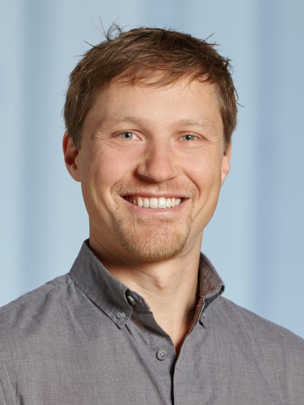
Kajsa Møllersen is an Associate Professor at the Department of Computer Science, UiT The Arctic University of Norway. Her research focuses on statistical approaches to image analysis and machine learning in medical and biological contexts. Research Interests: Histopathological image analysis, divergence functions, multi-instance learning, melanoma detection, breast cancer diagnostics Teaching Roles: PhD course leader in Statistical Models, Master's lecturer in Biostatistics, Medical Statistics, and research methodology Recent publications highlight trends in applying machine learning to histopathology, omics data integration, and educational innovations in statistical pedagogy. She actively contributes to research on data sharing ethics and gender diversity in STEM fields. Key Collaborations: Computational Pathology Group, Systems Epidemiology, Realfagsdidaktikk i høyere utdanning Outreach: Public data ethics discussions, teaching methodology research, and medical informatics dissemination
Professor Christopher Poulton serves as Discipline Leader for Physics at the School of Mathematical and Physical Sciences of the University of Technology Sydney (UTS). With a PhD from the University of Sydney (2000) and postdoctoral experience at institutions including the Karlsruhe Institute of Technology and Max Planck Institute for the Science of Light, he specializes in numerical and analytical methods in photonics, particularly focusing on electromagnetic/elastic wave propagation and light-sound interactions via Brillouin scattering. PhD, University of Sydney (2000) BSc (Hons), University of Sydney (1996) His research explores Brillouin scattering for applications in optical data storage, ultrafast acoustic dynamics, and nonlinear waveguide phenomena. He develops analytical models for photonic crystal fibers, metasurfaces, and optoacoustic devices, with recent work emphasizing machine learning integration for Brillouin microscopy data analysis and on-chip signal processing. Recent publications highlight trends in Brillouin-based memory systems , metasurface design , and nonlinear optoacoustic interactions . Methodologies span principal component analysis for biological imaging to quasi-soliton pulse dynamics in photonic waveguides, often combining numerical simulations with experimental validation. Professor Poulton actively supervises honors projects and teaches advanced mathematical topics including Complex Analysis , Vector Calculus , and Numerical Methods . He has contributed to On-chip photonics: principles, technology and applications as a book chapter author. His laboratory collaborates on projects like optoacoustic isolation and 3D hydrogel engineering under grants including ARC Discovery Projects DP200101893 and DP160101691 . Current infrastructure includes chalcogenide waveguide fabrication and Brillouin response measurement systems.
Brian Meckes serves as an Assistant Professor in Biomedical Engineering at the University of North Texas, with his laboratory situated in Discovery Park (K240D). His academic role involves cutting-edge research in nanobiotechnology and biomaterials, alongside teaching responsibilities in the biomedical engineering curriculum. Dr. Meckes actively contributes to the scientific community through numerous high-impact publications in leading journals. Dr. Meckes' research program centers on engineered nanomaterials for biomedical applications , particularly spherical nucleic acids and light-responsive hydrogels. His work explores how nanoscale interactions govern cell behavior, with applications in targeted cancer therapy, tissue regeneration, and biosensing. Key methodologies include DNA nanotechnology, polymer chemistry, and advanced microscopy. This interdisciplinary approach integrates principles from materials science, molecular biology, and engineering to address challenges in drug delivery and cellular microenvironment control. Analysis of his 15 most recent publications (2020-2025) reveals a consistent focus on dynamic biomaterial systems . Major themes include light-activated cell assembly (2023-2025), biomimetic lung models (2023), and enzyme-responsive nanoparticles for cancer (2022). His work increasingly emphasizes spatiotemporal control in biological systems, using DNA as a programmable tool for precision medicine applications. This trajectory positions his research at the forefront of nanomedicine and regenerative engineering. Based at UNT's Discovery Park, Dr. Meckes leads a research group developing innovative platforms for cellular engineering. His team utilizes advanced nanofabrication facilities and collaborates across disciplines to translate fundamental discoveries into therapeutic strategies, particularly for cancer and fibrotic diseases.
Yongho Bae, PhD, is an Associate Professor in the Department of Pathology and Anatomical Sciences at the Jacobs School of Medicine & Biomedical Sciences, University at Buffalo. He is also an Affiliated Associate Professor in the Department of Biomedical Engineering and an affiliate of the NSF Science and Technology Center for Engineering MechanoBiology. His research bridges cell biology, bioengineering, and mechanomedicine, with a focus on how mechanical forces regulate cellular function in health and disease. Department: Pathology and Anatomical Sciences School: Jacobs School of Medicine & Biomedical Sciences University: University at Buffalo Affiliation: Biomedical Engineering (Affiliated Associate Professor) NSF Science and Technology Center for Engineering MechanoBiology (Affiliate Faculty) Education: PhD, Bioengineering, University of Pittsburgh (2010) MS, Chemical and Biochemical Engineering, Rutgers University (2005) BS, Biotechnology, Ajou University (1998) Dr. Bae's research centers on cell mechanics and mechanotransduction , particularly in cardiovascular biology and stem cell regulation. His work investigates how extracellular matrix stiffness influences vascular smooth muscle cell (VSMC) behavior in diseases like atherosclerosis and arterial stiffness. He employs advanced techniques including atomic force microscopy, traction force microscopy, optogenetics, and machine learning to study cellular responses. His lab also develops nanophotonic platforms to control stem cell differentiation using light, offering non-invasive strategies for regenerative medicine. His recent publications reveal a strong trend in understanding Survivin (BIRC5) as a central mediator of stiffness-induced cell proliferation, motility, and ECM remodeling in VSMCs. He also explores lamellipodin , FAK , and Rac signaling in mechanosensitive pathways. His work integrates transcriptomics, computational modeling, and high-resolution imaging to decode mechanobiological networks. Scientific Awards: Excellence in Mentoring Award for Research (2025) Louis Sklarow Award (2025) Professional Development Award (2023) LeapRx Drug Discovery Incubator Program Award (2022) Buffalo Blue Sky Silver Coin (2019) American Heart Association Career Development Award (2018) Eugene Mindell & Harold Brody Clinical Translational Research Award (2017) Cell and Molecular Bioengineering Meeting: Fellow Travel Award (2015) American Heart Association Postdoctoral Fellowship (2013) Dr. Bae actively mentors students and has served as a faculty mentor for undergraduate research programs. He has been the Principal Investigator on multiple NIH, NSF, and industry-funded grants, including projects on corneal endothelium, vascular matrix, and drug discovery for arterial stiffening. He is also a co-PI on NSF-funded collaborative research in neuronal network programming. His lab, the Bae Mechanobiology and Mechanomedicine Laboratory, focuses on translating mechanistic insights into therapeutic strategies. He serves on editorial boards and as a peer reviewer for journals such as APL Bioengineering , Scientific Reports , and Cellular and Molecular Bioengineering , and is involved in faculty governance and admissions committees.
Constantino Reyes-Aldasoro is a Senior Lecturer in Biomedical Image Analysis at the Department of Computer Science, School of Mathematics, Computer Science and Engineering, City, University of London. His research focuses on the analysis, interpretation, and visualization of biomedical data, particularly in the context of cancer, inflammation, and neurodegenerative diseases. PhD in Computer Science – University of Warwick (2004) MSc in Electrical Engineering – Imperial College London (1994) Bachelor’s in Mechanical and Electrical Engineering – Universidad Nacional Autónoma de México (1993) His research spans image analysis, machine learning, and computational modeling applied to biomedical imaging, with emphasis on electron microscopy, histopathology, and radiology. He has developed algorithms for cell segmentation, vessel tracing, and tumor microenvironment analysis, contributing significantly to open-source tools in the field. His recent publications show a strong trend toward integrating deep learning with traditional image analysis, especially in cancer diagnostics and Alzheimer’s disease assessment. He has also explored topological data analysis and persistent homology for evaluating dataset consistency in colorectal cancer research. Senior Member, IEEE Member Level 1, Sistema Nacional de Investigadores CONACYT (Mexico) He has supervised multiple PhD students in areas such as HeLa cell analysis, coronary plaque detection, and Alzheimer’s imaging. He has secured grants from the Leverhulme Trust, Australian Research Council, and Cancer Research UK. He is an academic editor for journals including PLOS ONE and Journal of Imaging , and has chaired conferences like MIUA and BMVA. He is part of the giCentre research group at City, and actively promotes interdisciplinary collaboration in AI for healthcare.
Brandon Helfield is an Assistant Professor at Concordia University with cross-appointments in Physics and Biology. He holds the Tier II Canada Research Chair in Molecular Biophysics in Human Health and serves as a Junior Scientist at the Physical Sciences Platform of Sunnybrook Research Institute. His research bridges biomedical ultrasound, microbubble dynamics, and therapeutic applications. Education : PhD in Medical Biophysics (University of Toronto), BSc in Physics (McGill University), Postdoctoral Fellowship in Cardiology (University of Pittsburgh) Research Interests : Dr. Helfield focuses on biomedical ultrasound imaging and therapy, particularly microbubble-based technologies for targeted drug/gene delivery to treat cardiovascular diseases. His recent work explores CRISPR-Cas9 delivery to stem cells, immunomodulation via focused ultrasound, and super-resolution imaging algorithms. Publication Trends : His research spans ultrasound physics, microbubble dynamics, and clinical translation. Key areas include cavitation mechanics, vascular delivery optimization, and machine learning applications for imaging analysis. Awards : Burroughs Wellcome Fund Career at the Scientific Interface Award, Arthur E. Weyman Young Investigator Award, European Symposium on Ultrasound Young Investigator Award
Mart Krupovic is a researcher at the Institut Pasteur in Paris, France, where he leads projects in the Department of Cell Biology and Virology of Archaea. He serves as Principal Investigator (PI) for multiple research initiatives, including the ANR-ENVIRA and Emergence(s) MEMREMA projects, focusing on archaeal membrane remodeling and virus egress mechanisms. His research interests include archaeal virology, extracellular vesicles, membrane dynamics, structural biology, and viral evolution . His work leverages advanced techniques such as cryo-electron microscopy (cryo-EM), genomics, and biophysical analysis to study extremophilic archaea and their viruses. Recent publications highlight discoveries in viral capsid malleability, DNA-carrying extracellular vesicles, and the structural evolution of archaeal motility appendages. The trends in his recent publications (2022–2025) reflect a strong emphasis on structural characterization of archaeal systems , particularly viral and surface appendage architectures. His work often bridges evolutionary biology and molecular mechanics, revealing deep homologies between archaeal and bacterial systems, and uncovering novel mechanisms in DNA transfer, membrane remodeling, and virus-host coevolution. Scientific Awards: No awards mentioned in the provided text. Advising and Grants: Dr. Krupovic has supervised or collaborated with several PhD students and postdoctoral researchers, including Emmanuelle Quemin, Sofia Medvedeva, and Miguel Gomez-Raya-Vilanova. He leads externally funded projects such as ANR-ENVIRA, ANR-FunVesi, and the Emergence(s) MEMREMA project, indicating active grant support for his research. Labs and Teams: He is a key member of the 'Evolution & Ecology Moléculaire Microbienne' research group at the Institut Pasteur, specifically within the 'Cell Biology and Virology of Archaea' team. His lab employs interdisciplinary approaches combining virology, structural biology, and microbial genomics to study archaeal systems.
Jean-Yves Tinevez is the Head of the Image Analysis Hub (IAH) at the Institut Pasteur in Paris, France. He serves as a Principal Investigator for key image analysis software projects and is a central figure in the institute's bioimage analysis community. His research is fundamentally centered on bioimage analysis and open-source software development . His primary interests include single-particle tracking , machine and deep learning for image segmentation , the creation of custom analysis pipelines , and the development of extensible software platforms like TrackMate, MaMuT, and JDLL. His work bridges the gap between complex biological imaging data and quantitative scientific discovery. The recent publications he is associated with highlight a strong trend in methodological innovation for bioimage analysis. The focus is on creating powerful, accessible software tools (e.g., SAMJ, CellTracksColab, JDLL, GeNePy3D) and enhancing existing ones (e.g., TrackMate 7) to leverage state-of-the-art AI and computational techniques. While his core expertise is in developing the tools, these tools are applied to diverse biological problems, such as host-pathogen interactions , single-cell analysis in microfluidics , and cellular and tissue morphometrics . Head of Facility, Image Analysis Hub, Institut Pasteur (Current) Principal Investigator, TrackMate, Institut Pasteur (Completed) Principal Investigator, MaMuT, Institut Pasteur (Completed) Member, Advanced Light Microscopy initiative, Institut Pasteur Member, Artificial Intelligence at the Institut Pasteur initiative Steering Committee Member, NEUBIAS Tinevez plays a vital role in training and knowledge dissemination . He is a regular instructor for the Institut Pasteur's PhD training programs and has led numerous workshops on Fiji/ImageJ, Python for image analysis, and advanced bioimage analysis. The IAH, under his leadership, operates as a collaborative core facility with a strong commitment to open science and quality management (ISO-9001:2015 certified). He fosters extensive collaborations with research units across the Institut Pasteur and beyond. The facility provides infrastructure, walk-in support, and develops custom tools to empower researchers, effectively acting as a grant-funded service that enables a vast amount of research across the campus. The Image Analysis Hub, led by Tinevez, is a core technological facility within the Institut Pasteur's Center for Technological Resources and Research (C2RT) and the Research and Resource Centre for Scientific Informatics (C2RI). It operates a dedicated analysis room with specialized workstations and offers remote access via virtual machines. The hub is involved in several transversal projects, including the Advanced Light Microscopy initiative and the application of Artificial Intelligence in biomedical research at the institute.
Christian L. Vestergaard is a CNRS researcher (Researcher rank) in the Decision and Bayesian Computation group within the Department of Neuroscience at the Institut Pasteur, Paris. He holds a PhD in theoretical biophysics from the Technical University of Denmark and has held postdoctoral positions at the Institut Pasteur, Center for Theoretical Physics (Marseille), and DTU Nanotech. He currently leads research projects on neural connectomes and numerical methods for network analysis. Education: PhD in Theoretical Biophysics, DTU Nanotech, Technical University of Denmark (2008–2012) Master’s in Physics and Biophysics, Niels Bohr Institute, University of Copenhagen (2003–2008) Exchange Semester, UPMC, Paris (2007) Second-Year Master’s in Complex Systems His research lies at the intersection of statistical physics, biophysics, and computational neuroscience. He focuses on data-driven modeling of complex dynamical systems, particularly in biological contexts such as neural networks, single-molecule dynamics, and behavioral analysis. His work emphasizes the development of statistical and computational methods to infer structure-function relationships in biological systems. The analysis of his recent publications reveals a strong trend toward methodological innovation in network science, Bayesian inference, and machine learning applied to neuroscience and biophysics. Key themes include temporal network modeling, stochastic processes, information maximization in decision-making, and high-throughput behavioral phenotyping in Drosophila. His work combines theoretical rigor with practical applications in biological data analysis. Scientific Awards and Grants: ANR JCJC grant — SiNCoBe (2021–2024) ANR Inception grant — BitesGoingViral (2022–2024) ANR PR[AI]RIE Springboard Chair (2021–2023) ACIP Inter-Institut Pasteur Concerted Actions grant — ETHOMOS (2019–2021) Pasteur-Roux-Cantarini postdoctoral fellowship (2017–2019) Copenhagen Graduate School pre-doc stipend (2008) Christian L. Vestergaard actively collaborates with principal investigators such as Jean-Baptiste Masson and François Laurent. He has been involved in advising PhD students and postdoctoral researchers within the lab, contributing to interdisciplinary projects that integrate physics, computer science, and biology. His group utilizes advanced computational tools and software such as TRamWAy and LarvaTagger for analyzing large-scale biological datasets. Laboratories and Research Teams: Decision and Bayesian Computation Lab, Institut Pasteur (current) Statistical Physics and Complex Systems team, CPT Marseille (former) Stochastic Systems and Signals group, DTU Nanotech (former)
Patrik Vagovic is a Staff Scientist at the European XFEL GmbH, affiliated with the Center for Free-Electron Laser Science (CFEL), a collaborative research center between DESY, the University of Hamburg, and the Max Planck Society. He leads research in the Coherent Imaging Team, focusing on advanced X-ray imaging techniques using X-ray free-electron lasers. His work bridges the gap between fundamental physics and practical applications in materials science, biology, and fluid dynamics. Dr. Vagovic's research interests center around developing and applying cutting-edge X-ray imaging methodologies, particularly high-speed and phase-sensitive techniques. His work encompasses X-ray phase contrast imaging, coherent diffractive imaging, tomography, and advanced data processing methods. He has pioneered MHz frame rate X-ray imaging capabilities at the European XFEL, enabling unprecedented observation of ultrafast phenomena previously impossible to capture with conventional X-ray sources. Analyzing his recent publication record reveals a strong focus on pushing the temporal and spatial boundaries of X-ray imaging. His work demonstrates a consistent progression from developing fundamental imaging techniques to applying them to complex scientific problems across multiple disciplines. The research shows increasing sophistication in both hardware development (optical systems, detectors) and computational methods (phase retrieval, machine learning). Dr. Vagovic actively collaborates with international research teams across Europe and beyond, contributing to numerous high-impact publications in top journals including Optics Express, Journal of Synchrotron Radiation, and Nature Communications. His work on MHz X-ray microscopy has particularly advanced the field of time-resolved imaging of irreversible phenomena. As part of the Coherent Imaging Team at European XFEL, Dr. Vagovic works with state-of-the-art instrumentation including the SPB/SFX instrument, where he has developed pump-probe capabilities and advanced diagnostics for megahertz pulse trains. His research group utilizes advanced computational approaches alongside experimental innovations to solve complex imaging challenges.
Valentine Ananikov is a Professor at Moscow State University and Head of the Division of Structural Studies at the Zelinsky Institute of Organic Chemistry. Elected as a Member of the Russian Academy of Sciences in 2008 and Academia Europaea in 2018, his work focuses on catalysis, nanoparticle chemistry, and sustainable processes. Research Interests : Mechanistic studies of chemical reactions, development of nanoscale and molecular catalysts, biomass conversion, and AI integration in chemistry. His team combines experimental and theoretical methods to advance green chemistry. Scientific Awards : 2024 International Markovnikov Prize 2023 "Gravity" International Prize for AI research 2016 Organometallics Distinguished Author Award 2016 Hitachi High-Technologies Award 2004 Russian State Prize for Young Scientists Notable Contributions : Pioneering hybrid catalytic systems (homogeneous/heterogeneous), sustainable catalyst design from biomass, and AI applications in reaction discovery. His work addresses environmental safety and carbon-neutral chemical processes.
Wally Block is a full Professor in the Department of Biomedical Engineering at the University of Wisconsin–Madison, where he has led an MRI-focused laboratory since 2000. Previously, he was a systems engineer at GE Healthcare on the first commercial MRI scanners, and he earned his PhD from Stanford University in 1998. Education PhD 1998 – Stanford University MS 1988 – Stanford University BS 1986 – University of Illinois, Urbana-Champaign Research Focus Professor Block’s laboratory pioneers ultra-fast MRI acquisition and reconstruction techniques that dramatically shorten scan times and simplify workflows. Central to his current agenda is image-guided, minimally invasive brain therapy , particularly leveraging intraparenchymal drug-delivery routes to bypass the blood–brain barrier. This highly interdisciplinary program integrates signal processing, machine learning, mechanical engineering, biophysics, and advanced image processing to enable transformative treatments for neurological diseases. Scientific Recognition Fellow, American Institute for Medical and Biological Engineering Senior Fellow, International Society for Magnetic Resonance in Medicine Multiple Distinguished Reviewer awards from Magnetic Resonance in Medicine and Journal of Magnetic Resonance Imaging Vilas Associate Professorship, UW-Madison Honored Instructor Award, UW Housing Whitaker Foundation Grantee Teaching & Mentorship Professor Block regularly teaches cornerstone courses such as Biomedical Engineering Capstone Design (B M E 400/402), Medical Devices Ecosystem: The Path to Product (B M E 640), and directs graduate research credits ( MED PHYS 990 ). He also offers advanced independent study opportunities through B M E 799 , fostering the next generation of engineers and physician-scientists. Laboratory & Collaborative Environment His lab, located in the Wisconsin Institute for Medical Research (WIMR), hosts a multidisciplinary team of graduate students, post-doctoral researchers, and clinical collaborators. The group maintains active partnerships with neurosurgeons, radiologists, and industry leaders to translate novel MRI methods into first-in-human trials.
Edward S. Boyden is the Y. Eva Tan Professor in Neurotechnology at MIT, where he holds appointments in the Department of Brain and Cognitive Sciences, Media Arts and Sciences, and Biological Engineering. He is a full member of the McGovern Institute for Brain Research, co-director of the Center for Neurobiological Engineering and the K. Lisa Yang Center for Bionics, and an investigator at the Howard Hughes Medical Institute. Boyden joined the MIT faculty in 2007 and was awarded tenure as a full professor seven years later. Boyden's research spans multiple areas of neurotechnology, with groundbreaking contributions in optogenetics, expansion microscopy, deep brain stimulation, and multiplexed imaging. His work has transformed neuroscience by providing researchers with powerful tools to observe and manipulate brain activity at unprecedented resolution. Boyden's research integrates principles from physics, engineering, chemistry, and biology to develop novel approaches for understanding and treating brain disorders. His publications reveal a consistent focus on developing innovative technologies that push the boundaries of what's possible in neuroscience. The trend shows increasing clinical translation of his basic science discoveries, with recent work moving from fundamental tool development toward therapeutic applications, particularly in vision restoration through optogenetics and non-invasive brain stimulation for memory enhancement. Breakthrough Prize in Life Sciences (2016) The Brain Prize (2013) Rumford Prize (2019) National Academy of Sciences (2019) Gairdner Foundation International Award (2018) Warren Alpert Foundation Prize (2019) Wilhelm Exner Medal (2020) Boyden has mentored numerous students who have gone on to establish their own research programs, including Deblina Sarkar, Christian Wentz, and Kate Adamala. His research has been supported by significant grants from the NIH, NSF, and private foundations. Through his leadership of the Synthetic Neurobiology Group, Boyden has fostered interdisciplinary collaborations across multiple institutions and fields. Boyden leads the Synthetic Neurobiology Group at MIT, which brings together researchers from diverse backgrounds including neuroscience, engineering, physics, and computer science. The group operates state-of-the-art facilities for developing and testing new neurotechnologies, with close connections to clinical researchers for translational work.
Gabriel Neurohr serves as Assistant Professor in the Department of Biology at ETH Zurich, Switzerland, where he leads research on the physiological consequences of cell size dysregulation. His work bridges fundamental cell biology with implications for aging and cancer, investigating how deviations from species-specific cell size norms disrupt cellular homeostasis. Academic training includes a BSc in Biochemistry from ETH Zurich (2003-2006), PhD at Barcelona's Center for Genomic Regulation (2008-2012), and postdoctoral fellowship at MIT's Koch Institute (2013-2020). His research program addresses four core questions: (1) functional size limits of cells, (2) size-senescence relationships, (3) macromolecule-volume coordination, and (4) density-dependent functional impacts, employing genetic, live-imaging, and computational approaches in yeast and mammalian systems. Recent publications reveal a methodological evolution toward quantitative analysis, with machine-learning-enhanced imaging (2023) complementing mechanistic studies linking genome instability to size-induced senescence (2023). His 2024 work establishes cell enlargement as a primary driver of senescence through cytoplasmic dilution and organelle dysfunction, demonstrating conserved principles across eukaryotes. Major recognitions include: SNF Eccellenza Fellowship (2020-2025) EMBO Long Term Fellowship (2013-2014) ETH Medal for Master's excellence (2008) Neurohr's lab develops innovative tools like holotomography-based vacuole quantification while maintaining focus on fundamental size-regulation mechanisms. He teaches advanced courses on biomolecular condensates and cellular matter properties, mentoring through ETH's structured graduate programs. Current work explores therapeutic targeting of size-dependent senescence pathways in age-related diseases.
Prof. Dr.-Ing. Tim Wilhelm Nattkemper leads the Biodata Mining Group at the Faculty of Engineering , Universität Bielefeld , while holding affiliations with the Center for Biotechnology (CeBiTec) and the Institute for Bioinformatics Infrastructure . His work bridges bioinformatics with marine environmental monitoring , focusing on machine learning and computer vision applications. The group specializes in multivariate bioimage analysis , developing platforms like BioIMAX for web-based high-dimensional data exploration. Research spans from MALDI imaging to deep-sea megafauna classification , integrating information visualization and web technologies . Recent projects address seafloor macrolitter monitoring , coral stress response analysis , and self-supervised learning for diatom classification. Their 15 most recent publications (2023-2025) highlight advancements in marine imaging , automated annotation systems , and AI-driven biodiversity assessment , particularly in polymetallic nodule fields. The group also tackles technical challenges like data imbalance in marine image classification and FAIR data principles implementation. As module responsible for courses like Information Visualization and Introduction to Bioinformatics , Nattkemper contributes to academic training in bioinformatics and data science . His interdisciplinary collaborations span physics , chemistry , and ecology within Bielefeld's Material World strategic research area.