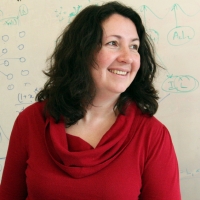
Polina Golland is a Professor in the Department of Electrical Engineering and Computer Science (EECS) at MIT and a Principal Investigator in the Computer Science and Artificial Intelligence Laboratory (CSAIL). Her research focuses on developing novel techniques for biomedical image analysis and understanding, particularly in medical vision, AI/ML, and health care applications. She leads the Medical Vision Group and collaborates with the Vision Group at CSAIL. Her work emphasizes statistical modeling of medical images, shape modeling, and predictive analytics for biological processes. Current projects include fetal MRI analysis, cardiac MRI segmentation, and quantitative assessment of pulmonary edema in chest X-rays. She has secured grants from NIH, MIT-IBM Watson AI Lab, and other institutions to support her research. Dr. Golland teaches courses on inference, probability, and probabilistic systems. She advises graduate students in MIT's EECS program and has mentored numerous postdocs and researchers. Her lab focuses on translating advanced imaging techniques into clinical workflows, with applications in neuroimaging, fetal health monitoring, and cardiovascular disease analysis. Notable collaborations include work with Harvard Medical School affiliates, Brigham and Women's Hospital, and the MIT Jameel Clinic. Her research aims to bridge computational methods with clinical needs, improving diagnostic tools and treatment planning through machine learning and medical imaging innovation.
Dr. Saree Alnaghy is an Honorary Associate Professor at the University of Wollongong's Faculty of Engineering and Information Sciences, affiliated with the Centre for Medical Radiation Physics. She holds a Bachelor of Medical and Radiation Physics (Honours) and a PhD in Physics from the University of Wollongong (2009–2017). Her research focuses on advanced medical imaging and radiation therapy technologies, including photon counting detectors, dosimetry systems, and robotic motion phantoms for quality assurance. Key research areas include: Development of novel X-ray detectors for radiotherapy guidance High-resolution dosimetry techniques using silicon and polymer-based systems Integration of real-time imaging in radiation therapy Robotic systems for motion management in oncology Her work has been supported by grants such as 'Sharper Targeting, Brighter Future' (2024) and 'Bringing Colour to Radiotherapy' (2021–2025). She currently supervises PhD and MRes students on projects involving photon counting CT scanners and radiotherapy imaging. Alnaghy also serves as a Radiation Oncology Medical Physics Registrar at the Nelune Comprehensive Cancer Centre.
Adrien Desjardins is a Professor at the University of British Columbia, jointly appointed in the Department of Mechanical Engineering and Department of Electrical and Computer Engineering within the Faculty of Applied Science. He joined UBC in 2024 after serving as a Full Professor at University College London from 2019-2024, following 13 years on faculty there. His educational background includes a B.Sc. from UBC (2001) and a Ph.D. from MIT and Harvard University (2007). Dr. Desjardins' research program focuses on interdisciplinary development of imaging and sensing modalities and autonomous robotics with marine and biomedical applications. His work integrates photonics, ultrasound, machine learning, and robotics to create innovative diagnostic tools and sensing systems, particularly in optical coherence tomography, diffuse optical spectroscopy, and photoacoustic imaging. His publication record reveals an evolution from foundational neuroimaging work (2001) toward increasingly sophisticated optical systems culminating in breakthroughs like ultrasensitive optical microresonators for ultrasound sensing (2017), demonstrating consistent innovation in biomedical optics with growing emphasis on machine learning integration and real-world applications. His scientific contributions have been recognized through prestigious awards: Research Chair from the Royal Academy of Engineering Healthcare Technologies Challenge Award from EPSRC Starting grants from ERC, EPSRC, and Royal Society World Economic Forum Young Scientist (2015) UCL Provost Teaching Prize (2013) Dr. Desjardins actively mentors graduate students and secures major research funding through competitive grants, with current openings for January/September 2025 intakes. His program involves close industry collaboration indicating strong translational focus, though specific lab names aren't mentioned. The interdisciplinary nature of his work suggests teams spanning engineering, computer science, and medical disciplines working on next-generation imaging systems for healthcare and marine exploration.
Michael McAlpine is a Professor in the Mechanical Engineering department at the University of Minnesota . He also holds affiliations with the Biomedical Engineering and Electrical and Computer Engineering departments. His research focuses on 3D printing functional materials & devices , Nanoscale inks , Biomedical devices , Bioelectronics , and Flexible Microsystems . Research Interests : 3D Printing, Biomedical Engineering, Nanotechnology, Flexible Electronics, Microfluidics Labs : ME 361/363 Contact : mcalpine@umn.edu , (612) 626-3303, ME 117 Recent Research Trends include 3D Printed Biomedical Devices , Flexible Electronics , and Bioprinting Applications . His work spans from Spinal Organoid Formation to Programmable Drug Release Capsules . Scientific Award : Circulation Research 2020 Best Manuscript Award
Jeremy Dahl is a Professor of Radiology (Pediatric Radiology) at Stanford University School of Medicine. He directs the Ultrasound Imaging & Instrumentation Lab and serves as Director of Research Academic Affairs in the Department of Radiology since 2020. He holds multiple affiliations across Stanford including Bio-X, the Cardiovascular Institute, Wu Tsai Human Performance Alliance, Maternal & Child Health Research Institute, Stanford Cancer Institute, and Wu Tsai Neurosciences Institute. Dr. Dahl received his B.S. in Electrical Engineering from the University of Cincinnati (1999) and Ph.D. in Biomedical Engineering from Duke University (2004). His research focuses on developing ultrasonic beamforming and image reconstruction methods for diagnostic imaging applications, particularly techniques that generate high-quality images in difficult-to-image patients. His laboratory specializes in B-mode and Doppler imaging techniques that utilize additional information from ultrasonic wavefields to improve image quality and develop real-time imaging systems for clinical applications including cardiac, liver, and fetal imaging. Dr. Dahl's research has led to significant advancements in ultrasound molecular imaging platforms, sound speed estimation, aberration correction, and reverberation noise suppression. His work often bridges engineering innovation with clinical applications for cancer detection and other diseases. His recent publications demonstrate strong focus on machine learning applications in ultrasound, distributed aberration correction, and molecular imaging techniques. Fellow, American Institute of Ultrasound in Medicine (2021) Senior Member, Institute of Electrical and Electronics Engineers (2020) Distinguished Investigator Award, The Academy for Radiology & Biomedical Imaging Research (2018) Outstanding Paper Award, IEEE Ultrasonics, Ferroelectrics, and Frequency Control Society (2011) Dr. Dahl serves in editorial roles for major journals including IEEE Transactions on Medical Imaging (2017-2024) and IEEE Transactions on Ultrasonics, Ferroelectrics, and Frequency Control (2013-Present). His laboratory has successfully translated numerous innovations into clinical applications, with multiple patents including recent developments in pulsed focused ultrasound therapy and speed of sound quantification.
Dr. Merry Mani is an Associate Professor in Radiology and Imaging Sciences and Biomedical Engineering, specializing in biomedical imaging and signal processing. Her work focuses on advancing MRI-based imaging technologies to study neurological disorders such as Alzheimer's, Autism, and Epilepsy. She holds a Ph.D. in Electrical and Computer Engineering from the University of Rochester (2014) and completed a postdoctoral fellowship at the University of Iowa School of Medicine (2018). Her research combines biophysical modeling with machine learning to explore brain microstructures. Key achievements include the NNARSAD Young Investigator Grant and NIH-funded projects like 'Fast Multi-dimensional Diffusion MRI with Sparse Sampling'. Her lab develops cutting-edge reconstruction methods like qModeL and MUSSELS, prioritizing high spatio-temporal resolution imaging. Major contributions span diffusion MRI acquisition, model-based deep learning, and clinical applications in neurodegenerative diseases. Notable grants include NIH R01EB031169 for Alzheimer’s neurodegeneration studies and projects on rTMS for depression. Her work bridges imaging innovation with clinical impact, aiming to improve diagnosis and treatment through advanced imaging biomarkers.
Carlos Fernandez-Granda is an Associate Professor of Mathematics and Data Science at New York University, holding joint appointments at the Courant Institute of Mathematical Science and the Center for Data Science. He currently serves as the Interim Director of the Center for Data Science. His academic career spans over a decade at NYU, where he has taught probability and statistics to data-science students. Dr. Fernandez-Granda's research focuses on designing and analyzing data-science methodology, with current emphasis on machine learning applications in medicine, climate science, and scientific imaging. His work bridges theoretical foundations with practical applications across multiple domains. His notable contributions include: Development of the COBRA (COnfidence-Based chaRacterization of Anomalies) score for automatic assessment of impairment and disease severity Applications of machine learning to improve climate projections through the M2LInES project Research on magnetic resonance fingerprinting for quantitative tissue parameter estimation Development of AI systems for medical diagnostics including Alzheimer's detection and breast cancer diagnosis Dr. Fernandez-Granda is the author of the book "Probability and Statistics for Data Science," published by Cambridge University Press. The book serves as a comprehensive guide to the two pillars of data science, featuring real-world datasets and addressing fundamental challenges like overfitting, the curse of dimensionality, and causal inference. His research has been supported by grants from the National Science Foundation (Division of Mathematical Sciences, grants 1616340 and 2009752) and the Alzheimer's Association (grant AARG-NTF-21-848627). Dr. Fernandez-Granda is actively involved in several collaborative projects: M2LInES project: An international collaboration focused on improving climate projections using machine learning to capture unaccounted physical processes at the air-sea-ice interface Math and Data group: Exploring the intersection of mathematical theory and data science applications
Professor Sylvia Urban is a distinguished academic at RMIT University, serving as a Professor of Chemistry in the School of Science. She leads the Marine and Terrestrial Natural Product (MATNAP) research group and is the Program Manager for the Bachelor of Science degree, the largest and flagship program in the School of Science. Professor Urban also holds significant leadership roles including Reconciliation and Responsible Practice Facilitator in the School of Science (STEM College) and member of the Nugulu Committee at RMIT University. Her expertise spans natural products chemistry and separation science, with particular focus on chromatography for purification and instrumental analysis for structural characterisation and elucidation. Professor Urban's research interests encompass natural product chemistry isolation and structural elucidation, NMR spectroscopy and mass spectrometry for characterisation of natural products, High Performance/Pressure Liquid Chromatography (HPLC) and other chromatographic techniques for natural product purification, hyphenated spectroscopic techniques such as HPLC-NMR and HPLC-MS for natural product profiling, and biological evaluation of natural products for drug discovery applications. Her work primarily focuses on exploring the biodiversity of Australian marine and terrestrial organisms including plants, fungi, sponges, and algae to discover new compounds with therapeutic potential. She has developed various dereplication and chemical profiling strategies to expedite the discovery process. Professor Urban's publication record demonstrates a strong focus on natural products derived from Australian flora and marine organisms, with particular emphasis on their chemical characterisation and biological evaluation. Her research spans ethnobotanical studies of Indigenous Australian medicinal plants, phytochemical profiling of Australian species, anthelmintic and antimicrobial assessments of natural compounds, and development of analytical methodologies for natural product research. The interdisciplinary nature of her work connects chemistry with pharmacology, ethnobotany, and sustainable development goals related to health, education, and gender equality. STEM College Learning & Teaching Award (Award for Values in Action) 2024 STEM College Athena Swan Award 2023 Top STEM College Media Star 2022 School of Science Reconciliation Champion Award for 2021 School of Science Associate Dean's Impact Award (Applied Chemistry) for 2021 STEM Female Educator of the Year Award in the STEM College in 2021 Fellow of the Royal Australian Chemical Institute (RACI) in 2020 2019 Australian Award for University Teaching (AAUT) Citation for Outstanding Contributions to Student Learning Professor Urban actively supervises Masters and PhD students, with recent projects focusing on nanoparticle synthesis, natural product evaluation from Australian plants and marine organisms, food science applications, and biomedical imaging agents. She has received numerous teaching grants including the SteLR Grant 2017 for Pen-enabled, Real-time Student Engagement for Teaching in STEM Subjects, SteLR Plus Learning and Teaching Grant 2016 for Contextualizing Learning Chemistry, and Global Learning by Design (GLbD) Learning and Teaching Grant 2014. As the leader of the MATNAP research group, Professor Urban oversees a team focused on exploring Australian biodiversity for drug discovery. She has been instrumental in establishing the VICS Molecular Resolution Facility (chromatography node at RMIT University) as part of "The Pipeline – An Integrated Approach to Drug Design and Development." Her research involves collaborations both within and external to RMIT University, including Australian and international university and industry partners.
Essa Yacoub is a Professor in the Department of Radiology at the University of Minnesota, affiliated with the PhD Program in Medical Physics and the Center for Magnetic Resonance Research. His work focuses on advancing MRI and fMRI technologies, particularly at ultrahigh magnetic fields (e.g., 10.5 T), to achieve unprecedented spatial and temporal resolution in brain imaging. He leads projects in RF coil design, noise reduction algorithms, and developmental neuroimaging. Roles: Professor, Medical Physics Program Faculty Affiliations: Center for Magnetic Resonance Research, Department of Radiology Research emphasizes high-resolution fMRI applications, including layer-specific brain mapping, pediatric neurodevelopment studies (e.g., Baby Connectome Project), and translational tools like BIBSNet for infant brain segmentation. His innovations bridge hardware engineering (RF coils) and software (denoising pipelines) to tackle challenges in mesoscopic-scale imaging. Key contributions include optimizing imaging protocols at 7T/10.5T, developing NORDIC denoising for submillimeter data, and advancing understanding of brain networks in aging and neurological disorders. His work is foundational for large-scale initiatives like the Human Connectome Project and non-human primate neuroimaging collaborations. Grants and collaborations focus on translational imaging technologies, while educational contributions include training through the Medical Physics PhD Program. Ongoing efforts aim to refine ultra-high field MRI applications for clinical and basic neuroscience research.
Zhi-Pei Liang is the Franklin W. Woeltge Professor in the Department of Electrical and Computer Engineering at the University of Illinois at Urbana-Champaign, with joint appointments in the Department of Bioengineering, Beckman Institute for Advanced Science and Technology, and Coordinated Science Laboratory. His research spans biomedical engineering, medical imaging, and signal processing with a focus on advancing magnetic resonance imaging and spectroscopy technologies. His educational background includes a Ph.D. in Biomedical Engineering from Case Western Reserve University (1989) and a B.S. in Electrical Engineering from South-China University of Technology (1982), followed by postdoctoral training at UIUC (1989-1991). Professor Liang's research interests center on magnetic resonance imaging and spectroscopy , with particular emphasis on ultrafast imaging techniques , model-based reconstruction methods , and the integration of physics-based modeling with machine learning . His pioneering work on SPICE (SPectroscopic Imaging by exploiting spatiospectral CorrElation) has revolutionized high-resolution metabolic brain imaging by enabling label-free molecular imaging through the marriage of spin physics and machine learning. His research spans pattern recognition, parameter estimation, image formation theory, and algorithms for medical imaging applications. Analysis of his recent publications reveals a strong focus on high-resolution metabolic imaging , particularly using SPICE methodology to map brain metabolism with unprecedented detail. His work bridges fundamental physics of magnetic resonance with advanced computational methods to overcome traditional limitations in imaging speed and resolution. Current research directions include J-resolved spectroscopic imaging, deuterium-based metabolic mapping, and multimodal integration of PET and MRSI for studying neurological disorders. Elected to International Academy of Medical and Biological Engineering (2012) Gold Medal, International Society for Magnetic Resonance in Medicine (2022) Technical Achievement Award, IEEE Engineering in Medicine and Biology Society (2014) Fellow, National Academy of Inventors (2021) Author of influential book 'Principles of Magnetic Resonance Imaging' (1999) President of IEEE Engineering in Medicine and Biology Society (2011-2012) Professor Liang has advised numerous students and postdocs in biomedical imaging research and has received multiple teaching honors including the Ronald W. Pratt Outstanding Teaching Award (2005) and multiple listings among UIUC's Excellent Teachers. His research has been supported by various grants from NIH, NSF, and other funding agencies. He leads the SPICE (Spectroscopic Imaging by exploiting spatiospectral Correlation) research group which focuses on developing novel imaging techniques that combine physics-based modeling with machine learning for ultrafast metabolic imaging. His laboratory, part of the Beckman Institute's Integrative Imaging Theme, collaborates extensively with clinical researchers at Carle Illinois College of Medicine and other institutions to translate advanced imaging techniques into clinical applications for neurological disorders, cancer, and metabolic diseases. Current projects focus on high-resolution mapping of brain metabolism in Alzheimer's disease, stroke, and brain tumors using novel MR spectroscopic imaging techniques.
Daniele Loiacono is an Associate Professor at Politecnico di Milano's Department of Electronics, Information, and Bioengineering (DEIB), affiliated with the Artificial Intelligence and Robotics Lab (AIRLab). His research focuses on interdisciplinary applications of Artificial Intelligence, Machine Learning, and Deep Learning in medical imaging, radiation therapy, and procedural content generation for games. He leads projects in synthetic image generation for radiotherapy quality assurance, automated treatment planning, and bias analysis in medical AI systems. Key research areas include medical image synthesis using GANs, radiation therapy optimization, and algorithmic game design. His contributions span clinical applications such as total marrow irradiation (TMI) planning and lymph-node segmentation, alongside innovations in shader generation and interactive evolutionary tools for game development. Loiacono collaborates on multi-center studies to validate AI-driven workflows in healthcare and has pioneered methods combining lean Six Sigma with machine learning for treatment process improvement. His work bridges clinical medicine and computer science, addressing challenges in radiation oncology, anatomical imaging, and procedural content automation. The AIRLab serves as a hub for his research, integrating AI advancements into real-world medical and engineering solutions.
Hee Kwon Song, Ph.D. is an Associate Professor of Radiology at the University of Pennsylvania , affiliated with the Institute for Translational Medicine and Therapeutics and the Penn Center for Musculoskeletal Disorders . His expertise lies in MRI physics and MRI engineering , focusing on novel dynamic imaging strategies. Education: BS in Electrical Engineering, Northwestern University (1991) MS in Bioengineering, University of Pennsylvania (1994) PhD in Bioengineering, University of Pennsylvania (1999) Master of Law, University of Pennsylvania Carey Law School (2021) Dr. Song’s research specializes in dynamic contrast-enhanced MRI (DCE-MRI) using radial data acquisition and KWIC reconstruction for ultra-fast imaging. His work enables retrospective respiratory motion compensation and flexible resolution adjustments in tumor treatment monitoring ( lung, liver, breast, kidney, ovarian ). Applications extend to non-contrast MRA and tissue T1 mapping . Selected Publications demonstrate his focus on accelerated MRI , bone-selective imaging , and deep learning reconstruction , with collaborations spanning Medicine, Obstetrics and Gynecology, Neurology , and Radiation Oncology . Contact: heekwon.song@pennmedicine.upenn.edu
James S. Duncan is the Ebenezer K. Hunt Professor of Biomedical Engineering at Yale University, with additional appointments in Electrical & Computer Engineering and Radiology & Biomedical Imaging. His research focuses on biomedical image processing, quantitative image analysis using geometrical models, and applications in cardiac function and neuro-structure analysis. He has pioneered image-guided interventions and developed computational frameworks for medical imaging challenges. He holds a Ph.D. from the University of Southern California. His work integrates AI, deep learning, and statistical decision-making to advance medical imaging technologies. Notable contributions include advancements in 3D image segmentation, deformable models, and MRI-based tumor response assessment. Dr. Duncan has received prestigious awards, including IEEE Fellow (2001) and induction into the American Institute for Medical and Biological Engineering (2000). His recent research spans AI-driven hemodynamics modeling, trustworthy healthcare AI guidelines, and molecular MRI innovations in immunotherapy monitoring. He collaborates across disciplines to address challenges in cardiovascular, neuroimaging, and oncological applications.
Hunor Kertész is a Research Fellow at the University of Sydney's Image X Institute, affiliated with the Discipline of Medical Imaging Sciences in the School of Health Sciences. His research focuses on advancing medical imaging technologies, particularly in PET (Positron Emission Tomography) and CT (Computed Tomography) systems, with an emphasis on improving image quality, reducing radiation exposure, and developing novel imaging tools. His work spans several key areas: optimizing positron range correction in PET/CT for cardiac and oncology applications, exploring low-dose imaging techniques for pediatric patients, and creating open-source software tools like Dvgardener for medical image processing. He has contributed to the development of anthropomorphic phantoms for clinical simulations and portable CT systems for lung cancer screening. Recent publications highlight advancements in PET/MRI dose reduction for epilepsy patients, high-resolution total-body PET/CT through positron range correction, and the validation of Rubidium-82 myocardial imaging methods. His research often intersects with radiation dosimetry, image reconstruction algorithms, and 3D-printed anatomical models for quality assurance. While not explicitly mentioned, his contributions likely involve collaborations with clinical teams to translate imaging innovations into practical diagnostic solutions.
Volker J Schmid is a Professor of Bayesian Imaging and Spatial Statistics at the Department of Statistics, Ludwig Maximilian University of Munich. He leads the Bayesian Imaging and Spatial Statistics group and contributes to interdisciplinary initiatives like the Munich Center of Machine Learning. His work bridges statistical theory with applications in medical imaging and biology. PhD in Statistics (2004), LMU Munich Diploma in Statistics (2000), LMU Munich Abitur, Joseph-von-Fraunhofer-Gymnasium Cham (1993) His research focuses on Bayesian computational methods for high-dimensional data, particularly in medical imaging (MRI, DCE-MRI) and biological microscopy (e.g., 3D nuclear architecture analysis via super-resolution microscopy). Key applications include disease mapping , image segmentation , and spatio-temporal modeling . His software tools (e.g., nucim , bioimagetools , BAMP ) enable quantitative analysis in nuclear imaging and age-period-cohort modeling. His 15 most recent publications span Bayesian modeling for medical imaging , spatio-temporal epidemiology , and computational biology . Topics include co-localization metrics in fluorescence microscopy, nuclear architecture analysis, and dynamic Bayesian frameworks for MRI data. Collaborations extend to neuroimaging, oncology, and nuclear biology.