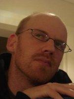
Henrik Nils Latter is a Professor at the Department of Applied Mathematics and Theoretical Physics (DAMTP), University of Cambridge, and a Fellow of Girton College. His research focuses on astrophysical fluid dynamics, particularly in protoplanetary disks, Saturn's rings, and galaxy cluster plasmas. Doctorate in Astrophysics (2006), University of Cambridge Master of Science (2003), University of Sydney Bachelor of Arts and Science (2000), University of Sydney Latter's research spans instabilities, waves, and turbulence in astrophysical disks. Key areas include the vertical shear instability (VSI) in protoplanetary disks, gravitoturbulence, and magnetothermal instability (MTI) in galaxy clusters. His work combines analytical methods with large-scale numerical simulations. His recent publications (2022-2025) address topics such as streaming instability in debris disks, thermal hysteresis in planetary rings , and MHD dynamos in gravitoturbulent systems . These studies often appear in journals like MNRAS and A&A, reflecting his expertise in disk dynamics and magnetic plasma behavior. Scientific Awards: Adams Prize Latter has supervised numerous PhD and Master's students on disk turbulence, planetary ring instabilities, and magnetic field dynamics. He contributes to outreach through the Faculty of Mathematics' Astrophysical Fluid Dynamics group and maintains active collaborations in computational astrophysics.
Dr. Mingyan Li is an Adjunct Research Fellow at The University of Queensland's School of Electrical Engineering and Computer Science. Their research focuses on advanced imaging and sensing technologies with applications in biomedical engineering, particularly in MRI system development, RF coil design, and medical signal processing. They hold a PhD from The University of Queensland (2015). Research interests include high-field MRI systems, rotating RF coil technologies, MRI-Linac integration, and electrical properties tomography (EPT). Key contributions include innovations in MRI-Linac distortion correction, RF shielding for SAR reduction, and deep learning approaches for cardiac arrhythmia classification. Publications span MRI hardware optimization, image reconstruction algorithms, and biomedical signal analysis. Collaborations include work on metamaterial-inspired RF shielding and multi-modal antenna systems for body MRI.
Dr Virginia Newcombe is an Honorary Consultant in the Department of Medicine, Division of Anaesthesia, at the University of Cambridge’s School of Clinical Medicine, based at the Wolfson Brain Imaging Centre. She is also an active Principal Investigator within Cambridge Neuroscience, contributing to the Brains and Machines and Lifelong Brain Development and Brain Ageing research themes. Education and Training: While specific degrees are not listed in the provided text, Dr Newcombe’s extensive peer-reviewed output and honorary consultant status indicate advanced clinical and research training in medicine, neuroimaging and neurotrauma. Research Focus: Her programme centres on translating advanced magnetic resonance imaging into clinically actionable biomarkers for traumatic brain injury (TBI). Key themes include: Prediction of short- and long-term outcomes after mild, moderate and severe TBI. Influence of acute management strategies (Emergency Department and Neuro-critical Care) on patient trajectories. Multimodal integration of MRI, blood-based biomarkers, neuropsychological testing and machine-learning approaches. Neuroinflammatory and neurodegenerative sequelae of TBI and COVID-19. Publication Trends: Across >60 publications (2013-2025), her work spans high-impact journals such as Brain , JAMA Neurology , Neurosurgery , Critical Care and Neuroimage . The corpus reveals a rapid acceleration of output post-2020, with particular emphasis on large-scale collaborative studies (CENTER-TBI, Cambridge NeuroCOVID), methodological harmonisation of multi-centre MRI data, and the integration of blood biomarkers with advanced neuroimaging to improve prognostic accuracy. Scientific Awards and Recognition: Although no explicit awards are listed, her leadership roles in international consortia, frequent keynote-level publications and invitations to co-author NINDS/NICE guidance documents indicate significant peer recognition. Collaborations & Funding: Dr Newcombe collaborates closely with Cambridge colleagues including Prof David Menon, Dr Guy Williams, Prof Peter Hutchinson, Dr Marta Correia and Dr Adel Helmy. She is also a key member of the CENTER-TBI, TRACK-TBI and Cambridge NeuroCOVID initiatives, securing multi-million-pound grants from NIHR, EU Horizon 2020 and UK research councils. Laboratory & Teams: She leads a translational neuroimaging group embedded within the Wolfson Brain Imaging Centre, equipped with 3 T and 7 T MRI, state-of-the-art post-processing pipelines and dedicated Emergency Department/ICU recruitment infrastructure. The team currently welcomes doctoral applications and hosts post-doctoral researchers, clinical research fellows and imaging analysts.
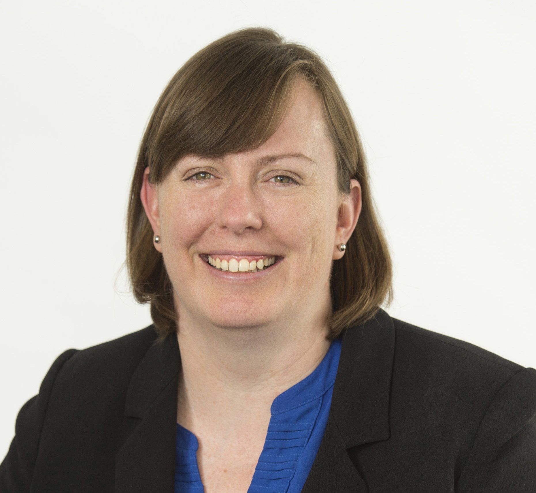
Rebecca Feldman is an Assistant Professor in Medical Physics and Physics at the Irving K. Barber Faculty of Science, University of British Columbia Okanagan. Her research integrates MR physics, engineering, and medical research to advance MRI pulse sequences and hardware for clinical translation, particularly in neurological disorders. University: University of British Columbia Okanagan Academic Rank: Assistant Professor PhD: University of Western Ontario Research Interests: Dr. Feldman specializes in technical innovation in MRI (accelerated imaging, spectroscopic imaging, non-proton imaging) and translational research applying MRI to neurological disease detection, characterization, and treatment. Her work leverages ultra-high-field (7T) MRI for enhanced resolution of brain structures like hippocampal subfields and perivascular spaces. Publications: Recent work includes advancements in self-supervised medical imaging backbones (MedMAE), segmentation of venous structures in epilepsy, automated MRI pulse design via neural networks, and clinical applications of 7T MRI in neurosurgical planning and psychiatric disorders like major depressive disorder. Teaching: Currently teaches courses in physics, including physics of waves.
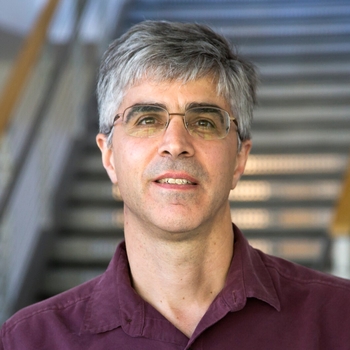
Douglas Noll is the Ann and Robert H. Lurie Professor in the Department of Biomedical Engineering at the University of Michigan , with additional appointments as Professor of Radiology and affiliate of multiple research institutes including the Michigan Alzheimer’s Disease Research Center, Michigan Neuroscience Institute, and Michigan Institute for Imaging Technology and Translation (MIITT). He co-directs the Functional MRI Laboratory and leads the NeuroImaging Core at the Michigan Alzheimer’s Disease Research Center. Prof. Noll’s research focuses on MRI technology development , particularly for functional MRI (fMRI) . His group specializes in high-speed MRI acquisition , signal processing , and image reconstruction to map brain function. Key areas include artifact elimination , physiological modeling , and quantitative imaging for neurological and psychiatric disorders. Recent collaborations apply MRI to histotripsy therapy monitoring. The 15 most recent articles reflect his work in dynamic MRI reconstruction , artifact correction , and cross-vendor protocol standardization . Topics span B0 shimming , histotripsy targeting , and non-Cartesian sampling , emphasizing spatiotemporal modeling and machine learning integration for accelerated imaging. Scientific Awards : Ann and Robert H. Lurie Professor. Research Collaborations : Functional MRI Laboratory, Michigan Alzheimer’s Disease Research Center, Michigan Institute for Imaging Technology and Translation (MIITT).
Ahmet Tekalp is a Professor in the Department of Electrical and Computer Engineering at Koc University's College of Engineering since 2001. He holds dual citizenship in Turkey and the USA, with prior academic roles at the University of Rochester (1986-2005) and research positions at Eastman Kodak (1984-1987) and Rensselaer Polytechnic Institute (1981-1984). He chairs the Electronics and Informatics Group at TUBITAK since 2004 as a part-time position. B.S. (1980) in Electrical Engineering & Mathematics, Bogaziçi University M.S. (1982) and Ph.D. (1984) in Electrical, Computer, and Systems Engineering, Rensselaer Polytechnic Institute His research focuses on digital image and video processing, including video compression, motion-compensated filtering for high-resolution applications, video segmentation, object tracking, content-based video analysis, multi-camera surveillance processing, and digital content protection. He has led numerous European and U.S. grants, including FP7 STREP projects and NSF awards, emphasizing applications in sensor networks, visual databases, and medical imaging. His scholarly work spans diverse areas such as superresolution reconstruction, head gesture animation, 3DTV streaming, and reversible data hiding. He has played pivotal roles in editorial boards, including serving as Editor-in-Chief of Signal Processing: Image Communication, and has contributed to major standards bodies like ISO MPEG and ANSI NCITS. Member, Turkish Academy of Sciences (TUBA) Fellow, IEEE Fulbright Senior Scholarship (1999) TUBITAK Science Award (2004) IEEE Signal Processing Society Distinguished Lecturer (1998) He has led multiple international research collaborations and projects, including European FP6/FP7 networks and NATO programs, with substantial grant funding from NSF, NYSTAR, and industry partners like Eastman Kodak, Xerox, and Siemens.
Attila Keresztes is an Assistant Professor in the Department of Cognitive Psychology at Eötvös Loránd University (ELTE), Budapest, Hungary. He serves as Principal Investigator of the Lifespan Memory Development Lab and Head of the Hippocampal Circuit and Code for Cognition Lab (HCCCL), a Max Planck Partner Group. His research focuses on hippocampal structure-function relationships across the lifespan, employing high-resolution MRI and experimental methods to study memory development, stress impacts, and cognitive aging. Keresztes holds a PhD in Cognitive Science from Budapest University of Technology and Economics (2014) and completed postdoctoral training at the Max Planck Institute for Human Development in Berlin. Key interests include hippocampal maturation’s role in memory specificity, longitudinal brain development, and the interplay between stress hormones (e.g., cortisol) and hippocampal morphology. His work has been funded by grants such as FK128648 from Hungary’s National Research, Development, and Innovation Office. In 2022, he received the prestigious Bolyai János Research Scholarship from the Hungarian Academy of Sciences. Current lab activities involve collaborations with the Max Planck Institute and the ELTE Babylab, including the NeMO study on early childhood memory development. The HCCCL team includes PhD students (Alex Ilyés, Zsuzsanna Nemecz, Hunor Kis) and student researchers focusing on neuroimaging, semantic memory, and pattern separation mechanisms. Public engagement initiatives include participation in events like the European Researchers' Night and science outreach for children.
Devid Maniglio is an Associate Professor at the Department of Industrial Engineering, University of Trento. His research focuses on bioengineering, biomaterials, and tissue engineering, with a particular emphasis on bioprinting, surface modification, and functional materials. He has contributed to advancements in silk fibroin and hydrogel-based systems for medical applications. Research Interests Bioengineering for personalized medicine Biomaterials and surface engineering 3D bioprinting and tissue regeneration Molecular imprinting and biosensors Drug delivery and cell encapsulation Teaching Diagnostic and therapeutic technologies for personalized medicine Engineered materials for precision medicine Fundamentals of biomedical technologies Functional surfaces laboratory Labs & Collaborations Devid Maniglio is affiliated with the Functional Surfaces Laboratory at the University of Trento, collaborating with researchers such as Stefano Rossi and Flavio Deflorian. His work integrates interdisciplinary approaches in biomedical engineering and sustainable medical technologies.
Spencer L. Bowen, Ph.D., serves as Assistant Professor of Radiology at UT Southwestern Medical Center where he leads PET research within the Radiology Research section. His work develops nuclear tomographic imaging tools to advance precision medicine for oncology, neurology, and cardiology applications through innovative scanner technologies and quantitative imaging methodologies. Dr. Bowen earned his bachelor's degree in biomedical engineering from the University of Washington and doctoral degree from the University of California at Davis, followed by a research fellowship at Massachusetts General Hospital. His academic journey includes prior appointment as Research Assistant Professor at the Fralin Biomedical Research Institute and Virginia Tech-Wake Forest University School of Biomedical Engineering. His research program investigates advanced acquisition techniques, reconstruction algorithms, and post-processing methods for quantitative hybrid PET-CT/MR systems. Key focus areas include attenuation correction methodologies, partial volume correction, dedicated breast imaging systems, and cardiac PET quantification. This work bridges engineering innovation with clinical applications to improve diagnostic accuracy and treatment monitoring across multiple disease domains. Dr. Bowen's publication record demonstrates consistent contributions to medical imaging science, with recent emphasis on quantitative cardiac PET, attenuation correction techniques, and dedicated breast imaging systems. His 2016 study on partial volume correction methods in aging research and 2023 work on cardiac PET attenuation correction represent significant methodological advances in the field. As an active scientific contributor, Dr. Bowen serves as reviewer for leading journals including the Journal of Nuclear Medicine, Medical Physics, Physics in Medicine and Biology, and IEEE Transactions on Nuclear Science and Medical Imaging. His work has received notable recognition including a cover feature in the Journal of Nuclear Medicine. The Bowen Lab maintains active collaborations across medical imaging disciplines and currently recruits PhD graduate students to advance nuclear tomography research. Dr. Bowen's mentorship extends through his role as faculty advisor and his participation in graduate training programs focused on biomedical imaging technologies.
Harald E. Möller is a Professor and Head of the Nuclear Magnetic Resonance Research and Development Unit at the Max Planck Institute for Human Cognitive and Brain Sciences in Leipzig. With a career spanning over four decades, he has held academic positions including Honorary Professor at the University of Leipzig and leadership roles in institutions like Duke University Medical Center and the University of Münster. His research focuses on advancing MRI methodologies, biophysical imaging principles, and their applications in neurology and neuroscience. Education: 1979-1985: Chemistry & Physics studies at Universities of Dortmund and Münster 1985: M.Sc. (Diploma) in Chemistry 1988: PhD in Physical Chemistry (summa cum laude) 2000: Habilitation in Physical Chemistry 2002: Habilitation in Biophysical Chemistry Research Interests: Development of novel MRI methods Quantitative tissue characterization Myelin sheath imaging Cerebral blood flow dynamics High-field MRI hardware
Naveed Mahmud is an Assistant Professor at the Department of Electrical Engineering and Computer Science, Florida Institute of Technology. He specializes in quantum computing, hybrid quantum-classical systems, and reconfigurable computing architectures. His research focuses on optimizing quantum algorithms, data encoding/decoding techniques, and secure communications using quantum technologies. Research interests include quantum-classical integration, algorithm emulation on high-performance reconfigurable computers, and applications of quantum computing in pattern recognition and cryptography. Key areas of exploration are hybrid quantum-classical machine learning, quantum wavelet transforms, and securing free-space optical communications with quantum key distribution. His recent work emphasizes scalability and efficiency in quantum computing frameworks, including frameworks like QASM-to-HLS for quantum circuit acceleration, and decoherence-optimized quantum circuits. Articles highlight advancements in quantum data decoding, algorithm emulation, and secure communication systems. No scientific awards or formal advisees are listed. His profile includes links to ORCID, Google Scholar, and ResearchGate for further details on publications and collaborations.
Dr. Daniel Tward is an Assistant Professor at the University of California, Los Angeles (UCLA), holding joint appointments in the Department of Neurology and the Department of Computational Medicine. He earned his Ph.D. in Biomedical Engineering from Johns Hopkins University and completed postdoctoral training at the Kavli Neuroscience Discovery Institute. His research integrates neuroimaging, machine learning, and differential geometry to analyze brain structure changes in neurodegenerative diseases like Alzheimer's, with a focus on bridging molecular pathology and clinical imaging. Research Interests: Dr. Tward's work addresses challenges in neuroimaging data complexity, developing computational tools to map brain anatomy across scales (from centimeters to microns). Key areas include neurodegeneration in the medial temporal lobe, multi-modal image registration, and spatial transcriptomics. His lab emphasizes high-dimensional statistics and geometry-driven analysis to improve diagnostic accuracy and clinical trial design. Grants & Projects: Secured NIH funding for: A 3D multimodal human brain atlas integrating MRI and histology. CloudReg—a distributed framework for massive neuroimage registration. Contributions to the BRAIN Initiative Cell Census Network (BICCN) for mouse/rat brain atlases. Students & Training: Mentors undergraduate researchers via the BIG Summer program, with projects on neural networks, spatial transcriptomics, and MRI analysis. No PhD/Master's advisees listed.
Dr. Brad Oborn is a Research Fellow at the School of Physics, University of Wollongong. His work focuses on advancing radiation therapy techniques, particularly in MRI-guided radiotherapy and proton therapy. He specializes in high-resolution dosimetry, magnetic field effects on radiation beams, and the integration of medical imaging with therapeutic systems. His research interests include radiation dosimetry in magnetic fields, proton beam characterization, and the development of novel detectors like the 'MagicPlates' silicon array. He collaborates on projects such as the Australian MRI-Linac Program, aiming to improve cancer treatment through real-time adaptive radiotherapy. Dr. Oborn has supervised numerous higher-degree students, focusing on topics like skin dosimetry in MRI-linacs, ion chamber response in magnetic fields, and 4D dosimetry modeling. He has secured grants from institutions like the National Health and Medical Research Council (NHMRC) and Cancer Council, supporting his work on high-resolution dosimetry and MRI-guided therapies. His publications reflect contributions to medical physics, including studies on proton dosimetry, electron streaming in MRI systems, and the clinical challenges of MRI-guided proton therapy. His work bridges fundamental physics and clinical applications, aiming to enhance precision and safety in radiation oncology.
Associate Professor Kai-Hsiang Chuang is a Principal Research Fellow at the School of Biomedical Sciences within the Faculty of Health, Medicine and Behavioural Sciences at the University of Queensland. He is also affiliated with the Queensland Brain Institute and the Centre for Advanced Imaging. His research focuses on understanding brain networks, developing advanced imaging techniques, and translating these findings to improve diagnosis and intervention for neurological disorders. Dr. Chuang received his Ph.D. in electrical and biomedical engineering from the National Taiwan University, Taiwan, in 2001. His doctoral research focused on improving the detection of brain activity using functional magnetic resonance imaging (fMRI). Ph.D. in Electrical and Biomedical Engineering, National Taiwan University (2001) Dr. Chuang's research spans multiple areas of brain imaging and neuroscience. His primary focus is on functional brain mapping , where he develops in vivo imaging techniques including functional MRI and multimodal integration with optogenetics, calcium imaging, and electrophysiology. He applies these techniques in both humans and animal models to improve understanding and intervention of brain function, disease processes, and treatment effects. Another key area is brain networks in learning, memory, and dementia . His work explores how brain network wiring and activity underpin cognition and behavior, with particular focus on understanding the causal relationship between brain network activity and memory formation. He develops techniques to modulate behavior by manipulating brain network activity. More recently, Dr. Chuang has expanded into brain waste clearance research, studying the brain's fluid drainage system that clears waste and toxic molecules like amyloid plaques. His lab is developing imaging techniques to track this system's function and understand its regulatory mechanisms, which could provide new treatment targets for dementia. Analysis of Dr. Chuang's recent publications reveals a strong focus on advancing functional MRI techniques for brain network analysis, particularly in rodent models. His work consistently bridges basic neuroscience with clinical applications, especially in understanding memory formation and dementia. A notable trend is the development of multimodal approaches that combine fMRI with optogenetics, calcium imaging, and electrophysiology to establish causal relationships in brain networks. His research increasingly addresses the translation of preclinical findings to human applications, with growing emphasis on Alzheimer's disease mechanisms and potential interventions. Dr. Chuang serves on the editorial boards of multiple prestigious journals including Frontiers in Neuroscience: Brain Imaging Methods , Imaging Neuroscience , and Scientific Reports , reflecting his standing in the field. Editorial Board Member, Frontiers in Neuroscience: Brain Imaging Methods Editorial Board Member, Imaging Neuroscience Editorial Board Member, Scientific Reports Dr. Chuang is actively involved in research supervision, currently serving as Principal Advisor for one PhD student working on "Developing imaging and neuro-technologies for decoding memory formation" and Associate Advisor for two other PhD projects. He has successfully completed supervision of three PhD students on topics related to resting-state networks, memory consolidation, and functional MRI. ARC Discovery Projects (2024-2028): "Decoding the brain network of memory formation" ARC Training Centre for Innovation in Biomedical Imaging Technology (2017-2024) NHMRC-NIH BRAIN Initiative Collaborative Research Grants (2016-2023) Universities Australia - Germany Joint Research Co-operation Scheme (2017-2018) Mater Medical Research Institute Limited grant for mindfulness-based cognitive therapy research (2017-2020) Dr. Chuang leads the Functional and Molecular Neuroimaging Group at the Queensland Brain Institute. His laboratory focuses on understanding the functional connectome of the brain and developing functional and molecular imaging techniques to study brain connectivity associated with behavior. The group has developed various MRI techniques to track neuronal connections, map large-scale brain synchrony, and quantify cerebral blood flow and metabolism in vivo. His research team collaborates extensively with other experts at UQ and internationally, including collaborations with Associate Professor Darryl Eyles, Professor Jürgen Götz, Professor Tianzi Jiang, Dr. Fatima Nasrallah, Professor Linda J. Richards, Professor Pankaj Sah, Professor Elizabeth Coulson, Dr. Patricio Opazo, Professor Feng Liu, and Professor Markus Barth.
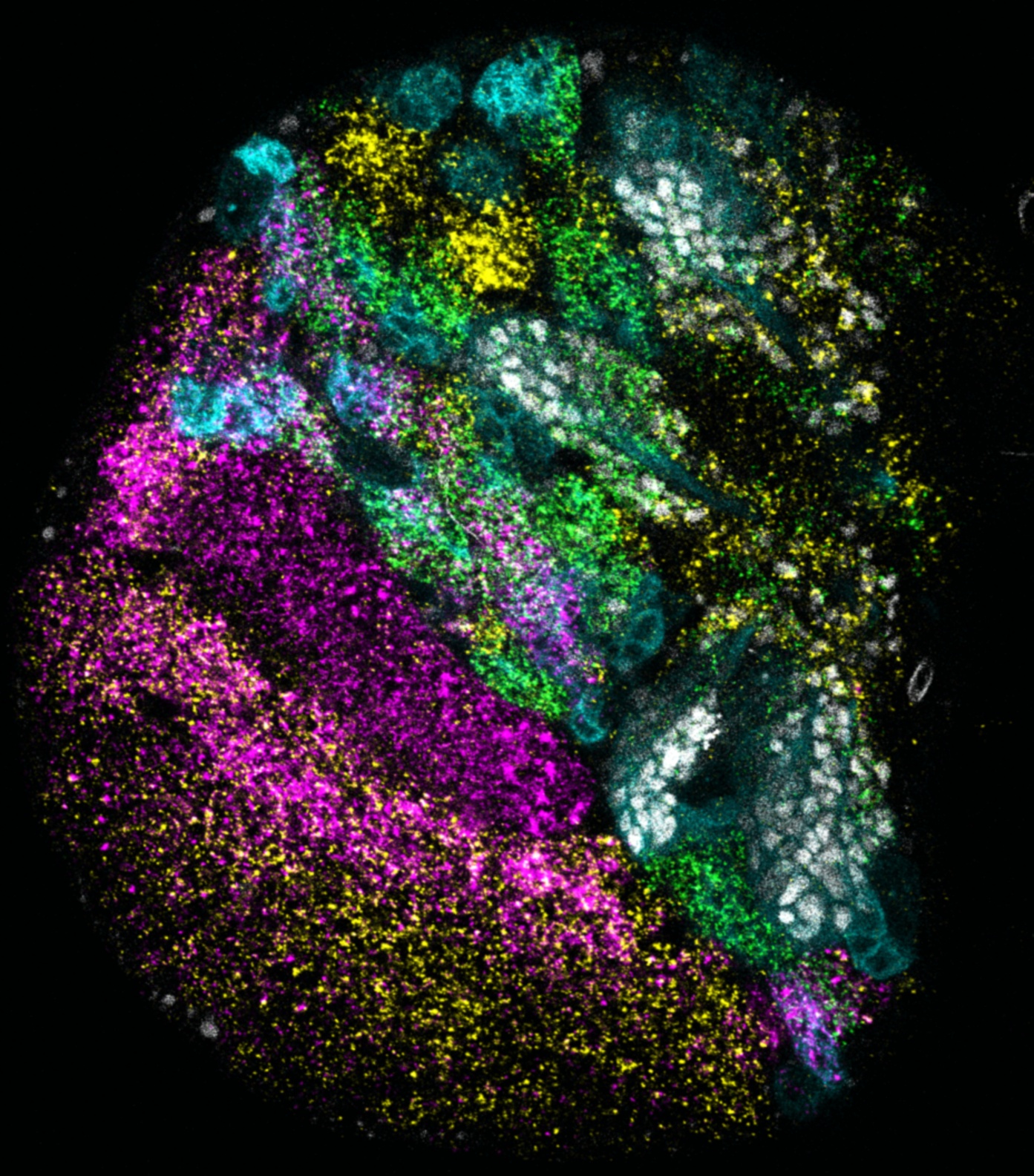
Dawen Cai, Ph.D., is an Associate Professor at the University of Michigan Medical School in the Department of Cell and Developmental Biology , with a secondary affiliation in the Biophysics Department under the College of Literature, Science, and the Arts (LS&A). He is also affiliated with the Neuroscience Graduate Program at the Medical School. His research focuses on integrating computational and experimental approaches to study neuronal subtype determination using scRNA-seq and in situ analysis. His research explores the intersection of RNA biology, neuroscience, and bioinformatics. He develops tools for multispectral imaging and lineage tracing to decode neural development and connectivity in Drosophila and mammalian models. His work combines single-cell transcriptomics with advanced microscopy to identify marker genes and model neuronal architecture. The articles reflect a strong interdisciplinary focus on neuroscience and biomedical imaging. Recent publications highlight innovations in 3D imaging technologies, image compression algorithms, and machine learning applications for medical image segmentation. These works emphasize scalable solutions for high-resolution data analysis, advancing tools for neurophysiology, and leveraging RNA sequencing to map neural development. No scientific awards were explicitly mentioned in the text. Dawen Cai actively recruits PhD students and postdoctoral fellows for the Cai Lab, prioritizing candidates with wet-lab skills, bioinformatics expertise, and experience in quantitative image processing. His lab emphasizes training in interdisciplinary research, paper/grant writing, and critical thinking.