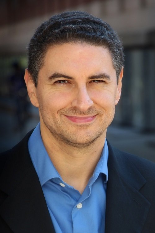
Zhi-Xun Shen is the Paul Pigott Professor in Physical Sciences at Stanford University, holding dual appointments in the Physics and Applied Physics Departments. He is a senior fellow at the Precourt Institute for Energy and serves on advisory boards for the Knight-Hennessy Scholars and Stanford Science Fellows programs. His research focuses on condensed matter and materials physics, particularly the electronic structures of superconductors, topological insulators, and novel materials. Dr. Shen pioneered advanced spectroscopic techniques, including photon-based imaging and scattering methods, and has authored over 600 publications with significant citation impact. His honors include the Kamerlingh Onnes Prize (2000), E.O. Lawrence Award (2010), and Oliver E. Buckley Prize (2011). He co-founded PrimeNano Inc., commercializing technologies from his lab, such as microwave impedance microscopy. His work bridges fundamental physics with energy-related applications, emphasizing the interplay between electronic structure and material properties. Dr. Shen’s research group explores cutting-edge topics like topological surface states, electron-phonon interactions, and superconductivity mechanisms. His inventions, such as non-resonance microwave imaging, have found applications in materials characterization. He remains active in advancing instrumentation and fostering interdisciplinary collaborations through his academic and industry roles.
California Institute of Technology (Caltech)United States
Antonio Rangel is the Bing Professor of Neuroscience, Behavioral Biology, and Economics at the California Institute of Technology (Caltech), where he also serves as Head Faculty in Residence. He is a faculty member in the Division of Humanities and Social Sciences (HSS) with research focusing on the computational and neurobiological basis of value-based decision-making. Dr. Rangel received his educational training at prestigious institutions: B.Sc. from Caltech in 1993 M.S. from Harvard University in 1996 Ph.D. in 1998 Professor Rangel's research lies at the intersection of neuroscience, economics, and psychology, with a focus on understanding how the brain makes decisions. His work investigates the neural mechanisms underlying value computation, choice processes, self-control, and social decision-making. Using a multidisciplinary approach that combines functional magnetic resonance imaging (fMRI), eye-tracking, computational modeling, and behavioral experiments, his lab has made significant contributions to the field of neuroeconomics. His research has revealed how value signals are represented in the brain, how attention influences choice, and the neural basis of self-control failures. Professor Rangel has pioneered methods for studying decision processes with high temporal resolution using eye-tracking data, demonstrating how fixation patterns relate to value computations and choice outcomes. His work spans from theoretical frameworks of value-based decision-making to practical applications in behavioral public economics. Professor Rangel's work has been recognized with prestigious awards: 2019 NOMIS Distinguished Scientist Award 2018 Fellow of the Association for Psychological Science As an academic leader, Professor Rangel has mentored numerous students and researchers who have gone on to make their own contributions to neuroscience and economics. His lab, the Rangel Neuroeconomics Laboratory at Caltech, serves as a hub for interdisciplinary research, bringing together students and scholars from neuroscience, economics, psychology, and computer science. The lab has received significant funding to support its research on the neural basis of decision-making, including support from the NOMIS Foundation. The Rangel Neuroeconomics Laboratory is equipped with state-of-the-art facilities including fMRI analysis capabilities, eye-tracking systems, and computational resources for modeling decision processes. The lab fosters a collaborative environment where researchers apply methods from experimental economics and cognitive neuroscience to unravel the complexities of human decision-making.
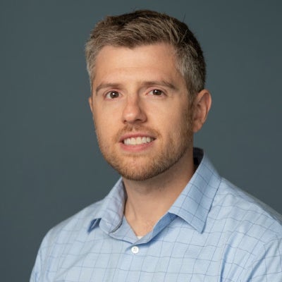
Prof. Dr. Dennis Säring is a faculty member at the University of Applied Sciences Wedel , specifically affiliated with the School of Engineering. His academic and research activities focus on Deep Learning , Medical Image Analysis , and applications of Artificial Intelligence in healthcare and biomedical imaging. He has led seminars on Deep Learning topics and supervised student projects in Autonomous Driving at Audi's AADC 2018 competition. Research Highlights : Cardiovascular imaging, forensic age estimation via MRI, neural network-based bone segmentation, and cerebrovascular aneurysm analysis. Technical Expertise : Cardiac MRI, 3D/4D image processing, parametric mapping, and spatiotemporal data fusion. His recent publications (2018-2023) emphasize 3D MR segmentation for age assessment, CMR strain analysis in athletes, and T1/T2 mapping for myocarditis. Key collaborations include institutions like the University Medical Center Hamburg-Eppendorf and Wedler Hochschulbund, with funding for autonomous vehicle research. While no explicit scientific awards are listed, his work spans clinical cardiology, forensic radiology, and AI-driven medical diagnostics.
Michael McAlpine is a Professor in the Mechanical Engineering department at the University of Minnesota . He also holds affiliations with the Biomedical Engineering and Electrical and Computer Engineering departments. His research focuses on 3D printing functional materials & devices , Nanoscale inks , Biomedical devices , Bioelectronics , and Flexible Microsystems . Research Interests : 3D Printing, Biomedical Engineering, Nanotechnology, Flexible Electronics, Microfluidics Labs : ME 361/363 Contact : mcalpine@umn.edu , (612) 626-3303, ME 117 Recent Research Trends include 3D Printed Biomedical Devices , Flexible Electronics , and Bioprinting Applications . His work spans from Spinal Organoid Formation to Programmable Drug Release Capsules . Scientific Award : Circulation Research 2020 Best Manuscript Award
Dr. Cassandra Sampaio Baptista is a Lecturer at the University of Glasgow's School of Psychology & Neuroscience. Her research focuses on brain plasticity in adulthood, particularly exploring how experiences like skill learning or rehabilitation influence structural and functional changes in the brain. She employs neuroimaging techniques such as fMRI neurofeedback and MRI to investigate mechanisms of myelin and white matter plasticity. Her work emphasizes translational applications, including stroke rehabilitation and promoting healthy aging. Key contributions include demonstrating myelin's role in motor learning and developing MRI protocols for white matter analysis. She collaborates on projects funded by the BIAL Foundation (2025–2026) and has supervised multiple postgraduate students. Recent publications highlight studies on oligodendrocyte dynamics, neurofeedback interventions for stroke survivors, and cross-species neuroscience approaches. While no specific awards are listed, her extensive publication record reflects her leadership in neuroplasticity research.
Dr. Akhilesh Jaiswal serves as Assistant Professor of Electrical and Computer Engineering at the University of Wisconsin-Madison, where his research pioneers device-circuit co-design for next-generation computing systems. His work focuses on enabling extreme-edge intelligence through processing-in-pixel technology, in-memory computing architectures, and bio-inspired neuromorphic systems. His academic credentials include: PhD in Nano-electronics from Purdue University (2019) MS from the University of Minnesota (2014) Bachelor of Technology from Shri Guru Gobind Singhji Institute of Engineering and Technology (2011) Dr. Jaiswal's research program centers on revolutionizing edge computing through hardware innovations that integrate sensing and processing. His device-circuit co-design approach leverages alternate state variables to create energy-efficient systems for real-time applications, with particular emphasis on retina-inspired sensors and photonic memory architectures. This work bridges semiconductor physics with AI acceleration needs, targeting applications from autonomous systems to biomedical devices. Analysis of his 2023-2025 publications reveals dominant themes in photonic SRAM-based in-memory computing (40% of output), retina-inspired motion processing (30%), and secure hardware architectures (20%). His team consistently develops novel bitcell designs that enable XOR logic execution within memory arrays while maintaining compatibility with CMOS fabrication processes. Recent work shows increasing focus on biomedical applications of processing-in-pixel technology. His distinguished recognition includes: Three consecutive ISI Exploratory Research Awards (2020-2023) IEEE Brain Community Best Paper Award (2022) 27 issued US patents with multiple pending applications Nomination for USC Moore Inventor Fellowship (2022) Dr. Jaiswal actively mentors graduate researchers through ECE 790/890/990 courses while securing exploratory funding through ISI and Keston Foundation awards. His patent portfolio demonstrates exceptional translational impact, with industry game-changer classifications from USPTO. The 2022 VLSI-SoC nomination and multiple research highlights in major outlets validate his contributions to hardware security and neuromorphic vision sensors. His laboratory develops integrated hardware platforms combining magnetic tunnel junctions, photonic memory, and CMOS image sensors to create unified processing-in-sensor systems. Current projects include retina-inspired motion segmentation for event cameras and electro-optic frequency transducers for quantum computing interfaces, with strong industry collaboration through patent licensing.
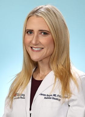
Carmen Bergom, MD, PhD, is an Associate Professor of Radiation Oncology at Washington University School of Medicine (WashU Medicine), where she joined the faculty in 2020. She holds secondary appointments in the Division of Cancer Biology and the Siteman Cancer Center. Her research focuses on leveraging genetic models to improve radiation therapy efficacy while mitigating radiation-induced cardiotoxicity , particularly in breast cancer patients. Key areas include radiation biology , tumor microenvironment , and cardio-oncology . Recent publications highlight her work in genetic mapping of radiation sensitivity (e.g., rat chromosome 3 variants), innovative imaging techniques (e.g., integrin-targeted PET), and clinical trial design for cardiovascular risk stratification. Her laboratory has pioneered the first genetic studies of radiation-induced cardiac dysfunction. Scientific awards include the Michael Fry Research Award (2021) and recognition as a Top Doctor in America (2018, 2019). She serves as Chair of the ASTRO Science Council Scientific Review Panel and co-chair of the Clinical and Experimental Research in Radiation Oncology meeting at ESTRO. Dr. Bergom treats breast cancer patients clinically and mentors students across all levels (undergraduate to postdoctoral). Her work bridges basic research with clinical applications , aiming to develop biomarkers and therapeutic targets for radiation oncology.
Professor Guy Williams is a leading academic at the University of Cambridge with a focus on imaging science and clinical neurosciences, affiliated with Downing College and the Wolfson Brain Imaging Centre . Holding a PhD in Physics from his initial Natural Sciences degree, he specializes in nuclear magnetic resonance (NMR) and MRI techniques for brain imaging. Education: BA, PhD in Physics His research centers on non-invasive imaging of brain structure and function, particularly in traumatic brain injury (TBI) and dementia. His work involves developing novel MRI pulse sequences and advanced data analysis algorithms, including AI-based diagnostic tools. He leads studies on white matter integrity post-trauma, longitudinal dementia assessment, and applications of MRI in disorders of consciousness and addiction. Recent publications highlight collaborations in traumatic brain injury outcomes, AI-guided dementia prediction, and neuroimaging of post-COVID cognitive deficits. His team's work on ultra-high field laminar fMRI and distortion correction methods has advanced clinical neuroscience applications. Key techniques include diffusion tensor imaging (DTI), 7 Tesla MRI, and positron emission tomography (PET/MR). His research spans from basic NMR physics to clinical translation, with a strong emphasis on multi-site studies and real-world diagnostic implementation.
Professor Hala Zreiqat AM is a leading biomedical engineer at The University of Sydney , serving as the Director of the ARC Training Centre for Innovative BioEngineering . A Fellow of all major Australian academies (AAS, ATSE, FAHMS, FRSN), she develops 3D printed bioceramics for bone regeneration while championing diversity through initiatives like the IDEAL Society and BIOTech Futures mentorship program. Her work bridges academia, clinical practice, and industry in musculoskeletal research . Research Focus: Her lab creates synthetic bone scaffolds that mimic natural bone architecture, strength, and porosity, enabling non-rejected bone regeneration via patient-matched implants. Key applications include orthopaedic, dental, and maxillofacial repair , with over $18M in competitive funding and multiple patents. Current projects explore AI-driven scaffold performance prediction and anti-senescence strategies for aging-related bone loss. Scientific Trends: Recent publications highlight 3D printed nanovoxelated ceramics , antisenescence biomaterials , and multifunctional theranostic platforms . Her team integrates machine learning for scaffold design, atom probe tomography for interface analysis, and two-photon imaging for cellular monitoring in 3D environments. 2021-2022 Fulbright Senior Scholar 2018 NSW Premier's Woman of the Year 2019 Eureka Prize for Innovative Use of Technology Fellow of Australian Academy of Science (2021) Over $18M in research funding Teaching & Leadership: She designed core courses like Tissue Engineering and Nanomaterials in Medicine , mentoring 158 students in 2020 alone. As Chair of CAAR (2020-2023), she strengthens Australia-Arab collaborations. Her lab trains early-career researchers , with alumni now in academia and industry.
Dr. Lukas Frey is a Researcher at ETH Zurich, affiliated with the Chair of Physical Chemistry and the Institute of Molecular Physical Sciences (IMPS). His work focuses on biophysical studies of membrane proteins, lipid dynamics, and protein aggregation mechanisms. Key research areas include structural biology of ion channels, NMR spectroscopy of membrane proteins in nanodiscs, and the role of lipid environments in modulating protein dynamics. Frey employs advanced techniques like mass photometry and solid-state NMR to investigate molecular mechanisms in biological systems. His recent studies address amyloid fibril formation, pH-dependent α-synuclein polymorphism, and cholesterol-mediated modulation of membrane protein behavior. Based at the HCI F 228 facility in Zurich, Frey collaborates on projects involving lipid bilayer environments, ion channel function, and the structural basis of protein aggregation. His email is lukas.frey@phys.chem.ethz.ch, and he holds an ORCID identifier 0000-0002-1052-1104. Research contributions span from fundamental biophysical insights to methodological advancements in membrane protein analysis.
Dr. Daniel Roxbury is an Associate Professor and Graduate Director at the Department of Chemical, Biomolecular and Materials Engineering within the University of Rhode Island's College of Engineering. With expertise in nanoscience and carbon nanomaterials, his research focuses on nano-bio interactions, developing functionalized nanotubes for biomedical applications and environmental monitoring through his NanoBio Engineering Laboratory. His work spans multiple disciplines including: Biomedical nanosensors Smart wearable biomaterials Targeted drug delivery systems Environmental nanotechnology Single-molecule imaging Nanotoxicology Recent publications emphasize machine learning-enhanced spectral analysis, coral reef conservation nanotechnology, and wearable stress monitoring textiles. His 2024 ACS Nano study introduces AI-driven macrophage phenotyping, while 2023 Nature Nanotechnology work explores coral reef restoration strategies using nanomaterials. Awarded the 2019 NSF CAREER grant for cellular nanometrology, he leads multiple NIH-funded projects including: $820,000 NSF CAREER: Spectral Imaging for Sub-Cellular Nanometrology $140,000 Miriam Hospital COBRE: Cortisol Detection Textiles $700,000 NSF EAGER: Multiplexed Wound Biomarker Detection His laboratory houses state-of-the-art equipment including: Near Infrared Hyperspectral Microscope Custom NIR Fluorescence Spectrometer Jasco UV/VIS/NIR Spectrophotometer Biosafety Cabinet Cell Culture Incubator Cryo-Storage System
Prof. Dr. Raphael Sznitman serves as Director of the ARTORG Center for Biomedical Engineering Research and Head of the Artificial Intelligence in Medical Imaging group at the University of Bern, Switzerland, holding a Full Professor position in AI for Medical Imaging since 2015. Education: PhD in Computer Science, Johns Hopkins University (2011) MSc in Computer Science, Johns Hopkins University (2009) BSc in Cognitive Systems, University of British Columbia (2007) Research Interests: Sznitman's work centers on computational vision , probabilistic methods , and statistical learning applied to medical imaging challenges. His group develops AI algorithms for ophthalmic diagnostics, surgical robotics, and medical image analysis, with emphasis on OCT, surgical phase recognition, and domain adaptation techniques. Key application areas include retinal disease detection and cataract surgery automation. Publication Trends: His 2021-2025 publications reveal concentrated efforts in deep learning for medical imaging , particularly in ophthalmology (OCT analysis) and surgical video understanding. Emerging themes include LLM applications for clinical monitoring, unsupervised out-of-distribution detection for surgical safety, and physics-informed AI for multimodal medical data fusion. Research Leadership: As ARTORG Center Director, Sznitman oversees interdisciplinary research bridging computer science and clinical medicine. His group collaborates extensively with Bern University Hospital clinicians on translational projects, securing funding for AI-driven diagnostic tools and surgical assistance systems. Current initiatives focus on real-time intraoperative guidance and spaceflight ophthalmology applications. Laboratory: The Artificial Intelligence in Medical Imaging group operates within ARTORG's dedicated facilities, maintaining partnerships with surgical robotics labs and ophthalmology departments for clinical validation of AI systems. Their work integrates multimodal data streams including OCT, VR perimetry, and surgical video feeds.
Evelyn Lake is an Associate Professor in the Department of Radiology and Biomedical Imaging at Yale University, with affiliations to the Wu Tsai Institute and Yale Biomedical Imaging Institute. Her research focuses on functional neuroimaging, particularly using rodent models to study brain connectivity, neurovascular coupling, and disorders like Alzheimer’s disease and autism spectrum disorder. PhD in Medical Biophysics, University of Toronto (2016) Postdoctoral Associate, Yale University (2019) BSc (Honors) in Biophysics, University of Guelph (2010) Her work spans multimodal imaging techniques (e.g., simultaneous Ca2+ and fMRI), examining how genetic mutations (e.g., PTEN, KATNAL2) affect cerebrospinal fluid dynamics and neuronal connectivity. She investigates longitudinal changes in Alzheimer’s models and methodological aspects of animal neuroimaging, such as sample size optimization and awake imaging protocols. Recent publications highlight her contributions to understanding autism-related white matter abnormalities, transdiagnostic connectome predictive modeling, and the impact of neurovascular uncoupling in awake mice. Collaborations include Todd Constable, Francesca Mandino, Xilin Shen, and Xenophon Papademetris.
Lawrence Staib is Professor of Radiology and Biomedical Imaging, Biomedical Engineering, and Electrical Engineering at Yale University. He serves as Director of Undergraduate Studies in Biomedical Engineering and is a member of Yale's Bioimaging Sciences division, Image Processing & Analysis Group, Yale Biomedical Imaging Institute, and Yale-BI Biomedical Data Science Fellowship program. Dr. Staib earned his A.B. in Physics from Cornell University (1982), followed by a Ph.D. in Engineering and Applied Science from Yale University (1990), and completed a postdoctoral fellowship at Yale School of Medicine (1991). His research focuses on developing advanced medical image analysis methods using machine learning and model-based approaches. Key research areas include neuroimaging applications for autism spectrum disorder classification, cardiac imaging analysis for strain and motion assessment, prostate cancer diagnosis and risk mapping, and innovative techniques for medical image segmentation with limited labeled data. Dr. Staib's work emphasizes uncertainty estimation in deep learning models, multi-modal image registration, and domain adaptation techniques to improve clinical decision support systems. His recent publications demonstrate a strong trend toward developing interpretable AI models for clinical applications, with particular emphasis on fMRI analysis for neurological conditions, cardiac motion analysis, and prostate cancer diagnosis. His work frequently addresses the challenge of limited labeled data in medical imaging through innovative self-supervised, semi-supervised, and few-shot learning approaches. Fellow of the American Institute for Medical and Biological Engineering (AIMBE) (2015) Distinguished Investigator Award from the Academy for Radiology & Biomedical Imaging Research (2017) MICCAI Fellow (2022) Medical Image Analysis Second Best MICCAI Paper Award (2005) ASNR Cum Laude Scientific Exhibit Award (2003) Dr. Staib serves on the editorial board of Medical Image Analysis and as Associate Editor of IEEE Transactions on Biomedical Engineering. His research is supported by NIH grants including the Autism Center of Excellence program. He leads the Image Processing & Analysis Group within Yale's Bioimaging Sciences division, collaborating extensively with James Duncan, John Onofrey, Xenophon Papademetris, and other Yale researchers on applications spanning neuroimaging, cardiology, and oncology. Current projects focus on developing robust AI models for clinical decision support with emphasis on uncertainty quantification and interpretability.
Duncan Astle is the Gnodde Goldman Sachs Professor of Neuroinformatics at the Department of Psychiatry, University of Cambridge. He serves as a Programme Leader at the Medical Research Council's Cognition and Brain Sciences Unit (MRC CBU) and is a Fellow of Robinson College. Astle heads the 4D Lab (Development, Dynamics, Disorders, Data Science), which provides a research home for approximately 15 Early Career Researchers working at the intersection of developmental cognitive neuroscience and advanced data science methodologies. Astle's research focuses on understanding childhood development through innovative analytical approaches. His work employs transdiagnostic methods to study children with attention, learning, and memory difficulties, moving beyond traditional diagnostic categories. He investigates how neural systems develop in childhood, how they relate to developmental disorders, and how they respond to intervention. His research integrates network science, machine learning, and generative modeling to capture the complexity of neurodevelopmental diversity, examining how cognitive skills, literacy, numeracy, and mental health interrelate over developmental time. His publication record reveals a strong focus on brain connectivity and organization across development. Recent work explores structural and functional neurodevelopmental trajectories, brain wiring economics, and the impact of environmental factors on neural development. Astle's research frequently employs advanced data science techniques to identify sub-populations of children with different cognitive or brain profiles, regardless of diagnosis, and to map non-linear relationships between brain organization and cognitive difficulties. His work has increasingly focused on transdiagnostic approaches to understanding developmental disorders and the application of computational models to developmental neuroscience. Astle actively supervises PhD students and has built a substantial research group that contributes to major projects including the Centre for Attention Learning and Memory (CALM) and Resilience in Education and Development (RED). His work has been supported by prestigious funding bodies including the Royal Society, the British Academy, the Medical Research Council, and the Economic and Social Research Council, as well as multiple charitable foundations. The 4D Lab, under Astle's leadership, utilizes state-of-the-art facilities at the University of Cambridge, including on-site magnetic resonance imaging and magnetoencephalography scanners. The lab contributes to building specialist cohorts such as CALM (800 children with cognitive difficulties plus 200 comparison children) and RED, which study children's development, resilience, and educational outcomes. Astle's team explores how growing up in adverse environments affects children's brains, behavior, and mental health, with the aim of identifying early markers of risk and resilience.