Chi Liu is a Professor of Radiology & Biomedical Imaging at Yale School of Medicine . He serves as Associate Director of Biomedical Imaging Technology at the Yale Biomedical Imaging Institute and Director for Research Faculty Affairs in the Radiology & Biomedical Imaging department. Education : PhD from Johns Hopkins University (2008) Postdoctoral Training : University of Washington (2010) Certification : American Board of Science in Nuclear Medicine (Nuclear Medicine Physics and Instrumentation) His research focuses on quantitative cardiac and oncological PET/CT and SPECT/CT imaging , emphasizing deep learning algorithms , reconstruction algorithms , data correction , and dynamic imaging . Key clinical applications include early detection of chemotherapy-induced cardiotoxicity , multimodality imaging of heart failure , and motion variability elimination in therapy response assessment . The 15 most recent publications reveal a strong emphasis on deep learning techniques for low-dose imaging , motion correction , and cross-tracer generalizability in PET/SPECT systems. These works span applications in cardiac imaging , neuroscience , oncology , and theranostics . Scientific Award : Bruce Hasegawa Young Investigator Medical Imaging Science Award (2012) Contact: chi.liu@yale.edu | ORCID 0000-0002-7007-1037
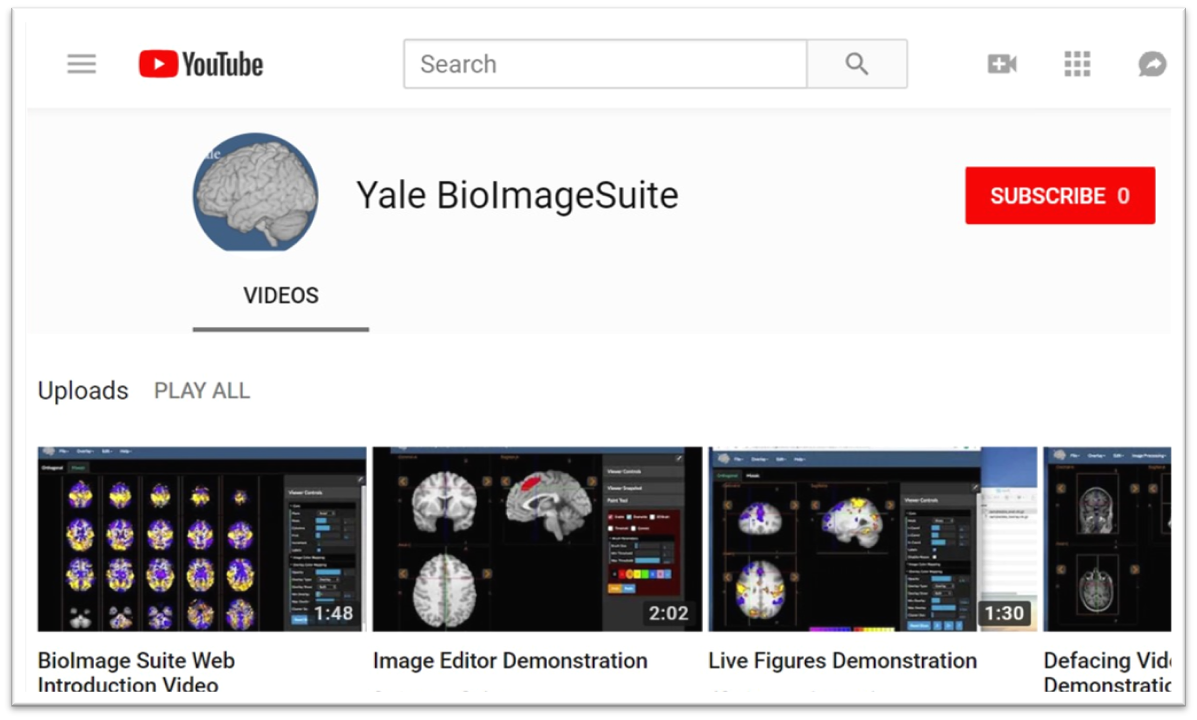
Xenophon Papademetris is a Professor of Biomedical Informatics & Data Science and Radiology & Biomedical Imaging at Yale School of Medicine. He serves as Associate Director of Biomedical Imaging Data Sciences at Yale Biomedical Imaging Institute and directs the Medical Software and Medical Artificial Intelligence Certificate Program. PhD in Electrical and Information Sciences from Yale University (2000) BA from Cambridge University (1994) Postdoctoral Fellowship at Yale University (2002) His research focuses on medical image analysis, machine learning, and biomedical software development. He has developed tools like BioImage Suite Web and contributed to standards committees at the Association for the Advancement of Medical Instrumentation (AAMI). His work spans modalities including MRI, CT, PET, and optical imaging. Recent publications emphasize neuroimaging analysis, explainable AI in healthcare, and multimodal data integration across species. He leads NIH-funded research under the BRAIN Initiative (R24 MH114805) and has authored a textbook on Medical Software published by Cambridge University Press. IEEE Senior Member Yale Brown-Coxe Postdoctoral Fellowship Harding Bliss Prize for Excellence in Engineering He directs the BioImage Suite Project, creating web-based image analysis tools using JavaScript and WebAssembly. His teaching includes both academic courses and a Coursera program on Medical Software with over 14,000 enrollments.
Steven Meikle is a Professor of Medical Imaging Physics and Head of the Imaging Physics Laboratory at the Brain and Mind Centre, University of Sydney. He also serves as Deputy Director (Preclinical) of Sydney Imaging and Deputy Director of the National Imaging Facility's Sydney node. His expertise spans advanced imaging technologies, with a focus on PET/SPECT instrumentation and molecular imaging. He holds a B.App.Sc.(Hons) from the University of Technology Sydney and a PhD from the University of New South Wales. Research focuses include developing novel PET systems like Open-field PET (for freely moving rodents) and Total Body PET, which enhance imaging sensitivity and enable real-time behavioral studies alongside brain function analysis. Collaborations include Tsinghua University (China) and UC Davis (USA). He leads projects on motion correction, quantitative imaging, and AI-driven analysis. Key achievements include over 180 peer-reviewed publications, editorial roles in Physics in Medicine and Biology , and leadership in professional societies. Awards include IEEE Senior Membership and Australian Institute of Physics Fellowship. Current student projects explore Total Body PET applications, motion correction, and radiopharmaceutical evaluation. Teaching roles include medical physics courses in diagnostic radiography and medical physics programs. He advises on imaging ethics, facility implementation, and translational research bridging basic science and clinical applications.
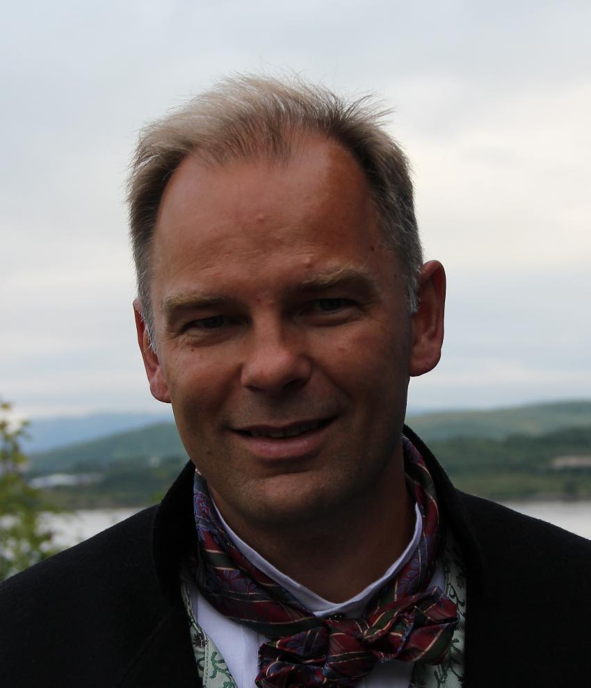
Trond Vidar Hansen is a Professor at the Department of Pharmacy, University of Oslo , and leads the LIPCHEM research group . He collaborates with institutions including the University of Bergen and Vestlandets Innovasjonsselskap through the VITADEL project, which recently received NOK 5,000,000 in verification support from the Research Council of Norway. His research focuses on the synthesis and biological evaluation of specialized pro-resolving lipid mediators derived from omega-3 fatty acids, with applications in inflammation resolution, neuroinflammation, and drug development. University : University of Oslo Department : Department of Pharmacy Research Group : LIPCHEM Collaborations : University of Bergen, Vestlandets Innovasjonsselskap Research Interests : H Hansen's work centers on the organic synthesis of bioactive lipid derivatives, particularly pro-resolving mediators from omega-3 polyunsaturated fatty acids. His team investigates their roles in inflammatory disease models , neuroinflammation , and PPAR receptor activation , aiming to develop therapeutic agents for conditions like chronic pain, diabetes, and neurodegenerative disorders. The research integrates stereoselective chemistry , biochemical profiling , and pharmacological evaluation to validate these mediators' clinical potential. Recent Awards : 2025: NOK 2,000,000 verification support from Research Council of Norway 2025: Co-leader of NOK 5,000,000 VITADEL project Publications : His articles (2015–2024) reveal a focus on stereoselective synthesis of resolvins, protectins, and maresins, with applications in anti-inflammatory and neuroprotective therapies . Key subfields include omega-3 metabolite profiling , PPAR agonist design , and biosynthetic pathway elucidation , often utilizing human cell models and mouse disease models . Collaborative projects emphasize commercialization of academic research and translational medicine .
Professor Jerome Liang is a distinguished faculty member at Stony Brook University's Renaissance School of Medicine, holding professorships in Radiology, Biomedical Engineering, Electrical and Computer Engineering, and Computer Science. He serves as Co-Director of Radiology Research and has established himself as a leading expert in medical imaging reconstruction techniques. Dr. Liang's educational background includes a Ph.D. in Physics from City University of New York, postdoctoral training at Duke University, and fellowship at Albert Einstein College of Medicine. His undergraduate degree in Modern Physics was obtained from Lanzhou University in China. His primary research interests focus on advanced medical imaging techniques, particularly low-dose computed tomography image reconstruction, quantitative SPECT reconstruction, high-resolution PET imaging, tissue segmentation from multi-spectral images, computer-aided diagnosis systems, and virtual colonoscopy development. His work bridges engineering principles with clinical applications to improve diagnostic imaging capabilities while reducing radiation exposure. Analysis of his recent publications reveals a strong focus on machine learning applications in medical imaging, particularly in polyp classification, dual-energy CT spectral analysis, and virtual endoscopy. His research consistently aims to enhance diagnostic accuracy while optimizing radiation dose and improving visualization techniques for various medical conditions. 1981 China-US Physics Examination and Application Program (CUSPEA) Winner (Top 25 among 250,000 candidates) 1990 NIH First Investigator Award 1996 American Heart Association Established Investigator Award 1996 Radiological Society of North America Certificate of Merit Award 2002 SUNY Chancellor's Entrepreneur Award 2007 IEEE Society Fellow 2011-2013 SBU, BNL and CSHL Certificates of Excellence in Research and Invention 2013 Stony Brook School of Medicine Award for Excellence in Translational Research Dr. Liang has secured significant research funding including NIH/NCI R01 grants for "Advanced Virtual Colonoscopy for Early Cancer Screening" and "Radiogenomics of Colorectal Polyps." He currently leads active protocols including IRB 93995-MODCR005 focused on integrating virtual and optical colonoscopies with pathological analysis. His laboratory (IRIS - Imaging Research and Informatics) continues to advance medical imaging technology while mentoring the next generation of researchers in this critical field.
Associate Professor Andre Kyme is an academic staff member in the School of Biomedical Engineering at The University of Sydney. His research focuses on developing enabling technologies for biomedical imaging, including motion compensation in MRI/PET, robotic platforms for image-guided therapy, and cross-disciplinary applications like plant salt uptake analysis using PET. He collaborates with institutions globally and advises students on projects like lameness detection in horses and AI-based motion correction. Research Interests: Kyme's work spans motion correction in medical imaging modalities, medical robotics integration with imaging systems, and innovative applications of imaging technologies in non-traditional fields. His team emphasizes leveraging advancements in computer vision, machine learning, and instrumentation to improve imaging performance and accessibility. Recent Projects: Current research includes MRI-compatible robotic platforms for therapy applications, AI-driven lameness detection in horses, and pediatric neuroimaging improvements. He leads the BREEZE initiative to enhance MRI accessibility for children with cerebral palsy through eye-gaze communication technology. Publications: His work spans 20+ years with over 50 peer-reviewed publications in journals like Physics in Medicine and Biology and IEEE Transactions. Key areas include PET/SPECT/CT motion correction algorithms, robotic systems for medical imaging, and novel imaging applications in plant science. Teaching: Kyme instructs core biomedical engineering courses including thesis supervision and capstone projects at both undergraduate and postgraduate levels. Labs/Teams: Active in the Brain and Mind Centre and Biomedical Imaging, Visualisation and Information Technologies groups at Sydney. Collaborates with industry partners like TeleMedVet and academic institutions including University of California Davis and Chinese University of Hong Kong.
Paul O'Callaghan is a Researcher at Uppsala University's Department of Medical Cell Biology within the Faculty of Medicine. His work spans multiple disciplines at the intersection of cell biology, neuroscience, and biomedical engineering. Based at the BMC campus in Uppsala, he maintains an active research program with publications spanning from 2008 to the present, demonstrating sustained scholarly productivity. Dr. O'Callaghan's research interests focus on cellular mechanisms relevant to neurodegenerative diseases, particularly Alzheimer's disease, with emphasis on amyloid-beta pathology, heparan sulfate proteoglycans, and glial cell biology. His recent work has expanded into biomedical engineering applications including bioprinting, microfluidics, and biomaterials for neural applications. His publication record reveals a trajectory from fundamental cell biology toward translational applications, with consistent focus on cellular signaling mechanisms. O'Callaghan's recent publications (2021-2024) demonstrate a shift toward more engineering-focused approaches to biological problems, including 3D bioprinting, microfluidic cell culture systems, and biomaterial testing. His work maintains strong connections to both fundamental cell biology questions and potential clinical applications, particularly in neurodegenerative disease and cancer research. He leads his own research group within the Department of Medical Cell Biology, collaborating extensively with colleagues across Uppsala University including Johan Kreuger, Olle Eriksson, and other researchers in related fields. His work appears in high-impact journals across cell biology, neuroscience, and biomaterials disciplines.
Dag Linnarsson is a Professor of Physiology at Karolinska Institutet, Department of Physiology and Pharmacology, where he has held the position since 1986 and transitioned to Senior Professor in 2009. His research primarily focuses on environmental physiology, with an emphasis on the human body's response to extreme conditions such as microgravity, hypergravity, and spaceflight. He leads the Environmental Physiology Research Group and the Lars Karlsson team, investigating topics like pulmonary function in astronauts, musculoskeletal countermeasures during space missions, and the effects of artificial gravity. His work spans studies on bed rest as a spaceflight analog, lung diffusing capacity, and nitric oxide dynamics under varying environmental pressures. Recent research includes assessing pulmonary nitric oxide levels in astronauts during long-term space missions and exploring the physiological impacts of lunar dust exposure. Linnarsson has also contributed to standardizing bed rest protocols for spaceflight research and evaluating centrifugation as a countermeasure for muscle and bone loss. He maintains affiliations with Karolinska Institutet’s C3 Fysiologi och farmakologi team and collaborates on interdisciplinary projects addressing space medicine challenges. While no formal student listings or awards are explicitly mentioned, his extensive publication record highlights contributions to respiratory physiology, cardiovascular adaptation, and musculoskeletal health in extreme environments.
Rune Sundset is an Associate Professor at UiT The Arctic University of Norway, affiliated with the Nuclear Medicine and Radiation Biology research group. His work focuses on radiopharmaceutical development, molecular imaging, and liposomal drug delivery systems. Research themes: Radioligand design, PET/SPECT imaging optimization, and nanoparticle-biology interactions Key collaborations: European Journal of Nuclear Medicine and Molecular Imaging, BMC Research Notes, and Acta Physiologica Recent publications highlight his contributions to: Development of Cu-67 and Ga-68 radiotracers for tumor imaging Liposomal formulations for arginase inhibition in cancer therapy Machine learning applications in dynamic PET imaging Sphingolipid effects on liposome uptake by human phagocytes In the SECURE project, he investigates radioligand therapy and imaging biomarkers. His research group (NMRB) spans nuclear medicine, radiation biology, and biomedical engineering, with applications in glioblastoma, prostate cancer, and neurodegenerative diseases.
Dr. Elahe Alizadeh is an Assistant Professor (Adjunct) at Queen's University and a Research Scientist at the Queen's Centre for Particle Studies (QCPU). She holds a BSc in Applied Physics from Amirkabir University of Technology (Iran), an MSc in Medical-Radiation Physics from the same institution, and a PhD in Nano-Bio-Physics from the University of Innsbruck (Austria). Her career includes a postdoctoral fellowship at Université de Sherbrooke (Canada) and roles at the University of Saskatchewan, where she developed pre-clinical radiopharmaceutical programs and managed advanced imaging facilities. Currently, she oversees nuclear imaging projects using tri-modality µPET/SPECT/CT scanners and maintains radiation safety certifications (RSO-1). Research Focus : Radiation physics, medical imaging, radiobiology, and pre-clinical theranostics Awards : Radiation Research Society Jack Fowler Award (2013) Committees : Canadian Radiation Protection Association (CRPA), Queen’s Radiation Safety Committee Her work bridges radiation safety, imaging technology, and translational medicine. Key research areas include radiation damage mechanisms, targeted cancer therapies, and oxygen-sensing pathways in pulmonary diseases. Recent studies span pulmonary hypertension treatments, PET imaging biomarkers, and plasma-based cancer therapies.
Kaylena Ehgoetz Martens serves as an Associate Professor in the Department of Kinesiology and Health Sciences at the University of Waterloo, where she directs the Neurocognition and Mobility Lab. Her research program integrates movement kinematics, functional neuroimaging, psychophysiology, and cognitive neuroscience to investigate the neural basis of gait control and its disruption in neurodegenerative conditions, with particular emphasis on Parkinson's disease, dementia with Lewy bodies, and isolated REM sleep behavior disorder. She focuses on the complex interplay between cognition, emotion, and motor function to develop translational approaches for early diagnosis and intervention in mobility disorders. Dr. Martens' academic training includes a BSc in Kinesiology & Physical Education from Wilfrid Laurier University, an MA in Psychology from the University of Waterloo, a PhD in Cognitive Neuroscience from the University of Waterloo, and postdoctoral training at the Medicine, Brain and Mind Centre, University of Sydney, Australia. Her educational background established the foundation for her multidisciplinary approach to movement neuroscience. Her research program centers on three interconnected aims: (1) investigating cognitive-emotional interactions in gait and balance control; (2) leveraging gait complexity to identify subclinical predictors of neurodegeneration; and (3) developing technology-enhanced diagnostic and intervention tools using virtual reality and mobile recording devices. This work addresses critical gaps in understanding how anxiety, threat processing, and autonomic dysfunction contribute to movement impairments in aging and neurodegenerative diseases. Analysis of her recent publications (2023-2025) reveals a strong trajectory in subtype-specific characterization of freezing of gait, identification of sex-specific neurodegeneration patterns, and development of AI-driven detection methods. Her work increasingly incorporates machine learning for gait analysis while maintaining clinical relevance through biomarker discovery and therapeutic innovation, particularly in the prodromal phases of synucleinopathies. Scientific Awards: No specific awards were documented in the provided source material. Dr. Martens actively supervises graduate students across all levels including undergraduate theses, MSc, PhD, and postdoctoral fellows within her Neurocognition and Mobility Lab. She provides research opportunities for volunteers, coursework interns, and research coordinators, with a focus on translating laboratory findings to clinical applications. While specific grant details weren't provided, her extensive use of advanced neuroimaging, wearable sensors, and virtual reality technologies indicates substantial research funding supporting her program. The Neurocognition and Mobility Lab operates as a collaborative hub bridging basic neuroscience with clinical practice, working closely with healthcare providers to develop practical tools for early mobility impairment detection. Current projects emphasize translating gait complexity metrics into clinical biomarkers and developing anxiety-targeted interventions to prevent falls in neurodegenerative populations, with particular attention to preserving functional independence throughout the lifespan.
Professor Mark Lythgoe is a distinguished academic at University College London (UCL), where he serves as Professor of Biomedical Imaging in the Department of Imaging within the Faculty of Medical Sciences. He is the Founder and Director of the Centre for Advanced Biomedical Imaging (CABI) at UCL, a multidisciplinary research center hosting 12 state-of-the-art imaging modalities and 50 researchers. Additionally, he is Co-Director of the UCL Department of Imaging and Director of Biomedical Imaging Research at the Francis Crick Institute. Mark Lythgoe earned his Doctor of Philosophy from University College London in 1999 and his Master of Science from the University of Surrey in 1993. Professor Lythgoe has a long-standing track record in the development and application of biomedical imaging techniques, with research spanning from fundamental imaging science to clinical applications. His work focuses on advancing imaging technologies for biomedical research and clinical practice, with particular emphasis on neuroimaging, molecular imaging, and the application of imaging to understand neurological disorders and cancer. He has translated his research findings into clinical radiological practice and established training programs in biomedical imaging. His extensive publication record demonstrates consistent innovation across imaging modalities, with recent work increasingly focused on the glymphatic system in neurological disorders, advanced tumor imaging techniques, and the development of novel imaging technologies for both preclinical and clinical applications. His research bridges engineering, physics, biology, and clinical medicine to solve complex biomedical challenges. Professor Lythgoe's significant contributions to the field have been recognized with numerous prestigious awards: IET Achievement Medal (2023) - for major and distinguished contribution in Medical Imaging Royal Society of Medicine Ellison-Cliffe Award (2021) - for contribution of fundamental science to the advancement of medicine Davies Medal from the Royal Photographic Society (2013) - for significant contribution to imaging science Alumni Achievement Award from the University of Salford Neuroscience Prize for Public Understanding from the British Neuroscience Association Dorothy Hodgkin Award Biosciences Federation Science Communication Award Fellow of the British Science Association Professor Lythgoe has secured substantial research funding, with £45 million awarded for his collaborative imaging research program. He is deeply committed to training the next generation of imaging scientists, serving as Co-Director of the MSc in Advanced Biomedical Imaging and Co-Founder of the UCL Centre for Doctoral Training in Medical Imaging. His public engagement efforts include directing the Cheltenham Science Festival, which has become one of the largest science festivals in the world. As Director of the Centre for Advanced Biomedical Imaging, Professor Lythgoe leads a vibrant research team of 50 researchers working across 12 state-of-the-art imaging modalities. His center fosters interdisciplinary collaboration between engineers, physicists, biologists, and clinicians to develop and apply cutting-edge imaging technologies. He has published over 300 papers including publications in Nature, Nature Photonics, Nature Medicine and The Lancet, demonstrating the high impact of his research across multiple disciplines.
Roger Fulton is a Professor of Medical and Preclinical Imaging at the University of Sydney and a Principal Nuclear Medicine Physics Specialist at Westmead Hospital's Department of Medical Physics. He is part of the Brain and Mind Centre and leads the Physics and Biomodelling team. His roles include Conjoint Professor in Medical Radiation Sciences, RHD student supervision, and coordination of the MRTY5134 (Computed Tomography Theory) and MRTY2107 (Imaging Technology 2) units of study. He holds a PhD in Applied Physics. His career has spanned collaborations with institutions like the Research Center Julich (Germany), Massachusetts General Hospital (USA), and the International Atomic Energy Agency (Austria). Professional recognitions include Fellow of ACPSEM, Senior IEEE Membership, and Chair of the Nuclear Medical Imaging Sciences Council (IEEE). Research interests focus on motion tracking/correction for SPECT, PET, and CT imaging, including patent innovations and leadership in radiation dose reduction techniques. He has secured $9.2M in competitive grants since 2004 and contributed to global standards through IAEA initiatives. Grants and advising: Over $9.2M in grants since 2004, advising PhD student Bader ALOUFI on liver imaging kinetics. His work bridges hardware engineering (e.g., Intel RealSense depth cameras) with software solutions (neural networks, Bayesian methods) to enhance imaging accuracy and reduce patient motion artifacts. Scientific Awards: US Patent (2020) ARC College of Experts (2016–2018) ACPSEM Fellowship IEEE Senior Membership IEEE Nuclear and Plasma Sciences Society leadership Labs/Teams: Physics and Biomodelling team at the Brain and Mind Centre, collaborating with international groups on preclinical imaging systems and awake-animal PET methodologies.
David S. Wack is an Associate Professor at the University at Buffalo's Jacobs School of Medicine & Biomedical Sciences, Department of Radiology. His research focuses on medical image analysis and neuroimaging, particularly in auditory processing and parameter estimation from PET, MRI, and CT images. He has held academic positions since 1992, progressing from instructor roles to his current rank. His work includes NIH-funded projects on imaging technologies and collaborations with institutions like Canon Medical Systems and UBNS. Education: PhD in Communicative Disorders and Sciences (SUNY Buffalo, 2010) MA in Applied Mathematics (SUNY Buffalo, 1992) BA in Applied Mathematics and Music Performance (SUNY Buffalo, 1989) Research Interests: Development of parametric maps from medical images Machine learning applications in neuroimaging Neuroimaging of auditory and language processing Dynamic image noise reduction and segmentation algorithms Neural markers of top-down compensation in speech processing Recent Projects: NIH-funded self-collimating SPECT breast tomography system 4D flow MRI for intracranial atherosclerotic disease assessment Machine learning for neuroimage classification Grants & Service: Member of Jacobs School faculty council and UB Faculty Student Association board Editorial roles at Frontiers in Neuroscience (Brain Imaging Methods, Decision Neuroscience) Labs & Teams: Buffalo Neuroimaging Analysis Center Collaborations with UBNS, Canon Medical, and VA Western New York Health Care System
Walter Noordzij is an Assistant Professor at the University of Groningen within the Faculty of Medical Sciences , affiliated with the Basic and Translational Research and Imaging Methodology Development in Groningen (BRIDGE) group. His research focuses on nuclear medicine imaging technologies , particularly PET/CT applications in oncology, cardiology, and healthcare economics. He has contributed to studies on cardiac sympathetic innervation in amyloidosis, financial literacy of medical residents regarding imaging costs, and prostate cancer management using PSMA PET/CT. Recent work also addresses physician workload trends in Dutch nuclear medicine and innovative parametric imaging techniques. Research interests include positron emission tomography-computed tomography (PET/CT) , fluorodeoxyglucose (FDG) imaging , and multidisciplinary approaches to prostate cancer diagnosis . He actively collaborates internationally, evidenced by his participation in the 19th International Conference on Radiation Therapy and the EANM Focus Meeting on Prostate Cancer . No awards are explicitly listed, but his involvement in high-impact studies suggests active recognition in the field. He has supervised 3 students but their names are not disclosed. His work spans clinical imaging methodology, translational research, and healthcare system analysis, reflecting a commitment to bridging basic science and clinical practice.