Brian Christie is Professor at the University of Victoria's Division of Medical Sciences. His research examines how the brain's capacity for synaptic plasticity and neurogenesis can be harnessed to alleviate neuropathologies from trauma, substance abuse, genetic disorders, and aging processes. His laboratory employs convergent techniques including electrophysiology, imaging, molecular biology, and behavioral analysis to identify mechanisms of learning and memory affected by neuropathologies. Research focuses on: Hippocampal neuroplasticity mechanisms Exercise-induced neurogenesis Fetal Alcohol Spectrum Disorders Traumatic brain injury recovery Neurodegenerative protection strategies Neurodevelopmental disorder interventions His research spans preclinical and clinical investigations, with international collaborations at San Diego State University, University of British Columbia, Monash University, Harvard University, and Hong Kong Polytechnic University. Major funding sources include CIHR, NSERC, NIH, Fragile-X Research Foundations, Scottish Rite Charitable Foundation, and private donors. Laboratory trainees have secured academic positions globally, with over 30 becoming practicing physicians in Canada and consistent medical school admissions over 15 years. Honors include: Vespasian V. Pella Medal Gold Medal in Social Sciences Multiple endowed research chairs
Mark D Does is a Professor of Biomedical Engineering, Radiology and Radiological Sciences, and Electrical Engineering at Vanderbilt University’s School of Engineering. His research focuses on developing MRI methods to characterize tissue microstructure, composition, and function. Current projects include optimizing MRI tools for studying neuronal microstructure in small animal models, improving fracture risk assessment via UTE MRI of bone composition, and enhancing quantitative MRI precision using statistical models. Education: Ph.D., M.S., and B.S. in Biomedical and Electrical Engineering from the University of Alberta. Research interests emphasize advancing MRI technologies to address clinical challenges such as bone fragility and neurological disease. The Does Lab develops novel pulse sequences, analysis algorithms, and validation frameworks to translate imaging methods into practical clinical tools. Recent work explores axon diameter estimation, bioresorbable MRI sensors, and MRI-based bone quality assessment without X-rays. Publications highlight innovations in MRI parameter mapping, diffusion tensor imaging for nerve injury assessment, and applications in crocodilian phallic morphology and mammalian brain microstructure. His work bridges engineering and medicine, with a focus on improving diagnostic accuracy and non-invasive tissue characterization.
Kevin Harkins is a Research Assistant Professor in Biomedical Engineering and Radiology at Vanderbilt University’s School of Engineering. His research focuses on advancing magnetic resonance imaging (MRI) techniques, computational modeling, and optimization for biomedical applications. Key areas include diffusion MRI methodologies, microstructural analysis, and radiomic features for clinical diagnostics. He leads efforts in developing tools like the MATI toolbox, enhancing MRI data processing and simulation. His work bridges engineering and medicine, addressing challenges in musculoskeletal imaging, tumor microstructure analysis, and long-term post-traumatic outcomes. Research interests emphasize MRI advancements for preclinical and clinical studies, particularly in ex vivo imaging, in vivo small-animal models, and radiomics for osteoarthritis and knee injury assessment. His contributions include optimizing MRI protocols for reproducibility and applying AI-driven methods like reinforcement learning for gradient waveform optimization. Publications highlight interdisciplinary applications, such as MRI-derived bone analysis for fracture risk, bioresorbable implant design, and multi-site cohort studies. While no awards or grants are explicitly mentioned, his work aligns with Vanderbilt’s commitment to translational biomedical research. Collaborations likely span engineering, radiology, and orthopedics departments.
Veronica Mullins is an Associate Professor and Director of Nutrition and Human Performance at the University of Arizona's School of Nutritional Sciences and Wellness. A Fellow of the Academy of Nutrition and Dietetics (FAND), she holds a Ph.D. in Nutritional Sciences from UA, with certifications in Strength and Conditioning (CSCS). Her research spans sports nutrition, environmental nutrition, and nutrigenomics. Professional experience includes team nutritionist for the Arizona Cardinals, Olympic athlete consultation, and curriculum development for courses like NSC 315 (Sports Nutrition) and NSC 425L (Advanced Sports Nutrition Lab). Research publications focus on omega-3 supplementation for brain injury prevention in athletes, neuroimaging biomarkers, and personalized nutrition strategies. Awards: Fellow of the Academy of Nutrition and Dietetics (FAND) Notable projects include developing the Early Science Matters professional development series and leading studies on asynchronous online learning for educators.
Dr. Walter Schneider is a Full Professor in the Department of Psychology at the University of Pittsburgh, with appointments in Neurosurgery and Radiology. His research uses functional MRI (fMRI) and Diffusion Tensor Imaging (DTI) to study dynamic cortical processing in human learning, executive control, and attention. He investigates neural networks across cortical and subcortical regions, developing methods for mapping human brain connectivity at network levels. His lab focuses on High Definition Fiber Tracking (HDFT) technology, creating detailed cortical circuit diagrams for clinical applications. This work supports pre-surgical planning to minimize damage during tumor resection and improves diagnosis of traumatic brain injury (TBI). Current projects include multimodal characterization of brain networks through combined MRI and histology, and clinical translation of HDFT for neurological disorders. Dr. Schneider has contributed to understanding cognitive control networks and their dissociable functions. His team collaborates with neurosurgeons to visualize fiber tracts in 3D for tumor excision planning. Outreach includes featured segments on CBS 60 Minutes demonstrating HDFT's application in autism research. Recent publications analyze serotonergic axon distributions using supercomputing simulations and fractional Brownian motion models.
Liu Zhanli is a Full Professor at the Department of Engineering Mechanics, School of Aerospace, Tsinghua University. He holds dual B.S. and Ph.D. degrees in Engineering Mechanics from Tsinghua University (2004 and 2009 respectively), followed by postdoctoral research at Northwestern University, USA (2009-2012). His research focuses on computational mechanics methods for understanding material and structural deformation/failure, particularly in aerospace and biomedical engineering contexts. Key areas include impact loading dynamics, machine learning-driven reverse engineering, lightweight protective structures, and biological tissue mechanics. Professional roles include Regional Editor for the International Journal of Fracture , Editorial Board member of Applied Mathematics and Mechanics , and leadership positions in multiple Chinese mechanical engineering societies. Notable awards include the 2020 Aeronautical Society Science Prize and the National Science Fund for Excellent Young Scholars (2017). Major research initiatives span: Dynamic response modeling under impact loads Machine learning-based inverse design methodologies Biomedical tissue damage and repair Ballistic-resistant composite material design His work bridges computational theory with practical applications in defense, aerospace, and medical engineering domains.
Raphael Carandang is an Associate Professor in Neurology at UMass Chan Medical School, specializing in neurocritical care and stroke management. He also holds secondary positions in Anesthesiology and Perioperative Medicine, Surgery, and the Neurocritical Care Division. Bachelor of Science in Biology, Ateneo de Manila University Doctor of Medicine, University of the East, Manila His research focuses on neurological emergencies, including subarachnoid hemorrhage, traumatic brain injury, cerebral edema, and stroke epidemiology. Recent publications explore prognostic modeling in TBI, neurohumanities in residency training, and molecular mechanisms like nitric oxide signaling in neurological diseases. Key trends in his work include clinical outcome prediction in critical care, diagnostic concordance in CNS vasculitis, and procedural innovations in neurology. Collaborative efforts link him to co-authors like Susanne Muehlschlegel, Nils Henninger, and Marcey Osgood.
Alejandra Sierra Lopez is a Research Director at the A.I. Virtanen Institute for Molecular Sciences , University of Eastern Finland, Faculty of Health Sciences. Her work focuses on neuroimaging , traumatic brain injury , and epileptogenesis using advanced MRI and electron microscopy techniques. Research Highlights: Developed multiscale imaging frameworks combining MRI and EM for CNS analysis Validated white matter metrics through 3D ultrastructural correlation Explored lysosomal dysfunction in neuroinflammation from air pollution Created gACSON/DeepACSON software for automated axon segmentation Methodological Innovations: Nonparametric diffusion-relaxation modeling Orientation-selective DBS for circuit modulation SWIFT MRI for calcification detection Steerable3D ImageJ plugin for neurovascular segmentation Key Collaborations: Coordinated Neuro-Innovation (2021–2026) and NOVEL (2024–2029) projects Collaborated with University of Oulu , University of Helsinki , and University of Iowa researchers Contributed to MRI-histology correlations in epilepsy and TBI Integrated Fourier-based analysis and Multimodal MRI approaches
Nela Durisic is a Research Fellow at the University of Queensland's Queensland Brain Institute within the Faculty of Health, Medicine and Behavioural Sciences. Her work bridges molecular neuroscience and pharmacology, focusing on inhibitory neuroreceptors in disease contexts. PhD in Molecular Neuroscience (McGill University) Postdoctoral training at University of Pennsylvania Independent research since 2021 at UQ Research focuses on GABA-A and Glycine receptors, their molecular architecture in inhibitory synapses, and their role in neurological disorders like epilepsy, autism, and chronic pain. Key techniques include super-resolution microscopy (STORM/PALM) , electrophysiology , and "artificial synapse" models . Recent studies address synaptic actin remodeling, receptor tyrosine kinase interactions, and TBI modeling via microfluidics. Her publications reveal a focus on neuroreceptor stoichiometry , disease-associated mutations , and pharmacological interventions . Articles span topics from quantitative imaging methods to chemogenetic tools like ivermectin-activated receptors. UQ Postdoctoral Research Fellowship UQ Early Career Researcher Grant ARC Discovery Projects funding Supervises PhD candidates in seizure control strategies and GABA receptor roles in synaptic plasticity. Collaborates with labs studying neuronal injury and brain cell communication.
Cynthia DeBoy is the Clare Boothe Luce Associate Professor of Biology at Trinity Washington University, specializing in neuroimmunology and multiple sclerosis research. Her work focuses on microglia and oligodendrocyte functions in inflamed brains and T lymphocyte-neuron interactions. Education: B.S. in Biology and Chemistry from Bridgewater College; Ph.D. in Cell Biology, Neurobiology and Anatomy from Loyola University Chicago. Research Interests: Neuroimmunology, multiple sclerosis pathogenesis, and mechanisms of neuroinflammation through microglial cell analysis. She employs advanced imaging techniques like diffusion tensor imaging to study axonal damage in neurodegenerative models. Awards: Recipient of National Multiple Sclerosis Society fellowship FG 165-A-1 (2006-2009). Courses Taught: Microbiology for Health Professions (BIOL 130), Introductory Genetics (BIOL 241), Cell and Molecular Biology (BIOL 341), Ecology (BIOL 351), and General Microbiology (BIOL 441).
Dr. Daniel R Altmann is a senior researcher in the Department of Medical Statistics at the London School of Hygiene & Tropical Medicine, specializing in multiple sclerosis (MS) and neuroimaging. With over 148 publications, his work focuses on Quantitative MRI analysis for MS diagnosis and progression Gray and white matter atrophy quantification Blood-brain barrier disruption in non-enhancing lesions Cortical network reorganization in early MS Longitudinal studies of MS subtypes Research trends in his articles emphasize Advanced MRI techniques (DTI, MTR, FLAIR*) Clinical correlations of neurodegeneration Biomarker development for MS progression Spinal cord and brain volumetric analysis Treatment response monitoring via imaging Computational modeling of lesion dynamics
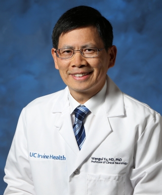
Dr. Wengui Yu serves as Director of the UCI Health Comprehensive Stroke and Cerebrovascular Center, Clinical Professor in the Department of Neurology, and Vice Chair for Hospital Affairs at the University of California, Irvine School of Medicine. Board-certified in Vascular Neurology, Neurology, and Neurocritical Care, he leads one of the nation's premier stroke programs with expertise in complex cerebrovascular disorders. His educational background includes: Medical Degree: Jiangxi Medical College, China PhD in Experimental Medicine: McGill University, Canada Neurology Residency: University of Missouri-Columbia Neurocritical Care & Stroke Fellowship: University of California, San Francisco Dr. Yu's research focuses on severe stroke outcomes, intracranial atherosclerotic stenosis, traumatic brain injury, and methamphetamine-related cerebrovascular disease. His work bridges clinical practice and translational research, particularly in optimizing endovascular interventions for intracranial stenosis and understanding socioeconomic impacts on stroke care. Recent publications demonstrate leadership in AI-assisted stroke detection and novel trial designs for intracranial artery stenosis. Key scientific recognition includes: 2013 Outstanding Contribution Award from Tiantan International Stroke Conference 2010 Honorary Award for International Exchange & Cooperation from China's Ministry of Health As Principal Investigator for the ASPIRE trial (Anticoagulation in ICH Survivors), he advances stroke prevention strategies while directing neurocritical care programs previously established at University of Texas Southwestern and Cedars-Sinai Medical Center. His clinical practice emphasizes patient-centered communication in both English and Mandarin Chinese.
Professor Mara Cercignani is Professor and Head of MRI within the School of Psychology at Cardiff University . Internationally recognised for developing and applying quantitative MRI techniques, she leads a multidisciplinary programme that links non-invasive microstructural imaging to brain function and pathology, with particular emphasis on Multiple Sclerosis, dementia and depression. Education & Career: Current appointment: Professor and Head of MRI, School of Psychology, Cardiff University. Previous roles and institutional affiliations have included collaborative posts across Europe and the UK, though full biographical dates are not given in the provided text. Research Interests: Prof Cercignani’s work exploits quantitative MRI —diffusion imaging, magnetisation-transfer imaging, and multi-parametric relaxometry—to map tissue microstructure in vivo. She integrates these data with EEG/MEG, TMS and behavioural paradigms to relate anatomy to function. A major strand investigates myelin density and axonal conduction velocity as determinants of cognitive and motor performance in health and disease. She has pioneered the use of VERDICT-MRI and diffusion-weighted MR spectroscopy to probe cellular and vascular micro-environment, and applies these tools to understand neuroinflammation, fatigue, and neurodegeneration. Publication landscape: Over the past five years her output (> 60 papers) has concentrated on three overlapping themes: (i) neuroimmune interactions —how peripheral inflammation and cytokine challenges remodel reward and punishment circuits; (ii) neurodegeneration and glymphatic dysfunction —early microstructural changes in Alzheimer’s disease, MS and ALS detected by diffusion and sodium MRI; and (iii) network-level plasticity —how cognitive reserve, sleep and memory reactivation alter structural connectivity. Scientific Awards & Recognition: Continuous major-project funding from UKRI (MRC, EPSRC), EU-FP7/H2020, Wellcome Trust and industry partners. Invited keynote speaker at International Society for Magnetic Resonance in Medicine (ISMRM) and Organisation for Human Brain Mapping (OHBM). Serves on editorial boards of NeuroImage , Human Brain Mapping and Multiple Sclerosis Journal . Supervision & Team Leadership: Prof Cercignani heads the Cardiff Neuroimaging Group , currently supervising 8 PhD students and 4 post-doctoral researchers working on projects spanning MR physics, computational modelling, and translational neurology. Recent major grants include a Wellcome Trust collaborative award on “Neuroimmune Imaging in Depression” and an MRC programme grant on “Quantitative Imaging of Myelin in MS”. Laboratory & Core Facilities: She directs the Cardiff University Brain Research Imaging Centre (CUBRIC) 3 T MRI Physics programme , managing state-of-the-art Siemens Prisma and Connectom systems, and co-leads the low-field 64 mT UNITY consortium aimed at deploying portable neuroimaging in low- and middle-income settings.
Dr. Swati Shah Mody serves as a Clinical Associate Professor of Radiology at the University of Michigan Medical School, based at C.S. Mott Children's Hospital in Ann Arbor, Michigan. Previously, she held the position of Director of Pediatric Neuroradiology and Fetal Imaging at Children's Hospital of Michigan until her transition to the University of Michigan in 2022. Her clinical expertise centers on advanced pediatric imaging with specialized applications in neonatal and fetal diagnostics. Her educational foundation includes: MBBS from University of Mumbai, Bombay (1987) DMRE from College of Physicians and Surgeons, Bombay (1990) MD from University of Mumbai (1991) Radiology Residency at Sir J J Group of Hospitals (1991) Senior Registrar in CT Scan/Neuroradiology at Bombay Hospital (1992) Radiology Residency at Wayne State University (1998) Pediatric Radiology Fellowship at Children's Hospital of Michigan/WSU (1999) Dr. Mody's research program focuses intensely on Pediatric Neuroradiology with neonatal imaging specialization and Fetal-Placental Imaging , leveraging advanced MRI techniques and artificial intelligence. Her work bridges clinical radiology with computational innovation, particularly through her active involvement in the University of Michigan's e-Health and Artificial Intelligence Initiative. Current investigations include deep learning applications for neonatal brain scan analysis, high-field fetal-placental MR angiography, and outcome prediction modeling in neonatal encephalopathy. Analysis of her 2022-2025 publications reveals a cohesive research trajectory prioritizing rare pediatric neurological conditions, advanced cardiac/fetal imaging, and AI-driven diagnostic solutions. Her work spans from case reports on infectious neuropathies to sophisticated 4D flow MRI characterization of vascular anomalies, demonstrating consistent methodological rigor while addressing critical gaps in pediatric diagnostic imaging. Dr. Mody actively contributes to institutional governance through multiple committee appointments and maintains an open mentoring posture for trainees. Her Center Membership in the e-Health and Artificial Intelligence Initiative positions her at the forefront of medical AI integration, where she collaborates on developing clinical decision support systems for pediatric imaging interpretation.
Asta Kristine Håberg serves as Professor at the Department of Neuromedicine and Movement Science within the Faculty of Medicine and Health Sciences at the Norwegian University of Science and Technology (NTNU). She concurrently holds leadership positions at St. Olav’s University Hospital as Head of the Norwegian National Advisory Unit for functional MRI and Director of the Center of Innovative Ultrasound Solutions (CIUS), a Norwegian Research Council-appointed Center for Research-based Innovation. Her research spans medical imaging and spectroscopy with emphasis on cognition across the lifespan and neurological conditions. Key focus areas include traumatic brain injury mechanisms, dementia risk factors, preterm birth neurodevelopmental outcomes, and advanced neuroimaging methodology development. Her work integrates population-based cohort studies (HUNT, Tromsø) with cutting-edge fMRI and diffusion MRI techniques. Professor Håberg leads major national initiatives including the Trondheim fMRI group and CIUS SFI center, driving translational research in ultrasound solutions. Her publication record demonstrates consistent high-impact contributions across neuroimaging, neurotrauma, and cognitive epidemiology with recent work emphasizing multi-cohort collaborations and methodological innovations in data harmonization. Notable professional roles include: Head, Trondheim fMRI Group National Advisory Unit Director for fMRI Director, Center of Innovative Ultrasound Solutions (CIUS) Principal Investigator in multiple HUNT Study dementia projects