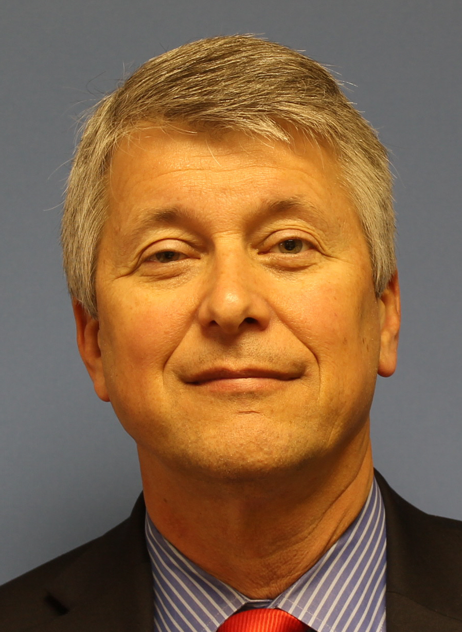
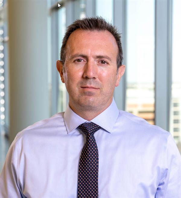
Allison Buchanan is an Associate Professor at the Dental College of Georgia , part of Augusta University . She serves as Interim Chair and Associate Chair for Oral Biology, while teaching at both predoctoral and graduate levels. Her work focuses on Cone Beam Computed Tomography (CBCT) and Digital Radiography , with a special emphasis on Quality Assurance and Obstructive Sleep Apnea imaging. Recent publications address radiation safety, disinfection methods for imaging plates, and software quality control in dental radiology. 2024 : Outstanding Faculty Award, Augusta University 2021 : Honorable mention awardee for Oral and Maxillofacial Radiology (OMR) section 2020 : Inducted into PHI KAPPA PHI Honor Society 2018 : Faculty Research and Scholarship Achievement Award Buchanan serves on multiple committees including the Standards Committee on Dental Informatics (2022-present) and the American Dental Association (2021-present), with editorial board memberships since 2018. Her teaching portfolio includes courses like Radiology Clinic II and Diagnostic Sciences Conference .
Craig Jones is an Assistant Professor of Computer Science at Johns Hopkins University's Whiting School of Engineering. He is affiliated with the Malone Center for Engineering in Healthcare and contributes to the Precision Medicine Analytics Platform's Imaging and Data Science Subcommittees. BSc in Computer Science and Mathematics from Simon Fraser University MSc in Medical Biophysics from the University of Western Ontario PhD in Physics from the University of British Columbia His research focuses on applying artificial intelligence and neural networks to medical image processing, particularly for MRI, CT, optical coherence tomography (OCT), and ultrasound datasets. Key areas include 2D/3D image processing, anomaly detection, segmentation, and uncertainty quantification, with clinical applications in neurosurgery, ophthalmology, and oncology. Projects span robotic imaging, neuroendoscopic guidance, and cancer boundary detection. Recent publications highlight advancements in vision-language models for 3D medical imaging, automated segmentation of venous malformations, and AI-guided neurosurgical tools. Articles emphasize multimodal data fusion, self-supervised learning, and federated learning for rare cancer analytics. He received a $310,000 Department of Defense grant in 2022 to develop AI-guided treatments for venous malformations. His work bridges clinical imaging domains and computer vision as a member of the Radiology AI Lab (RAIL), a collaborative effort across Johns Hopkins Hospital, the Whiting School of Engineering, and the Applied Physics Laboratory.
Jeffrey H. Siewerdsen, PhD, holds the John C. Malone Professorship in Biomedical Engineering at Johns Hopkins University, with joint appointments in Computer Science, Neurosurgery, and Radiology. As Vice-Chair for Clinical and Industry Collaboration, he bridges engineering, physics, and clinical practice to advance medical imaging technologies. His research focuses on image-guided interventions, surgical robotics, and CT innovations for radiation therapy and orthopedics. He leads the I-STAR Lab and collaborates with clinicians to translate imaging systems into clinical practice. Education: PhD in Physics (University of Michigan, 1998), MS in Physics (1994), BA in Physics & Astronomy (University of Minnesota, 1992). Research emphasizes 3D imaging systems, metal artifact reduction, and AI-driven reconstruction. He co-directs the Carnegie Center for Surgical Innovation and contributes to the Armstrong Institute for Patient Safety. Notable achievements include pioneering cone-beam CT (CBCT) for radiation therapy and intraoperative imaging. His work addresses clinical challenges through interdisciplinary collaboration. Awards and recognition include leadership in medical imaging societies and over 350 publications. His lab develops cutting-edge tools for surgical navigation, radiation oncology, and musculoskeletal imaging, with a focus on real-time 3D reconstruction and deformable motion correction. Labs/Teams: I-STAR Lab (Imaging Science for Technology and Applications in Research), Carnegie Center for Surgical Innovation.
University of Texas Southwestern Medical CenterUnited States
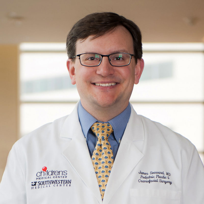
Dr. James Seaward is an Associate Professor of Pediatric Plastic and Craniofacial Surgery at UT Southwestern Medical Center and a pediatric plastic surgeon at Children’s Health℠. He specializes in cleft lip and palate surgery, craniofacial reconstruction, and vascular anomalies. His clinical practice focuses exclusively on pediatric cases, emphasizing minimally invasive techniques and multidisciplinary team care. Education: MBBS from Royal Free and University College Medical School (2000), Fellowship training in pediatric plastic surgery at UT Southwestern Certifications: Board-certified in plastic surgery (UK), Fellow of the Royal College of Surgeons of England Research interests include 3D imaging for facial asymmetry evaluation, cleft speech acoustics, MRI applications for speech disorders, and computer-aided surgical design. He leads the 22q Multi-Specialty Clinic and co-directs the Vascular Anomalies Team Clinic at Children’s Medical Center, Dallas. Publications emphasize surgical outcomes in craniofacial conditions, innovations in imaging, and multidisciplinary care models. Notable contributions include work on cleft palate speech analysis and minimally invasive craniosynostosis techniques. Awards: Royal College of Surgeons Fellowship He oversees surgical residency training as Associate Program Director and mentors craniofacial fellows. Active in global health, he previously volunteered with Operation Smile in Ghana.
Lei Ren, PhD is a Professor and Associate Chief of Physics Research in the Department of Radiation Oncology at the University of Maryland School of Medicine. Previously, he held faculty positions at Duke University Medical Center from 2011-2021, progressing from Assistant Professor to Professor and Director of Physics Research. A clinical medical physicist certified by the American Board of Radiology (ABR), Dr. Ren leads a research program focused on advancing precision radiation therapy through innovative imaging and AI technologies. Dr. Ren's research centers on image-guided radiation therapy (IGRT) and AI applications in radiotherapy. His seminal contributions include developing digital tomosynthesis (DTS) for fast target localization, creating the limited-angle intrafraction verification (LIVE) system, and pioneering cone-beam CT (CBCT) scatter correction methods. Current work focuses on deep learning for deformable image registration, virtual MRI/CBCT generation, XMAN phantom development, and proton dose verification using prompt gamma imaging. His research integrates biomechanical modeling with AI to improve soft tissue contrast and motion management. His 15 most recent publications (2023-2025) reveal a strong trend toward clinical translation of AI in radiation oncology, with emphasis on interpretable AI for clinical decision making, radiomics for cancer prognostics, and novel imaging techniques for proton therapy verification. Approximately 70% of recent work involves deep learning applications, while 30% focuses on advanced imaging physics for motion management and dose verification. Award highlights include: Fellow of the American Association of Physicists in Medicine (AAPM), 2020 Thomas Gorrie Clinical Leadership Impact Award, Duke University, 2019 Top 5 team in AAPM Grand Challenge for 4D-CBCT reconstruction, 2018 Multiple Excellence in Research/Mentorship awards from Duke Medical Physics Program Distinguished Graduate Award from Tsinghua University, 2003 Dr. Ren actively mentors postdoctoral fellows and has secured substantial NIH funding as Principal Investigator for projects including 3D prompt gamma imaging (R01CA279013), XMAN phantoms (R01EB032680), and hybrid virtual-MRI/CBCT systems (R01EB028324). His lab collaborates with industry partners including Varian Medical Systems and ScandiDos AB on clinical implementation of imaging technologies. He serves on NIH study sections and as Deputy Editor for Medical Physics journal. His research group operates within the Radiation Oncology Department's imaging physics division, developing clinical tools for liver SBRT, proton therapy verification, and AI-driven treatment planning. Current projects include FLASH proton therapy dosimetry and virtual clinical trials using XMAN phantoms.
University of North Carolina at Chapel HillUnited States
Professor Jianping Lu is a leading academic at the University of North Carolina at Chapel Hill, affiliated with the Department of Physics and Astronomy within the College of Arts and Sciences. His work focuses on advancing medical imaging technologies, particularly in X-ray and computed tomography (CT) systems, with a strong emphasis on carbon nanotube (CNT) X-ray sources. He holds a Ph.D. in Physics from the City University of New York (1988). Education: Ph.D. in Physics, City University of New York, 1988 Research Interests: Development of novel imaging systems, including stationary tomosynthesis and multisource CBCT Optimization of X-ray technology for clinical applications (e.g., oncology, cardiology, dentistry) Integration of artificial intelligence (AI) for diagnostic accuracy and automated analysis Portable and low-cost medical imaging solutions Recent Work Trends: His 2025 publications highlight advancements in AI-driven diagnostics for pancreatic cancer, improved contrast in adaptive radiation therapy, and stationary chest tomosynthesis systems. Key innovations include low-cost dual-energy CBCT and carbon nanotube-based X-ray arrays, which enhance image quality while reducing radiation exposure. His 2024 studies further explore cardiac imaging, dental tomosynthesis, and system optimizations for clinical adoption. Awards: None explicitly listed in the provided text. Advising & Grants: While student advisees are not listed, his research is likely supported by grants focusing on medical imaging innovation. Collaborations span physics, engineering, and clinical departments to bridge technical and clinical challenges. Labs/Teams: Likely affiliated with UNC’s imaging research groups, particularly those developing CNT X-ray technologies and clinical imaging systems for cancer and cardiovascular applications.
Robert Waugh is an Assistant Professor in the Department of Orthodontics at the Dental College of Georgia. His academic career focuses on advanced orthodontic diagnostics and treatment planning, integrating modern imaging technologies like Cone Beam Computed Tomography (CBCT) into clinical practice. Education: MS in Orthodontics Residency Program (Texas A&M University System), DMD in Dentistry (Medical College of Georgia), BS in Biology/Biological Sciences (Mercer University) Research Interests Diagnostic Imaging in Orthodontics CBCT Applications for Treatment Planning Self-Ligating Bracket Technology Herbst Appliance Evaluation Bond Retention Optimization Publications highlight his work in advanced imaging, dental technology, and clinical workflow improvements. His scholarship spans over a decade, with recent focus on CBCT integration and orthodontic appliance analysis. Professional Service Member, Academy of General Dentistry (2015–Present) Member, American Association of Dental Research (2015–Present) Member, American Association of Orthodontics (2015–Present) Member, American Dental Association (2015–Present) Member, American Society of Dentistry for Children (2015–Present)
Mark Oldham is Professor of Radiation Oncology and Professor of Physics at Duke University, where he also serves as Director of the Duke Medical Physics MS/PhD Graduate Program and is a Member of the Duke Cancer Institute. His academic career spans over three decades with progressive appointments from Instructor to full Professor in Radiation Oncology. Dr. Oldham received his Ph.D. from Newcastle University (United Kingdom) in 1991. His research interests focus on innovative approaches to radiation therapy, particularly FLASH radiation therapy using the Duke High Intensity Gamma Source (HIGS), radiation and immunotherapy utilizing mini-grids, Radiation Therapy Enhanced by Cherenkov photo-Activation (RECA), and comprehensive 3D dosimetry systems. His work has been recognized with multiple Paper of the Year awards from SEAAPM and the prestigious Farrington Daniel Award from AAPM. His recent publications demonstrate a strong focus on 3D dosimetry systems, radiation therapy verification techniques, and novel cancer treatment approaches. His research integrates advanced dosimetry methods with innovative radiation therapy techniques to improve cancer treatment outcomes. His work on polymer gel dosimetry, optical CT scanning, and radiation-immunotherapy combinations represents cutting-edge developments in the field. Dr. Oldham's scientific achievements have been recognized through multiple awards including the Paper of the Year (2018, 2017), Farrington Daniel Award (2016, 2002), Excellence in Teaching (2014), Excellence in Mentoring (2011), and Fellow of the AAPM (2010). He has also received the Director's Award for Exemplary Service multiple times. As a principal investigator and co-investigator, Dr. Oldham has secured significant research funding from the National Institutes of Health, National Institute of Dental and Craniofacial Research, and Ian's Friends Foundation. His current research portfolio includes exploring synthetic lethality with high energy electron FLASH radiation beams, super-FLASH therapy for pediatric brain tumors, and radiation protection mechanisms. He serves on the Board of Directors of the American Association of Medical Physics and has made significant contributions to medical physics education through his leadership of Duke's Medical Physics Graduate Program.
Moyed Miften is a Professor and Director of the Medical Physics Division in the Department of Radiation Oncology at the University of Colorado Anschutz Medical Campus School of Medicine. With extensive expertise in radiation oncology physics, he has made significant contributions to the field of medical physics and radiation therapy. Dr. Miften earned his MSc from the University of Michigan in 1990 and his PhD from the same institution in 1994. He completed a fellowship in Radiation Oncology at the University of Michigan Program in 1996, establishing a strong foundation for his academic and research career in medical physics. Dr. Miften's research focuses on several key areas within radiation oncology physics. His primary interests include medical physics, radiation therapy, image-guided radiation therapy (IGRT), stereotactic body radiation therapy (SBRT), functional lung avoidance radiation therapy, radiation dosimetry, and radiation treatment planning. His work has particularly emphasized the development and clinical implementation of 4DCT-ventilation functional avoidance techniques in radiation therapy, which aims to spare functional lung regions during treatment to minimize toxicity while maintaining tumor control. He has also made significant contributions to the field of CBCT (cone-beam computed tomography) imaging, particularly in developing 2D antiscatter grid technologies and scatter correction methods to improve image quality and enable more accurate dose calculations during radiation therapy delivery. Analysis of Dr. Miften's recent publications reveals a strong focus on advancing precision radiation therapy through innovative imaging techniques and functional avoidance approaches. His research spans multiple clinical applications including lung cancer, liver cancer, and head and neck cancer treatments. A significant portion of his recent work involves the development and validation of functional avoidance radiation therapy techniques, particularly using 4DCT ventilation imaging to guide treatment planning. He has also been actively researching advanced imaging techniques such as coded aperture scatter imaging, computer vision-assisted surface guidance, and novel CBCT methods with antiscatter grids to improve treatment accuracy and enable real-time adaptation. Fellow, American Society of Radiation Oncology (2023) Radiotherapy Technical Expert, IAEA (2016) Volunteer Service Award, American Board of Radiology (2014) Chancellor, Board of Chancellors, American College of Medical Physics (2011) Fellow, American Association of Physicists in Medicine (AAPM) (2011) Dr. Miften has been actively involved in numerous clinical trials and collaborative research projects, particularly focusing on functional avoidance radiation therapy. He has served as a principal investigator and co-investigator on multiple studies evaluating the clinical implementation of 4DCT-ventilation techniques in lung cancer treatment. His research has received support from various funding sources that enable the advancement of precision radiation therapy techniques. While the specific details of his current grant portfolio are not provided in the text, his extensive publication record suggests ongoing research funding supporting his work in medical physics and radiation oncology. As Director of the Medical Physics Division, Dr. Miften leads a team of medical physicists and researchers focused on advancing radiation therapy techniques. His division likely collaborates closely with radiation oncologists, dosimetrists, and therapists to implement cutting-edge treatment approaches in clinical practice. The research output suggests active collaborations with institutions involved in the PENTEC (Pediatric Normal Tissue Effects in the Clinic) initiative and other multi-institutional clinical trials focused on reducing radiation-induced toxicities.
Jyoti Mago serves as an Assistant Professor in Residence within the Department of Dental Medicine at the University of Nevada, Las Vegas, specializing in oral and maxillofacial radiology. Her clinical and research work focuses on advanced imaging diagnostics and the integration of artificial intelligence in dental practice and education. Her educational background includes: Certificate in Advanced Education Program in Oral and Maxillofacial Radiology from University of Connecticut School of Dental Medicine MS from University of Connecticut School of Dental Medicine MDS (Oral Medicine and Radiology) from Maharishi Markandeshwar College of Dental Sciences and Research, India BDS from Baba Jaswant Singh Dental College and Hospital, India Dr. Mago's research spans oral and maxillofacial radiology, dental imaging protocols, and transformative AI applications in dentistry. She investigates CBCT-based diagnosis of complex conditions like periodontal disease and oral cancers, while pioneering the use of generative AI for radiology reporting, curriculum development, and radiation safety education. Her work addresses critical gaps in geriatric oral healthcare and diagnostic accuracy through technological innovation. Analysis of her 2021-2025 publications reveals three dominant research vectors: (1) AI integration in dental radiology reporting and education using tools like ChatGPT, (2) advanced imaging diagnostics for oral pathologies and anatomical correlations, and (3) geriatric oral health challenges. Her recent work demonstrates increasing emphasis on AI's role in improving diagnostic precision, educational methodologies, and clinical workflows while maintaining rigorous investigation of traditional radiological techniques. No scientific awards were documented in the provided materials. Dr. Mago's academic contributions include mentoring dental students in radiology techniques and diagnostic interpretation, though specific advisees aren't listed. Her research program appears supported by institutional resources at UNLV's School of Dental Medicine, with recent projects focusing on AI implementation and imaging protocol optimization. Current initiatives likely involve expanding AI validation studies across dental specialties and developing standardized frameworks for AI-assisted diagnostics. She operates within UNLV's dental research ecosystem, collaborating on projects involving CBCT imaging analysis and AI tool development for clinical dentistry, though specific lab affiliations aren't detailed in available records.
Dr. Sajitha Kalathingal is a Professor and Director of Radiology in the Department of Oral Health and Diagnostic Sciences at the Dental College of Georgia, Augusta University, with additional appointments in the College of Allied Health Sciences for Dental Hygiene. She earned her Bachelor of Dental Surgery from Mangalore University (1995) and Master's in Oral and Maxillofacial Radiology from UNC Chapel Hill (2005). Her academic credentials include: MS in Oral and Maxillofacial Surgery, University of North Carolina (2005) DDS in Dentistry, Mangalore University (1995) Board Certification in Radiology (BCRD), American Board of Oral and Maxillofacial Radiology (2005) Georgia Dental License (2006) Dr. Kalathingal pioneers technology-integrated dental education while advancing digital radiography and 3D imaging applications. Her research focuses on quality assurance protocols, clinical implementation of cone beam CT, and diagnostic innovations for oral pathologies, establishing her as a leader in evidence-based radiographic techniques. Publication analysis reveals consistent contributions to dental imaging science, with emphasis on artifact reduction in digital radiography, 3D modeling for periodontal diagnostics, and early detection protocols for jaw pathologies. Her work bridges technical innovation with clinical practice across oncology, endodontics, and prosthodontics. Professional recognition includes: Councilor, Public Policy & Scientific Affairs (AAOMR, 2024) Outstanding Faculty Award (DCG/Augusta University, 2018) Oral Radiology Section Councilor (ADEA, 2018) P&T Committee MVP (DCG, 2017) OKU National Honor Society President (2017) As an educator, she teaches 12+ courses including Radiology, Oral Diagnostic Sciences, and Implantology while mentoring dental students and residents. Committee leadership spans faculty recruitment, clinical education policy, and radiation safety across departmental, college, and university levels since 2006. Dr. Kalathingal directs continuing education programs in Cone Beam CT and maintains active clinical practice, contributing to national standards through AAOMR and ADEA committees while advancing patient-centered imaging protocols.
Srijit Kamath serves as Assistant Professor of Therapeutic Radiology at Yale School of Medicine, Yale University, with additional responsibility as Regional Chief Physicist overseeing operations at Trumbull Radiation Oncology, Smilow Cancer Hospital Care Center at Greenwich, and Griffin Hospital Lawrence and Memorial Cancer Center. His clinical leadership ensures physics standards across Yale's integrated cancer care network including Smilow Cancer Hospital and Yale Gamma Knife Center. His academic foundation includes: Medical Physics Residency, University of Florida (2010) PhD, University of Florida (2005) MS, University of Florida (2000) Dr. Kamath's research program centers on computational optimization of radiation therapy delivery, with seminal contributions to intensity-modulated radiation therapy (IMRT) field splitting algorithms, multileaf collimator sequencing, and deformable image registration. His work addresses critical challenges in treatment delivery efficiency while maintaining dosimetric accuracy, particularly for large-field treatments where conventional approaches risk underdosage at field junctions. He has pioneered methods to eliminate tongue-and-groove effects and optimize feathering techniques for seamless dose distributions. Analysis of his 15 most recent publications (2003-2011) reveals a consistent trajectory from foundational work on leaf sequencing algorithms to sophisticated solutions for deformable image registration and CBCT system optimization. His comparative studies of XVI and OBI CBCT systems established critical benchmarks for image quality in radiation therapy guidance, while his field-splitting algorithms remain clinically implemented for complex treatment geometries. The research demonstrates exceptional focus on translating computational advances into practical clinical workflows. No scientific awards or honors were documented in the available information. As Regional Chief Physicist, Dr. Kamath directs medical physics services across multiple community cancer centers, ensuring compliance with safety standards and optimal implementation of advanced techniques like stereotactic body radiation therapy (SBRT). While student advising activities aren't specified, his role in Yale's residency training program involves mentoring medical physics residents through clinical rotations. The absence of grant documentation suggests primary focus on clinical service and technical innovation rather than externally funded research. He operates within Yale's comprehensive radiation oncology infrastructure, collaborating with multidisciplinary teams at Smilow Cancer Hospital and affiliated centers to integrate advanced imaging and treatment technologies. His work directly supports the clinical mission of delivering precision radiation therapy through physics-driven quality assurance and process optimization.
Steven J. Bartolac serves as an Assistant Professor in the Department of Radiation Oncology at the University of Maryland School of Medicine, applying expertise in medical physics to advance radiotherapy imaging and treatment protocols. His academic foundation includes dual undergraduate degrees from Queen’s University followed by specialized graduate training at the University of Toronto: BASc Hons in Engineering Physics (2000-2005) BSc in Geology (2001-2005) M.Sc. in Medical Physics (2006-2009) Ph.D. in Medical Physics (2009-2013) Residency in Radiation Oncology Physics (2013-2015) Dr. Bartolac’s research centers on optimizing CT imaging for radiotherapy applications, with particular focus on mitigating data insufficiencies in cone-beam CT acquisition and developing spatial fluence modulation techniques to balance radiation dose and image quality. His current work explores protocol optimization for radiotherapy and novel orthovoltage treatment methodologies, addressing critical challenges in pediatric imaging precision and equipment performance stability. Recent publications demonstrate evolving expertise from foundational fluence optimization (2011) to contemporary investigations of seasonal equipment variations and pediatric CBCT precision (2019), reflecting consistent contributions to medical physics with emphasis on dose reduction and image-guided therapy accuracy. No scientific awards were documented in available materials. Information regarding student mentorship, research funding, or laboratory affiliations was not provided in the source documentation.
Cem Altunbas, PhD is a Professor in the Department of Radiation Oncology at the University of Colorado School of Medicine . His work focuses on advancing Cone Beam Computed Tomography (CBCT) for radiation therapy applications, with a particular emphasis on scatter rejection, image reconstruction, and quantitative accuracy. Key Research Areas: Medical Physics, Radiation Oncology, CBCT Imaging, Scatter Correction, Tumor Imaging His research has led to significant innovations in 2D antiscatter grid design, cross-scatter suppression, and artifact reduction in CBCT systems. Recent publications highlight applications in pelvis imaging , brain imaging , and dual-energy material decomposition . His work often integrates collaborations with physicists and clinicians to improve image-guided radiation therapy workflows. Scientific Recognition includes board certification in Therapeutic Medical Physics by the American Board of Radiology (2010). His contributions span over a decade, with >55 publications addressing challenges in CBCT image fidelity and radiation dosimetry.
University of California, Los AngelesUnited States
John Neylon is an HS Assistant Clinical Professor in the Department of Radiation Oncology at the University of California, Los Angeles (UCLA). His research focuses on advancing radiation therapy techniques through medical physics innovations and AI-driven solutions. Key areas include image-guided radiotherapy, automated image review systems, and optimization of radiation delivery protocols. He collaborates extensively with peers in radiation oncology, medical physics, and computational sciences to enhance treatment precision and patient outcomes. His work integrates cutting-edge technologies such as deep learning models for medical imaging segmentation and AI-assisted decision-making in radiation therapy. Recent studies involve improving patient alignment accuracy through automated systems and analyzing motion management in prostate and gynecological cancers. He also explores the dosimetric impacts of anatomical changes during treatment, contributing to safer and more effective radiation protocols. Neylon’s publications highlight advancements in MRI-guided stereotactic body radiotherapy, cone beam CT applications, and cloud-based dose calculation frameworks. His research bridges clinical practice and computational innovation, aiming to reduce treatment side effects and improve treatment planning efficiency.