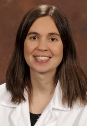
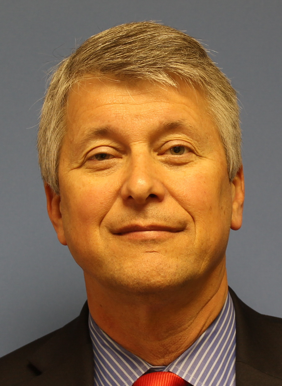
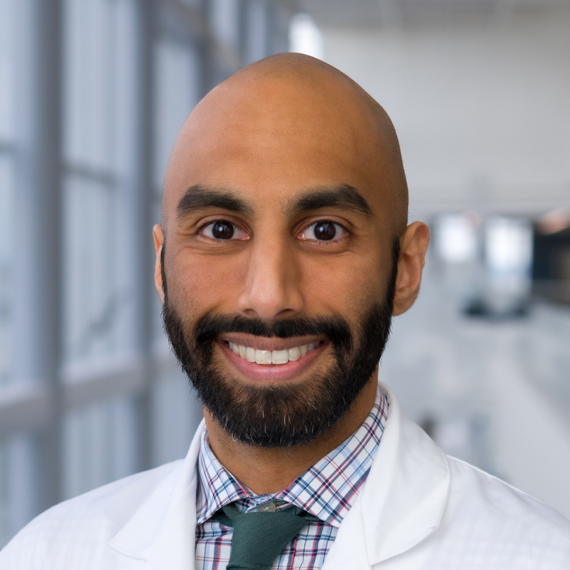
Allison Buchanan is an Associate Professor at the Dental College of Georgia , part of Augusta University . She serves as Interim Chair and Associate Chair for Oral Biology, while teaching at both predoctoral and graduate levels. Her work focuses on Cone Beam Computed Tomography (CBCT) and Digital Radiography , with a special emphasis on Quality Assurance and Obstructive Sleep Apnea imaging. Recent publications address radiation safety, disinfection methods for imaging plates, and software quality control in dental radiology. 2024 : Outstanding Faculty Award, Augusta University 2021 : Honorable mention awardee for Oral and Maxillofacial Radiology (OMR) section 2020 : Inducted into PHI KAPPA PHI Honor Society 2018 : Faculty Research and Scholarship Achievement Award Buchanan serves on multiple committees including the Standards Committee on Dental Informatics (2022-present) and the American Dental Association (2021-present), with editorial board memberships since 2018. Her teaching portfolio includes courses like Radiology Clinic II and Diagnostic Sciences Conference .
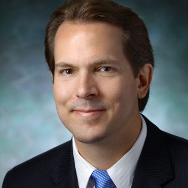
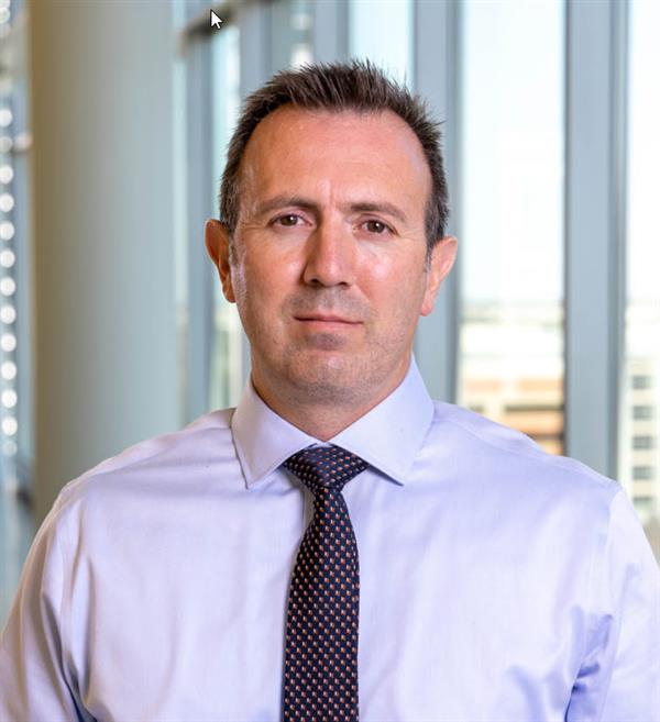
Jeffrey H. Siewerdsen, PhD, holds the John C. Malone Professorship in Biomedical Engineering at Johns Hopkins University, with joint appointments in Computer Science, Neurosurgery, and Radiology. As Vice-Chair for Clinical and Industry Collaboration, he bridges engineering, physics, and clinical practice to advance medical imaging technologies. His research focuses on image-guided interventions, surgical robotics, and CT innovations for radiation therapy and orthopedics. He leads the I-STAR Lab and collaborates with clinicians to translate imaging systems into clinical practice. Education: PhD in Physics (University of Michigan, 1998), MS in Physics (1994), BA in Physics & Astronomy (University of Minnesota, 1992). Research emphasizes 3D imaging systems, metal artifact reduction, and AI-driven reconstruction. He co-directs the Carnegie Center for Surgical Innovation and contributes to the Armstrong Institute for Patient Safety. Notable achievements include pioneering cone-beam CT (CBCT) for radiation therapy and intraoperative imaging. His work addresses clinical challenges through interdisciplinary collaboration. Awards and recognition include leadership in medical imaging societies and over 350 publications. His lab develops cutting-edge tools for surgical navigation, radiation oncology, and musculoskeletal imaging, with a focus on real-time 3D reconstruction and deformable motion correction. Labs/Teams: I-STAR Lab (Imaging Science for Technology and Applications in Research), Carnegie Center for Surgical Innovation.
Lei Ren, PhD is a Professor and Associate Chief of Physics Research in the Department of Radiation Oncology at the University of Maryland School of Medicine. Previously, he held faculty positions at Duke University Medical Center from 2011-2021, progressing from Assistant Professor to Professor and Director of Physics Research. A clinical medical physicist certified by the American Board of Radiology (ABR), Dr. Ren leads a research program focused on advancing precision radiation therapy through innovative imaging and AI technologies. Dr. Ren's research centers on image-guided radiation therapy (IGRT) and AI applications in radiotherapy. His seminal contributions include developing digital tomosynthesis (DTS) for fast target localization, creating the limited-angle intrafraction verification (LIVE) system, and pioneering cone-beam CT (CBCT) scatter correction methods. Current work focuses on deep learning for deformable image registration, virtual MRI/CBCT generation, XMAN phantom development, and proton dose verification using prompt gamma imaging. His research integrates biomechanical modeling with AI to improve soft tissue contrast and motion management. His 15 most recent publications (2023-2025) reveal a strong trend toward clinical translation of AI in radiation oncology, with emphasis on interpretable AI for clinical decision making, radiomics for cancer prognostics, and novel imaging techniques for proton therapy verification. Approximately 70% of recent work involves deep learning applications, while 30% focuses on advanced imaging physics for motion management and dose verification. Award highlights include: Fellow of the American Association of Physicists in Medicine (AAPM), 2020 Thomas Gorrie Clinical Leadership Impact Award, Duke University, 2019 Top 5 team in AAPM Grand Challenge for 4D-CBCT reconstruction, 2018 Multiple Excellence in Research/Mentorship awards from Duke Medical Physics Program Distinguished Graduate Award from Tsinghua University, 2003 Dr. Ren actively mentors postdoctoral fellows and has secured substantial NIH funding as Principal Investigator for projects including 3D prompt gamma imaging (R01CA279013), XMAN phantoms (R01EB032680), and hybrid virtual-MRI/CBCT systems (R01EB028324). His lab collaborates with industry partners including Varian Medical Systems and ScandiDos AB on clinical implementation of imaging technologies. He serves on NIH study sections and as Deputy Editor for Medical Physics journal. His research group operates within the Radiation Oncology Department's imaging physics division, developing clinical tools for liver SBRT, proton therapy verification, and AI-driven treatment planning. Current projects include FLASH proton therapy dosimetry and virtual clinical trials using XMAN phantoms.
University of North Carolina at Chapel HillUnited States
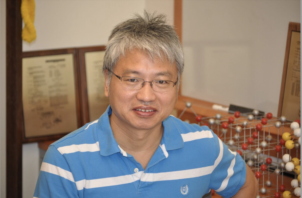
Professor Jianping Lu is a leading academic at the University of North Carolina at Chapel Hill, affiliated with the Department of Physics and Astronomy within the College of Arts and Sciences. His work focuses on advancing medical imaging technologies, particularly in X-ray and computed tomography (CT) systems, with a strong emphasis on carbon nanotube (CNT) X-ray sources. He holds a Ph.D. in Physics from the City University of New York (1988). Education: Ph.D. in Physics, City University of New York, 1988 Research Interests: Development of novel imaging systems, including stationary tomosynthesis and multisource CBCT Optimization of X-ray technology for clinical applications (e.g., oncology, cardiology, dentistry) Integration of artificial intelligence (AI) for diagnostic accuracy and automated analysis Portable and low-cost medical imaging solutions Recent Work Trends: His 2025 publications highlight advancements in AI-driven diagnostics for pancreatic cancer, improved contrast in adaptive radiation therapy, and stationary chest tomosynthesis systems. Key innovations include low-cost dual-energy CBCT and carbon nanotube-based X-ray arrays, which enhance image quality while reducing radiation exposure. His 2024 studies further explore cardiac imaging, dental tomosynthesis, and system optimizations for clinical adoption. Awards: None explicitly listed in the provided text. Advising & Grants: While student advisees are not listed, his research is likely supported by grants focusing on medical imaging innovation. Collaborations span physics, engineering, and clinical departments to bridge technical and clinical challenges. Labs/Teams: Likely affiliated with UNC’s imaging research groups, particularly those developing CNT X-ray technologies and clinical imaging systems for cancer and cardiovascular applications.
University of Texas Southwestern Medical CenterUnited States
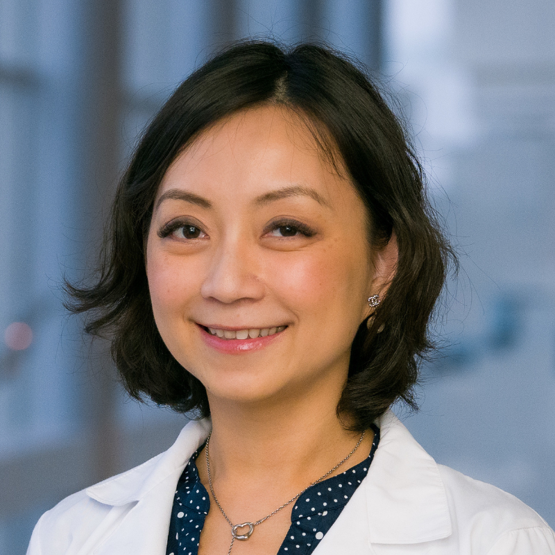
Mu-Han Lin, Ph.D., is a Professor in the Department of Radiation Oncology at UT Southwestern Medical Center, with leadership roles including Senior Director of Clinical Physics and lead physicist for head and neck radiation oncology. Her academic profile spans Medical Physics , Adaptive Radiation Therapy , and Artificial Intelligence (AI) in Radiotherapy . Education: Master’s and Ph.D. in Medical Physics from National Tsing Hua University, Taiwan Training: Medical Physics Residency at Fox Chase Cancer Center, Philadelphia Dr. Lin’s research focuses on online adaptive radiation therapy (oART) systems, particularly cone beam CT-guided workflows for gastric MALT lymphoma and head/neck cancers. She pioneered techniques for dose prediction , plan quality assurance , and deep learning integration to address anatomical variations during treatment. Her work demonstrates reduced PTV margins (0.5-0.7 cm) while maintaining target coverage and minimizing organ-at-risk (OAR) doses through auto-contouring and synthetic CT optimization . Recent publications highlight her leadership in adaptive treatment planning (15+ articles from 2024-2025) across journals like Medical Physics , Practical Radiation Oncology , and Radiotherapy and Oncology . Key contributions include: X-Ray Guided Ethos Platform workflows Plan Quality Review Checklists HyperSight CBCT for image quality enhancement Inter-patient adaptive strategies for spine SAbR AI-driven segmentation and dose adaptation She actively contributes to clinical education as an instructor in UT Southwestern’s Medical Physics Certificate Program and Biomedical Engineering Graduate Program , and chairs symposiums on X-ray Guided Adaptive RT . Dr. Lin serves on editorial boards for Therapeutic Radiation and Oncology and Medical Physics , and collaborates on grants with Varian Medical Systems and other industry partners.
Robert Waugh is an Assistant Professor in the Department of Orthodontics at the Dental College of Georgia. His academic career focuses on advanced orthodontic diagnostics and treatment planning, integrating modern imaging technologies like Cone Beam Computed Tomography (CBCT) into clinical practice. Education: MS in Orthodontics Residency Program (Texas A&M University System), DMD in Dentistry (Medical College of Georgia), BS in Biology/Biological Sciences (Mercer University) Research Interests Diagnostic Imaging in Orthodontics CBCT Applications for Treatment Planning Self-Ligating Bracket Technology Herbst Appliance Evaluation Bond Retention Optimization Publications highlight his work in advanced imaging, dental technology, and clinical workflow improvements. His scholarship spans over a decade, with recent focus on CBCT integration and orthodontic appliance analysis. Professional Service Member, Academy of General Dentistry (2015–Present) Member, American Association of Dental Research (2015–Present) Member, American Association of Orthodontics (2015–Present) Member, American Dental Association (2015–Present) Member, American Society of Dentistry for Children (2015–Present)
Mark Oldham is Professor of Radiation Oncology and Professor of Physics at Duke University, where he also serves as Director of the Duke Medical Physics MS/PhD Graduate Program and is a Member of the Duke Cancer Institute. His academic career spans over three decades with progressive appointments from Instructor to full Professor in Radiation Oncology. Dr. Oldham received his Ph.D. from Newcastle University (United Kingdom) in 1991. His research interests focus on innovative approaches to radiation therapy, particularly FLASH radiation therapy using the Duke High Intensity Gamma Source (HIGS), radiation and immunotherapy utilizing mini-grids, Radiation Therapy Enhanced by Cherenkov photo-Activation (RECA), and comprehensive 3D dosimetry systems. His work has been recognized with multiple Paper of the Year awards from SEAAPM and the prestigious Farrington Daniel Award from AAPM. His recent publications demonstrate a strong focus on 3D dosimetry systems, radiation therapy verification techniques, and novel cancer treatment approaches. His research integrates advanced dosimetry methods with innovative radiation therapy techniques to improve cancer treatment outcomes. His work on polymer gel dosimetry, optical CT scanning, and radiation-immunotherapy combinations represents cutting-edge developments in the field. Dr. Oldham's scientific achievements have been recognized through multiple awards including the Paper of the Year (2018, 2017), Farrington Daniel Award (2016, 2002), Excellence in Teaching (2014), Excellence in Mentoring (2011), and Fellow of the AAPM (2010). He has also received the Director's Award for Exemplary Service multiple times. As a principal investigator and co-investigator, Dr. Oldham has secured significant research funding from the National Institutes of Health, National Institute of Dental and Craniofacial Research, and Ian's Friends Foundation. His current research portfolio includes exploring synthetic lethality with high energy electron FLASH radiation beams, super-FLASH therapy for pediatric brain tumors, and radiation protection mechanisms. He serves on the Board of Directors of the American Association of Medical Physics and has made significant contributions to medical physics education through his leadership of Duke's Medical Physics Graduate Program.
Vinicius Dutra, DDS, PhD, MBA is a Clinical Associate Professor at the Department of Oral Pathology, Medicine and Radiology within the Indiana University School of Dentistry. His work focuses on innovative applications of 3D printing, virtual reality, and artificial intelligence in dental education and clinical practice. He specializes in oral radiology, implantology, and orthodontics, with a strong emphasis on improving diagnostic accuracy and surgical precision through advanced imaging and digital tools. Dr. Dutra holds a Doctor of Dental Surgery (DDS), a PhD, and an MBA, reflecting his interdisciplinary expertise in clinical practice, research, and healthcare management. His research explores topics such as guided implant surgery, 3D-printed anatomical models for training, and the integration of machine learning into dental diagnostics. His recent studies include evaluations of virtual reality in dental education, the accuracy of guided implant placement techniques, and comparisons between cone-beam CT scans and histological analyses. He has also contributed to advancements in complete denture fabrication workflows and bone thickness measurement methodologies. Despite his extensive publication record, no scientific awards were explicitly mentioned in the provided materials. Dr. Dutra’s clinical and educational roles involve mentoring students and contributing to the development of cutting-edge dental technologies. His work bridges traditional dental practices with modern digital innovations, aiming to enhance patient outcomes and streamline clinical workflows.
Steven J. Bartolac serves as an Assistant Professor in the Department of Radiation Oncology at the University of Maryland School of Medicine, applying expertise in medical physics to advance radiotherapy imaging and treatment protocols. His academic foundation includes dual undergraduate degrees from Queen’s University followed by specialized graduate training at the University of Toronto: BASc Hons in Engineering Physics (2000-2005) BSc in Geology (2001-2005) M.Sc. in Medical Physics (2006-2009) Ph.D. in Medical Physics (2009-2013) Residency in Radiation Oncology Physics (2013-2015) Dr. Bartolac’s research centers on optimizing CT imaging for radiotherapy applications, with particular focus on mitigating data insufficiencies in cone-beam CT acquisition and developing spatial fluence modulation techniques to balance radiation dose and image quality. His current work explores protocol optimization for radiotherapy and novel orthovoltage treatment methodologies, addressing critical challenges in pediatric imaging precision and equipment performance stability. Recent publications demonstrate evolving expertise from foundational fluence optimization (2011) to contemporary investigations of seasonal equipment variations and pediatric CBCT precision (2019), reflecting consistent contributions to medical physics with emphasis on dose reduction and image-guided therapy accuracy. No scientific awards were documented in available materials. Information regarding student mentorship, research funding, or laboratory affiliations was not provided in the source documentation.
Jeremy N. Kunz is an Assistant Professor in the Department of Radiation Oncology at the University of Utah and a Medical Physicist at Huntsman Cancer Hospital. He is board-certified in Therapeutic Medical Physics by the American Board of Radiology. BS in Physics and Applied Mathematics from Weber State University MS in Medical Physics from the University of Toledo PhD in Quantum Biophotonics from Baylor University His research focuses on clinical and theoretical aspects of radiation oncology, including: Brachytherapy (HDR, interstitial, gynecological) Stereotactic radiosurgery and VMAT techniques (CSI, TMI) Patient safety, quality assurance, and advanced imaging systems The 15 most recent articles highlight his work in cone-beam CT optimization, Acuros algorithm validation for TBI, HDR brachytherapy commissioning, and real-time linac quality assurance using novel detectors. Keywords span Medical Physics , Radiation Oncology , and Biophysics , with sub-fields like Image-Guided Radiotherapy and Dose Calculation Algorithms .
Cem Altunbas, PhD is a Professor in the Department of Radiation Oncology at the University of Colorado School of Medicine . His work focuses on advancing Cone Beam Computed Tomography (CBCT) for radiation therapy applications, with a particular emphasis on scatter rejection, image reconstruction, and quantitative accuracy. Key Research Areas: Medical Physics, Radiation Oncology, CBCT Imaging, Scatter Correction, Tumor Imaging His research has led to significant innovations in 2D antiscatter grid design, cross-scatter suppression, and artifact reduction in CBCT systems. Recent publications highlight applications in pelvis imaging , brain imaging , and dual-energy material decomposition . His work often integrates collaborations with physicists and clinicians to improve image-guided radiation therapy workflows. Scientific Recognition includes board certification in Therapeutic Medical Physics by the American Board of Radiology (2010). His contributions span over a decade, with >55 publications addressing challenges in CBCT image fidelity and radiation dosimetry.
Dr. Joshua P. Schiff is an Assistant Professor of Clinical Radiation Oncology at the Keck School of Medicine, University of Southern California (USC), with primary clinical practice at the Norris Comprehensive Cancer Center. He specializes in advanced radiotherapy techniques for diverse cancer types, including intensity-modulated radiation therapy (IMRT), volumetric-modulated arc therapy (VMAT), and motion management strategies. His research focuses on improving treatment outcomes through adaptive radiotherapy and biology-guided approaches. Education & Training: Bachelor’s in Biopsychology (University of California, Santa Barbara) M.D. (Tulane University School of Medicine, inducted into Alpha Omega Alpha Honor Society) Radiation Oncology Residency (Washington University School of Medicine, Chief Resident) Internal Medicine Internship (Cedars-Sinai Medical Center) His research emphasizes innovative radiotherapy technologies such as MR-guided adaptive radiotherapy (SMART) and CT-guided stereotactic adaptive radiotherapy (CT-STAR). He leads clinical trials exploring motion management and biology-guided strategies to enhance precision while minimizing toxicity. Key contributions include pioneering studies on pancreatic, liver, and thoracic cancers treated with advanced techniques. Awards: Roentgen Resident/Fellow Research Award (Radiological Society of North America, 2024) Alpha Omega Alpha Honor Medical Society (2019) Dr. Schiff’s work spans publications in high-impact journals like International Journal of Radiation Oncology and Radiotherapy and Oncology . He actively collaborates on grants focused on adaptive radiotherapy implementation and has contributed to professional organizations such as ASTRO and the American Medical Association.
University of California, Los AngelesUnited States
John Neylon is an HS Assistant Clinical Professor in the Department of Radiation Oncology at the University of California, Los Angeles (UCLA). His research focuses on advancing radiation therapy techniques through medical physics innovations and AI-driven solutions. Key areas include image-guided radiotherapy, automated image review systems, and optimization of radiation delivery protocols. He collaborates extensively with peers in radiation oncology, medical physics, and computational sciences to enhance treatment precision and patient outcomes. His work integrates cutting-edge technologies such as deep learning models for medical imaging segmentation and AI-assisted decision-making in radiation therapy. Recent studies involve improving patient alignment accuracy through automated systems and analyzing motion management in prostate and gynecological cancers. He also explores the dosimetric impacts of anatomical changes during treatment, contributing to safer and more effective radiation protocols. Neylon’s publications highlight advancements in MRI-guided stereotactic body radiotherapy, cone beam CT applications, and cloud-based dose calculation frameworks. His research bridges clinical practice and computational innovation, aiming to reduce treatment side effects and improve treatment planning efficiency.
University of Texas Southwestern Medical CenterUnited States
Zohaib Iqbal, Ph.D., is an Assistant Professor in the Department of Radiation Oncology at UT Southwestern Medical Center , where he leads the Vision RT surface-guided radiation therapy system. He is affiliated with the Division of Medical Physics & Engineering and has previously served as an Assistant Professor at the University of Alabama at Birmingham. B.S. in Physics from The College of New Jersey Ph.D. in Biomedical Physics from University of California, Los Angeles Residency in Radiation Oncology Medical Physics at UT Southwestern Dr. Iqbal’s research bridges radiation oncology and medical imaging , with a focus on: Deep learning applications for dose distribution prediction and spectroscopic imaging acceleration Adaptive radiation therapy for prostate, breast, and cervical cancers Validation of MRI-guided and CBCT-based treatment planning His recent publications (2024-2025) highlight trends in artificial intelligence-driven radiotherapy , peer-reviewed adaptive workflows , and innovative applicator designs for brachytherapy. He serves as a reviewer for journals like the International Journal of Radiation Oncology and is a member of the American Association of Physicists in Medicine (AAPM) .
Dr. Yannick Poirier is an Associate Professor in the Department of Radiation Oncology at the University of Maryland, specializing in medical physics and radiation dosimetry. He joined the institution in 2016 from the Tom Baker Cancer Centre (Calgary, Canada) and holds board certifications from both the Canadian College of Physicists in Medicine (CCPM) and the American Board of Radiology. His clinical expertise includes stereotactic radiation surgery, where he developed the University of Maryland's single-isocenter multiple-target treatment technique. Education: Ph.D. in Physics (2014), University of Calgary M.Sc. in Medical Physics & Applied Radiation Sciences (2009), McMaster University B.Sc. in Physics (2007), Université de Moncton Radiation Oncology Physics Residency (2015), CancerCare Manitoba Research Focus: Dr. Poirier's work centers on advancing dosimetric methodologies for radiation therapy, with emphasis on: FLASH radiotherapy (electron/proton) and ultra-high dose rate effects Kilovoltage x-ray source modeling and experimental validation Scintillator/radiochromic film dosimetry for preclinical studies CBCT imaging dose quantification and safety protocols Standardization of dosimetry reporting in radiobiology Publication Trends: His recent articles (2013-2023) demonstrate a consistent focus on FLASH radiotherapy innovation, DNA damage mechanisms from radiation, and practical solutions for clinical dosimetry challenges. Key themes include scintillator validation for FLASH, shielding requirements for high-dose-rate systems, and critical analyses of dosimetry reporting standards in radiobiology research. Awards & Honors: Fellow, Canadian College of Physicists in Medicine (CCPM) Best Paper in Imaging Physics 2016 (JACMP) Best Oral Presentation at ACRO/COMP/CRNA (2018) Leadership: Dr. Poirier chairs the AAPM Working Group for Conformal Small Animal Irradiators and serves on multiple task groups (TG-319, Veterinary Radiation Oncology). He is an Associate Editor for Medical Physics and International Journal of Radiation Biology , and reviews abstracts for AAPM/COMP annual meetings.