Dr. Remco Renken is a neuroimaging researcher at the University of Groningen's Faculty of Medical Sciences , specializing in advanced MRI and PET scan analysis. His work bridges clinical neuroscience and computational methods , with a focus on movement disorders, visual processing, and neurodegenerative diseases. Department of Radiology and Nuclear Medicine (UMCG) Clinical Cognitive Neuropsychiatry Research Program (CCNP) Perceptual and Cognitive Neuroscience (PCN) group Research Interests : Using fMRI , PET , and machine learning to study brain connectivity , visual cortex plasticity , and neurodegenerative patterns in Parkinson’s, multiple sclerosis, and glaucoma. His innovations include algorithms for 3D motion perception tracking and neuroimaging harmonization . Notable Contributions : Co-developed IRMA (Machine learning harmonization for multicenter PET scans) Co-inventor of a 3D motion perception evaluation system Co-supervisor for PhD research on auditory cortex responses and schizophrenia hallucinations Collaborations span neurology, psychiatry, and biomedical engineering, with datasets shared via Mendeley and DataverseNL.
Mark S. Smyczynski is an Assistant Professor in the Department of Radiology at UMass Chan Medical School. His work focuses on nuclear medicine imaging, particularly SPECT and 4D-CT techniques, with an emphasis on respiratory motion correction and lesion detection. Education: BS in Physics (University of Illinois), MD (Chicago Medical School), PhD in Medical Physics (Wayne State University) Affiliations: UMass Chan Medical School; T.H. Chan School of Medicine Research Interests: His research explores the intersection of medical physics and radiology, addressing technical challenges in: Respiratory motion modeling for improved imaging accuracy Iterative reconstruction algorithms in SPECT imaging Mediastinal lesion detection Cardiac motion analysis 4D-CT dataset processing Data standardization for translational science Publication Trends: His work spans 2007–2022, with a focus on nuclear medicine physics, pulmonary imaging, and optimization of diagnostic systems. Key collaborations include researchers like Michael King and Thomas FitzGerald. Technical Networks: Associated with concepts like Gallium Radioisotopes Mediastinal Neoplasms Tomography (SPECT) Computer-Assisted Image Processing Variance Analysis
Dr. Stamatios Sotiropoulos is an Associate Professor at the Sir Peter Mansfield Imaging Centre , University of Nottingham , with prior affiliations as Principal Investigator and Postdoctoral Research Associate at the FMRIB Centre , University of Oxford . He leads the Computational Neuroimaging (CoNI) Laboratory , focusing on advanced MRI methodology for brain connectome mapping. PhD in Brain Image Analysis (University of Nottingham, 2010) MSc in Biomedical Engineering (University of Minnesota, 2005) BSc in Electronics & Computer Engineering (Technical University of Crete, 2003) His research spans diffusion MRI , tractography , and connectome modeling , with applications in brain aging , neurodegenerative diseases , and cross-species neuroanatomy . Recent work includes AI-driven QC , probabilistic inference , and cross-modal integration . Grants include major awards from the European Research Council (2020), Wellcome Trust , and EPSRC . The lab has developed widely-used tools like XTRACT , CUDIMOT , and NFacT within the FSL framework. 2021 Clarivate Highly Cited Researcher ISMRM 2022 Merit Awards Notable students include Shaun Warrington (PhD, 2021) and José-Pedro Manzano-Patrón (PhD, 2022). Collaborations span the Human Connectome Project , Developing Human Connectome Project , and Centre for Mesoscale Connectomics .
Somsak Sittitavornwong is a Professor in the Department of Oral & Maxillofacial Surgery at the University of Alabama at Birmingham (UAB) School of Dentistry. He also holds secondary appointments in the UAB School of Medicine's Otolaryngology department, as well as research scientist roles at the Global Center for Craniofacial, Oral and Dental Disorders (GC-CODED), Comprehensive Arthritis, Musculoskeletal, Bone and Autoimmunity Center (CAMBAC), and Biomatrix Eng Regen Med (BERM) Center. Education: Doctor of Dental Medicine (DDS/DMD), University of Alabama at Birmingham (2010) MS in Biological and Biomedical Sciences, University of Alabama at Birmingham (2004) His research focuses on craniofacial surgical techniques, including biomechanical stability of bone fixation, nerve injury prevention, and computational modeling of airway dynamics in obstructive sleep apnea patients. He has pioneered cadaveric studies to improve mandibular fracture repair and lingual nerve preservation protocols. Publications demonstrate trends in biomechanical analysis , autogenous bone grafting , and computational fluid dynamics for surgical outcome prediction. Key subfields include titanium plate fixation, 3D airway reconstruction, and anatomic risk mapping for nerve preservation. Grants highlight industry collaborations like the Beta-Tricalcium Phosphate bone graft study with Synthes, Inc. Teaching activities show consistent instruction in Physical Diagnosis (PG511) and Maxillofacial Trauma (PG584) courses from 2005-2024. He is actively involved in multidisciplinary research teams across dental, medical, and engineering schools.
Tatiana Kelil, MD is an Associate Professor of Clinical Radiology at the University of California, San Francisco (UCSF), School of Medicine. She serves as Division Chief (Interim) of Breast Imaging and co-director of the UCSF Center for Advanced 3D Technologies. Dr. Kelil joined the UCSF faculty in 2018 after completing her Women's Imaging fellowship at the institution. Dr. Kelil's educational background includes: MD from Chicago Medical School (2012) Diagnostic Radiology Residency at Brigham and Women's Hospital, Harvard Medical School (2017) Women's Imaging Fellowship at University of California, San Francisco (2018) Undergraduate degree in Nursing prior to medical school Dr. Kelil's research focuses on innovative imaging techniques with particular emphasis on 3D printing applications in breast cancer management, machine learning for personalized risk assessment, and advanced visualization techniques including virtual and augmented reality. Her work bridges clinical practice with technological innovation to improve patient outcomes and surgical planning. She has pioneered the use of 3D-printed physical models converted from CT scans or MRIs for surgical planning and patient education in breast care. Analysis of Dr. Kelil's recent publications reveals a strong focus on the intersection of medical imaging technology and clinical applications. Her work spans breast imaging techniques, 3D printing applications, AI integration in radiology, and global health initiatives. The recurring themes across her publications include technological innovation in medical imaging, particularly in breast cancer diagnosis and management, with increasing emphasis on machine learning applications and global health applications of imaging technology. Dr. Kelil has received numerous awards including the RSNA Honored Educator Award (2017), RSNA Roentgen Resident/Fellow Research Award (2015), and Maker of Merit at the National Maker Faire (2015). Her other honors include the Glasgow-Rubin Certificate of Commendation from the American Medical Women's Association (2012), Alpha Omega Alpha recognition (2011), and the Academic Excellence Award from the American Society for Clinical Pathology (2010). Dr. Kelil's leadership extends to co-directing the UCSF Center for Advanced 3D Technologies, where her team converts two-dimensional imaging data into 3D-printed physical models for surgical planning. She also contributes to the Center for Intelligent Imaging (ci2), collaborating with data scientists on machine learning tools for breast cancer risk assessment. Her global health work includes establishing breast imaging services in Ethiopia through RAD-AID International and teaching in Tanzania through UC's Global Cancer Program. Dr. Kelil's laboratory and team structure includes her leadership role at the UCSF Center for Advanced 3D Technologies, where she provides imaging expertise for creating physical medical models. She collaborates extensively with the Center for Intelligent Imaging (ci2) on machine learning applications and works with global health teams in Ethiopia and Tanzania to establish breast imaging training programs in resource-limited settings.
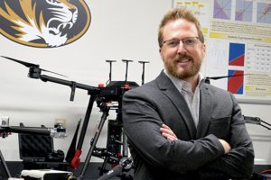
Derek Anderson is a Professor in the Department of Electrical Engineering and Computer Science at the University of Missouri, within the College of Engineering. He is a core faculty member of the MU Institute for Data Science & Informatics and leads the Mizzou INformation and Data FUsion Laboratory (MINDFUL). His research spans artificial intelligence, machine learning, information fusion, computer vision, and remote sensing, with applications in defense, healthcare, and humanitarian technology. Education: PhD, University of Missouri MS, University of Missouri BS, Wichita State University Anderson's research focuses on foundational AI challenges, particularly explainable AI (XAI) and the use of simulated data to train robust models under uncertainty. He investigates how AI systems make decisions and how to communicate those decisions clearly to humans, especially in high-stakes environments like medical imaging and autonomous vehicles. His work leverages tools like Unreal Engine and Infinite Studio to generate photorealistic and multispectral synthetic data, enabling broader and more diverse training sets than real-world data alone can provide. His recent publications and projects highlight trends in generative AI limitations , AI ethics , infrared sensor innovation , and AI for humanitarian demining . These works collectively emphasize transparency, robustness, and societal benefit in AI development. Scientific Awards and Leadership: Program Co-Chair, three national AI conferences (2023) Member, IEEE USA AI Committee Member, IEEE CIS Industry & Government (I&GA) Committee Member, IEEE Vertical on Societal Implications of AI Anderson is deeply committed to mentoring the next generation of AI scientists. He advises undergraduate, master’s, and PhD students, many of whom engage in hands-on research from their first year. His lab has secured approximately $29 million in funding across 25 projects from agencies like the U.S. Army ERDC and the National Science Foundation. He has published over 190 articles and is a sought-after leader in the AI research community. His lab, MINDFUL, focuses on information fusion and AI under uncertainty, with active projects in drone-based sensing, material design, and geospatial analytics. The team uses simulation as a core methodology to advance AI trustworthiness and performance across diverse domains.
Carolyn Reilly is a Professor and Director of the Division of Nursing at Berry College, where she leads academic and research initiatives grounded in caring, compassion, and creativity. She plays a pivotal role in shaping nursing education and advancing clinical research in cardiovascular health. Education: Ph.D., University of Nebraska Medical Center M.N., Emory University B.S.N., Brenau College Diploma, Georgia Baptist School of Nursing Her research focuses on heart failure, cardio-oncology, and cardiovascular disease prevention , particularly in cancer survivors. She explores biobehavioral interventions and self-care strategies to improve outcomes in chronic cardiovascular conditions. Her work bridges clinical practice, patient education, and health policy. Recent publications highlight trends in cardio-oncology, heart failure self-management, and the use of AI in cardiac imaging . Her research increasingly integrates interdisciplinary collaboration, health economics, and precision medicine approaches. She contributes to national scientific statements and clinical guidelines, influencing best practices in cardiovascular nursing. Scientific Awards and Honors: Fellow of the American Academy of Nursing (2021) Woodruff Leadership Academy Fellow (2019) Jean Thomas Award for Creativity in Teaching (2017) Nursing Investigator Award, Heart Failure Society of America (2014) Fellow of the American Heart Association (2012) Editorial Board Member, Heart & Lung Journal Dr. Reilly has secured significant grant funding from HRSA, AHA, CDC, and PCORI, serving as principal investigator and coinvestigator on projects related to rural healthcare expansion, congenital heart defect surveillance, and heart failure interventions. She mentors nursing students and leads high-impact research that improves patient care and health equity. Her leadership extends to national organizations and policy development in nursing and cardiovascular health. She directs the Nursing Education Program Expansion at Berry College and is involved in initiatives using high-fidelity simulation to increase nursing graduate capacity, particularly in rural Georgia. Her work emphasizes innovation in teaching and clinical training.
Francisco González García is a Professor in the Department of Surgery and Medical-Surgical Specialties at the University of Santiago de Compostela's Faculty of Medicine and Dentistry. He holds affiliations with the Center for Research in Molecular Medicine and Chronic Diseases (CIMUS) and the Vision Research Group in Ophthalmology and Visual Sciences. His academic career includes doctoral degrees from both the University of Santiago de Compostela (1982) and the University of La Laguna (2001), focusing on visuomotor neuroscience and otolaryngology. Research interests span ophthalmology, neurosciences, and biomedical data science with recent emphasis on retinal physiology, intraocular devices, and stem cell therapies. His work bridges clinical practice and engineering through innovations in tonometry techniques and smart lens development. Notable contributions include studies on prostaglandin-associated periorbitopathy and neuroprotective roles of erythropoietin in retinopathy. Publications (2021–2025) reflect multidisciplinary activity, combining clinical case reports with methodological advancements in neuronal activity modeling and orbital imaging. Collaborative projects like the Green Link initiative demonstrate environmental engineering applications of medical expertise in desertification reversal using fog capture technologies. He has advised numerous studies on ocular pharmacokinetics and thyroid eye disease management, though specific student names aren't listed. His lab work integrates computational tools like P-spline algorithms with traditional microscopy techniques.
Randi Rotne is a Senior Lecturer in Small Animal Surgery at Charles Sturt University, affiliated with the School of Agricultural, Environmental and Veterinary Sciences. She holds a BVSc and PhD, and has over 20 years of clinical experience in veterinary practice, including running her own clinic in the Hawkesbury region. Her academic work spans teaching, research, and clinical supervision, with a focus on surgical education and small animal orthopedics. BVSc, University of Sydney PhD, University of Sydney (2017) – Biomechanical study on locking plates and bone blood supply Her research interests center on small animal surgery , clinical skills training , orthopedic biomechanics , and perioperative complications . She has developed innovative surgical teaching models and investigated outcomes in desexing and fracture repair. Her work bridges clinical practice and academic rigor, emphasizing evidence-based veterinary medicine. The recent articles show a strong focus on small animal surgical outcomes , diagnostic imaging , and educational tools . Key themes include ultrasound-guided procedures, bone healing assessment via CT and histopathology, and neonatal immunity in camelids. Her research integrates clinical data with educational innovation, particularly in surgical training models and pain management strategies. Canine interdigital grass seed foreign body retrieval using ultrasonography (Grant, 2021) Determining the incidence of perioperative acute kidney injury in dogs undergoing routine surgery (External Award, 2017) Investigations into locking plates and bone blood supply (Internal Award, 2016) Winner of early stage HDR presentation, The University of Sydney (2010) Winner of mid stage HDR presentation, The University of Sydney (2012) Randi Rotne actively supervises HDR students and contributes to research grants focused on surgical outcomes and educational tools. She is involved in peer review for journals such as the Journal of Veterinary Medical Education and collaborates with national initiatives like VetCompass Australia. Her work extends to community engagement through public events and media interviews on veterinary health topics. She is actively involved in developing teaching models for ultrasonography and clinical skills training. Her research team collaborates on projects related to surgical innovation, diagnostic validation, and veterinary education. She participates in professional networks such as VetEd Down Under and contributes to curriculum development in veterinary surgical training.
Zhigang Deng is the Moores Professor of Computer Science and Director of Graduate Studies at the University of Houston's Department of Computer Science. He earned his Ph.D. in Computer Science from the University of Southern California (2006), with earlier degrees from Peking University (M.S. 2004) and Xiamen University (B.S. 1997). His research focuses on Human-Centered Computing, Computer Graphics, and Computational Surgery, with notable contributions to insect flight simulation, surgical telementoring, and AI-human collaboration in medical diagnosis. He has been funded by NSF, NIH, NASA, and industry partners like Google and EA. His work is featured in ACM TOG, IEEE TVCG, and SIGGRAPH. Awards include the ACM Distinguished Member (2021), Moores Professorship (2021), and multiple best paper awards. He directs the University of Houston's Computer Graphics and Interactive Media Lab, and serves on editorial boards for IEEE TVCG and Computer Graphics Forum. Teaching includes courses on 3D Graphics and Game Development. Current research projects include surgical simulation frameworks (HySim), farm scene modeling from satellite data, and multimodal conversation analysis in multi-party interactions.
Andreas Enqvist is a Professor in the Department of Materials Science and Engineering at the University of Florida, College of Engineering, where he leads the Enqvist Research Group (LOKE). The group is part of the Nuclear Engineering Program and actively contributes to national and international efforts in nuclear nonproliferation, safeguards, and radiation detection. They are members of the US Department of Energy's National Nuclear Security Administration (NNSA) Consortium for Monitoring, Technology, and Verification (NA-22) and collaborate with NASA on planetary protection research. Their research focuses on the detection and modeling of neutron and gamma-ray signatures from nuclear materials, particularly fresh and spent nuclear fuel. Key areas include nondestructive assay, neutron spectroscopy, detector development (especially He-4 scintillators), data fusion with vision systems, and simulation using tools like MCNPX-PoliMi and SCALE. The group conducts experiments at the University of Florida Training Reactor (UFTR) and the National Criticality Experiments Research Center (NCERC). The recent publications (2020–2025) reflect a strong emphasis on optimizing detector configurations, neutron scatter cameras, pulse shape discrimination using machine learning, dry cask monitoring, and advanced data fusion techniques. There is a clear trend toward integrating physics-based models with AI/ML methods and developing compact, field-deployable systems for nuclear security applications. Member, NNSA NA-22 Consortium for Monitoring, Technology, and Verification Collaborator, NASA Planetary Protection Research Program Principal Investigator, NEUP-funded projects The group mentors several students and researchers, including Cat, Brice, Sai, Jaylen, Nuo, and Joseph. They have secured funding from NNSA and DOE for projects related to nuclear safeguards, verification technologies, and training reactor applications. Their work bridges fundamental nuclear physics with practical engineering solutions for global security and space exploration. The Enqvist Research Group operates within the University of Florida’s Nuclear Engineering infrastructure, utilizing the UF Training Reactor and advanced simulation capabilities. They collaborate with national laboratories and other university teams within the NNSA consortium, forming a key part of a larger network dedicated to advancing nuclear monitoring technologies.
Anne Crijns is a Physician Scientist at the Faculty of Medical Sciences , University of Groningen. Specializing in radiation oncology and hematology, her research focuses on radiation-induced cardiac toxicity in breast cancer patients , with significant contributions to understanding long-term cardiovascular complications post-radiotherapy. Her work explores: Radiation dosimetry impacts on cardiac function Proton therapy for second primary cancer risk reduction AI applications in cardiac structure contouring Biological modeling of radiation-induced atherosclerosis Recent publications highlight collaborations with international experts across Radiology: Cardiothoracic Imaging , Radiotherapy and Oncology , and Clinical and Translational Radiation Oncology . Scientific recognition includes the 2020 Best of ASTRO Abstract Designation . Contact: a.p.g.crijns@umcg.nl
David Lalush is an Associate Professor in the Joint Department of Biomedical Engineering at North Carolina State University and The University of North Carolina at Chapel Hill. His research focuses on biomedical imaging, particularly in PET-MRI integration, X-ray imaging technologies, and applications of machine learning to medical imaging. He holds a Ph.D. in Biomedical Engineering from the University of North Carolina at Chapel Hill, an M.S. in Electrical Engineering from North Carolina State University, and a B.S. in Electrical Engineering from Lehigh University. Key research areas include simultaneous PET-MRI imaging for clinical applications in cancer and neuroscience, respiratory motion correction in upper abdominal PET imaging, and energy-resolved quantitative CT techniques. His lab emphasizes realistic imaging simulations and collaborations with clinical and research institutions. Recent publications highlight advancements in PET-MRI applications, machine learning-driven image synthesis (e.g., GANs), and neurobiological studies of dopamine and anhedonia. Courses taught include BME 412 (Biomedical Signal Processing) and BME 560 (Medical Imaging Systems). His work bridges engineering and clinical needs, with ongoing projects in multimodal imaging, motion correction algorithms, and translational biomedical research.
Dr Carl Roobottom is an Honorary Consultant in Radiology and a key academic figure at the Peninsula Medical School, Faculty of Health, University of Plymouth. He chairs radiology teaching and leads the Radiology Academy at Derriford Hospital, a state-of-the-art postgraduate training facility dedicated to developing future radiology consultants. His work focuses on integrating cross-sectional imaging into undergraduate anatomy education and advancing postgraduate radiology training through innovative methods, including simulation technology. The Radiology Academy has been nationally recognized as a model of excellence in specialist training. His clinical and academic research centers on improving patient care through advanced imaging techniques, particularly in cardiovascular diagnostics. A highlighted REF Impact Case Study demonstrates his contribution to enhancing safety, health outcomes, and cost-effectiveness in chest pain patients using Computed Tomographic Cardiac Angiography (CTCA). Dr Roobottom has played a pivotal role in shaping radiology education both at the undergraduate and postgraduate levels. His leadership in establishing a dedicated radiology training academy underscores his commitment to medical education and clinical excellence. The Radiology Academy, located at Derriford Hospital, features a 140-seat lecture theatre and employs novel teaching methodologies, earning praise from national health leaders, including Marin Marshall, Deputy Chief Medical Officer, who described it in 2008 as a "shining light for all specialities."
Alexander JE Raaijmakers is an Assistant Professor with a joint appointment at Eindhoven University of Technology (Department of Biomedical Engineering, Medical Image Analysis group) and University Medical Center Utrecht (7 Tesla Research group, Radiology Department). His research focuses on RF engineering for ultrahigh field MRI, including antenna design, body imaging, and RF safety protocols. He leads multiple projects involving 5 PhD students and 1 postdoc. Education: MSc in Applied Physics, University of Groningen (2004) PhD in Radiotherapy Physics, University Medical Center Utrecht (2008) Research Focus: His work bridges electromagnetic physics with clinical MRI applications, developing hardware/software solutions for ultrahigh field systems. Key innovations include dipole antenna arrays for MRI and safety frameworks for medical implants. Recent work integrates deep learning for image correction. Awards: Casimir Grant (2008-2010) for RF coil development at Philips Medical Systems Leadership: Directs the 7T Research Group at UMC Utrecht and collaborates on MRI education at TU/e. His team focuses on clinical translation for cardiac, prostate, and abdominal imaging.