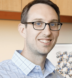
Prof. Knut Drescher is an Associate Professor at the Biozentrum, University of Basel , leading a research group focused on bacterial biofilms , swarming , and microbial multicellularity . Previously, he served as a Professor of Biophysics and Max Planck Research Group Leader at Philipps-Universität Marburg (2015-2021) and conducted postdoctoral research at Princeton University. Research Interests: Physical and biological mechanisms of biofilm formation Cell-cell interactions in microbial communities Antibiotic resistance in biofilms Hydrodynamics of bacterial swarms Evolution of cooperation in multispecies biofilms Development of bioimaging software (BiofilmQ, BacStalk) Scientific Awards: 2023: SNSF Consolidator Grant 2019: Heinz Maier-Leibnitz Prize (DFG), VAAM Research Prize, IUPAP Young Scientist Prize 2016: ERC Starting Grant Advising & Grants: Advises PhD and Master's students in microbiology, biophysics, and bioinformatics Secured major grants from ERC , HFSP , and DFG
Professor Hala Zreiqat AM is a leading biomedical engineer at The University of Sydney , serving as the Director of the ARC Training Centre for Innovative BioEngineering . A Fellow of all major Australian academies (AAS, ATSE, FAHMS, FRSN), she develops 3D printed bioceramics for bone regeneration while championing diversity through initiatives like the IDEAL Society and BIOTech Futures mentorship program. Her work bridges academia, clinical practice, and industry in musculoskeletal research . Research Focus: Her lab creates synthetic bone scaffolds that mimic natural bone architecture, strength, and porosity, enabling non-rejected bone regeneration via patient-matched implants. Key applications include orthopaedic, dental, and maxillofacial repair , with over $18M in competitive funding and multiple patents. Current projects explore AI-driven scaffold performance prediction and anti-senescence strategies for aging-related bone loss. Scientific Trends: Recent publications highlight 3D printed nanovoxelated ceramics , antisenescence biomaterials , and multifunctional theranostic platforms . Her team integrates machine learning for scaffold design, atom probe tomography for interface analysis, and two-photon imaging for cellular monitoring in 3D environments. 2021-2022 Fulbright Senior Scholar 2018 NSW Premier's Woman of the Year 2019 Eureka Prize for Innovative Use of Technology Fellow of Australian Academy of Science (2021) Over $18M in research funding Teaching & Leadership: She designed core courses like Tissue Engineering and Nanomaterials in Medicine , mentoring 158 students in 2020 alone. As Chair of CAAR (2020-2023), she strengthens Australia-Arab collaborations. Her lab trains early-career researchers , with alumni now in academia and industry.
Scott T. Acton is the Lawrence R. Quarles Professor and Chair of Electrical and Computer Engineering at the University of Virginia, with a courtesy appointment in Biomedical Engineering. He leads the VIVA lab, specializing in biological image analysis, machine learning, and AI for education. His research spans medical imaging, signal processing, and computer vision. Professor Acton holds a B.S. (Virginia Tech, 1988), M.S. (UT Austin, 1990), and Ph.D. (UT Austin, 1993) in Electrical Engineering. He has authored over 325 publications and served as Editor-in-Chief of IEEE Transactions on Image Processing and General Co-Chair of the IEEE International Symposium on Biomedical Imaging. His research interests include bioimage analysis, machine learning applications, and medical imaging technologies. The VIVA lab focuses on problems like cell tracking in bacterial biofilms, gait recognition using LiDAR, and AI-driven classroom activity analysis. Awards: IEEE Fellow (2013), All-University Teaching Award (2009), Outstanding Young Electrical Engineer (1996). Courses Taught: How the iPhone Works, Digital Image Processing, Signals and Systems. Labs/Teams: VIVA - Virginia Image and Video Analysis lab. Recent work emphasizes AI for education (e.g., automated classroom activity classification) and medical imaging advancements like 3D biofilm segmentation and LiDAR-based human identification. His contributions bridge engineering and healthcare, with applications in neuroscience and clinical decision support.
Ashis K. Patra is a full-time Professor in the Department of Chemistry at the Indian Institute of Technology Kanpur (IIT Kanpur). After obtaining his Ph.D. from the Indian Institute of Science (IISc) Bangalore in 2008, he carried out post-doctoral research at Harvard University and the University of Georgia before joining IIT Kanpur in 2012. Education & Academic Training Ph.D. (2008), Indian Institute of Science (IISc), Bangalore M.Sc. (2002), The University of Burdwan Research Interests Professor Patra leads an interdisciplinary program that sits at the interface of inorganic chemistry, chemical biology, and medicinal chemistry. His work is organized into three principal thrusts: Therapeutic Applications of Metal Complexes: Design and mechanistic evaluation of cytotoxic transition-metal complexes that target nucleic acids and proteins, aiming to overcome multidrug resistance in cancer. Nitric Oxide Delivery from Transition Metal Nitrosyls: Development of photo- and redox-triggered metal nitrosyl complexes for controlled NO release to biological targets such as hemoglobin, myoglobin, and glutathione. Luminescent Lanthanide Complexes: Synthesis and photophysical characterization of Eu(III) and Tb(III) complexes that serve as luminescent probes and theranostic agents. Research Output & Impact His group has published extensively in leading journals including Dalton Transactions , Inorganic Chemistry , Chemical Science , and Journal of Inorganic Biochemistry . The collective work demonstrates a clear trajectory from fundamental coordination chemistry to translational applications in drug delivery and bioimaging, with recurring themes of redox control, light activation, and biological targeting. Scientific Awards & Fellowships CSIR Junior & Senior Research Fellowships (2001, 2003) West Bengal SLET Qualification (2001) International Travel Grants from CSIR & INSA (2006) DST Fast-Track Fellowship for Young Scientists (2013) Erasmus Mundus NAMASTE Scholarship for Academic Staff (2014) JSPS Invitation Fellowship (2016) Teaching & Mentoring Professor Patra teaches core and advanced courses in inorganic chemistry, including Inorganic Chemistry Laboratory, Bioinorganic Chemistry, and Frontiers in Inorganic Chemistry. He actively mentors Ph.D. and master’s students (full list available on his lab website ) and has established the Laboratory of Inorganic Chemical Biology in the Old Core Lab complex at IIT Kanpur.
Aysegul Gunduz, Ph.D., is a Professor and Fixel Brain Mapping Professor at the University of Florida's Herbert Wertheim College of Engineering, Department of Biomedical Engineering. She leads the Brain Mapping Laboratory, focusing on neural networks and clinical translation for neurological disorders. Her work integrates electrophysiology, bioimaging, and neuromodulation to develop diagnostic and therapeutic systems for conditions like Parkinson’s disease, epilepsy, movement disorders, and stroke. Education: B.S., Electrical Engineering, Middle East Technical University (2001) M.S., Electrical Engineering, North Carolina State University (2003) Ph.D., Electrical Engineering, University of Florida (2008) Post-doctoral Fellowship in Neurology, Albany Medical College (2011) Research interests include human brain mapping, closed-loop deep brain stimulation (DBS), neuromodulation strategies for movement disorders, and wearable sensor technologies for neurological monitoring. Her lab emphasizes translational research, bridging basic science with clinical applications to improve patient outcomes. Awards include the BMES Fellowship (2024), AIMBE Fellowship (2022), and PECASE (2019), reflecting her leadership in neural engineering. Her articles explore cutting-edge topics like DBS efficacy, neural network dynamics, and ethical considerations in neural device research. Grants and collaborations focus on advancing adaptive DBS and brain-computer interfaces. She mentors students in neuroengineering and advocates for equitable participation in clinical research. The Brain Mapping Laboratory actively engages in multidisciplinary projects with neurologists, surgeons, and industry partners. Future work includes optimizing closed-loop systems for Tourette syndrome and Parkinson’s disease, developing open-source neuroimaging tools, and expanding wearable sensor applications for real-time neurological monitoring.
Nasir M. Rajpoot is a Professor in the Department of Computer Science at the University of Warwick, UK. His research focuses on computational pathology, medical image analysis, and deep learning applications in histology. He leads interdisciplinary projects integrating artificial intelligence with healthcare, particularly in cancer diagnostics and pathology workflows. Rajpoot’s work emphasizes developing robust algorithms for histology image analysis, including nuclear segmentation, tumor classification, and domain generalization in computational pathology. His contributions include the TIAToolbox, an open-source framework for tissue image analytics, and the CoNIC Challenge to advance nuclear detection and counting in histology images. He collaborates with clinicians and biologists to translate AI models into clinical practice, addressing challenges like tumor heterogeneity and staining variability. Rajpoot’s research spans colorectal, lung, and oral cancers, with a focus on predicting clinical outcomes via histological features and genomic data integration. Notable projects include the development of Handcrafted Histological Transformer (H2T) for unsupervised representations of whole slide images and the SAFRON framework for histology image synthesis. His work addresses domain adaptation, robustness evaluation, and explainability in AI-driven pathology systems.
Lawrence Staib is Professor of Radiology and Biomedical Imaging, Biomedical Engineering, and Electrical Engineering at Yale University. He serves as Director of Undergraduate Studies in Biomedical Engineering and is a member of Yale's Bioimaging Sciences division, Image Processing & Analysis Group, Yale Biomedical Imaging Institute, and Yale-BI Biomedical Data Science Fellowship program. Dr. Staib earned his A.B. in Physics from Cornell University (1982), followed by a Ph.D. in Engineering and Applied Science from Yale University (1990), and completed a postdoctoral fellowship at Yale School of Medicine (1991). His research focuses on developing advanced medical image analysis methods using machine learning and model-based approaches. Key research areas include neuroimaging applications for autism spectrum disorder classification, cardiac imaging analysis for strain and motion assessment, prostate cancer diagnosis and risk mapping, and innovative techniques for medical image segmentation with limited labeled data. Dr. Staib's work emphasizes uncertainty estimation in deep learning models, multi-modal image registration, and domain adaptation techniques to improve clinical decision support systems. His recent publications demonstrate a strong trend toward developing interpretable AI models for clinical applications, with particular emphasis on fMRI analysis for neurological conditions, cardiac motion analysis, and prostate cancer diagnosis. His work frequently addresses the challenge of limited labeled data in medical imaging through innovative self-supervised, semi-supervised, and few-shot learning approaches. Fellow of the American Institute for Medical and Biological Engineering (AIMBE) (2015) Distinguished Investigator Award from the Academy for Radiology & Biomedical Imaging Research (2017) MICCAI Fellow (2022) Medical Image Analysis Second Best MICCAI Paper Award (2005) ASNR Cum Laude Scientific Exhibit Award (2003) Dr. Staib serves on the editorial board of Medical Image Analysis and as Associate Editor of IEEE Transactions on Biomedical Engineering. His research is supported by NIH grants including the Autism Center of Excellence program. He leads the Image Processing & Analysis Group within Yale's Bioimaging Sciences division, collaborating extensively with James Duncan, John Onofrey, Xenophon Papademetris, and other Yale researchers on applications spanning neuroimaging, cardiology, and oncology. Current projects focus on developing robust AI models for clinical decision support with emphasis on uncertainty quantification and interpretability.
Prof. Dr. rer. nat. Lothar Elling is a University Professor and director at the Helmholtz Institute for Biomedical Engineering, RWTH Aachen University, Germany. His research focuses on biomaterials, glycoengineering, and enzymatic synthesis of carbohydrates and glycoconjugates. Institution: RWTH Aachen University Research Unit: Helmholtz Institute for Biomedical Engineering Academic Rank: Full Professor Prof. Elling's research interests include: Glycoengineering of biomaterials Enzymatic synthesis of glycans Glycosyltransferase immobilization Glycan-protein interactions Biocatalytic cascade reactions Biomedical applications of glycomaterials His recent publications demonstrate strong expertise in: - Automated enzymatic glycan synthesis - Multi-enzyme cascade systems for nucleotide sugar production - Glycosyltransferase engineering - Galectin-targeted glycomaterials - Microgel-based biosensors
John Capobianco, PhD, is a Professor in the Department of Chemistry and Biochemistry at Concordia University and holds the Honorary Concordia University Research Chair in Nanoscience. His research focuses on lanthanide-doped nanoparticles, upconversion luminescence, and biomedical applications. PhD, University of Geneva Key research areas include: Nanomaterials synthesis and spectroscopy Upconversion for biomedical imaging Drug delivery systems Photodynamic therapy for cancer treatment Optical thermometry and sensing Recent publications highlight advancements in: X-ray detection via photochromic nanoparticles Lipid-coated nanoparticles for lung permeation Cooperative energy transfer in Yb3+/Eu3+ complexes Biocompatible nanomaterials for secure information storage Pr3+-doped radiosensitizers for glioblastoma therapy Scientific recognitions: Honorary Concordia University Research Chair in Nanoscience Teaching includes undergraduate and graduate courses in inorganic chemistry and spectroscopy. His work bridges fundamental material science with applied biomedical engineering, emphasizing optical properties and therapeutic applications of lanthanide-based nanomaterials.
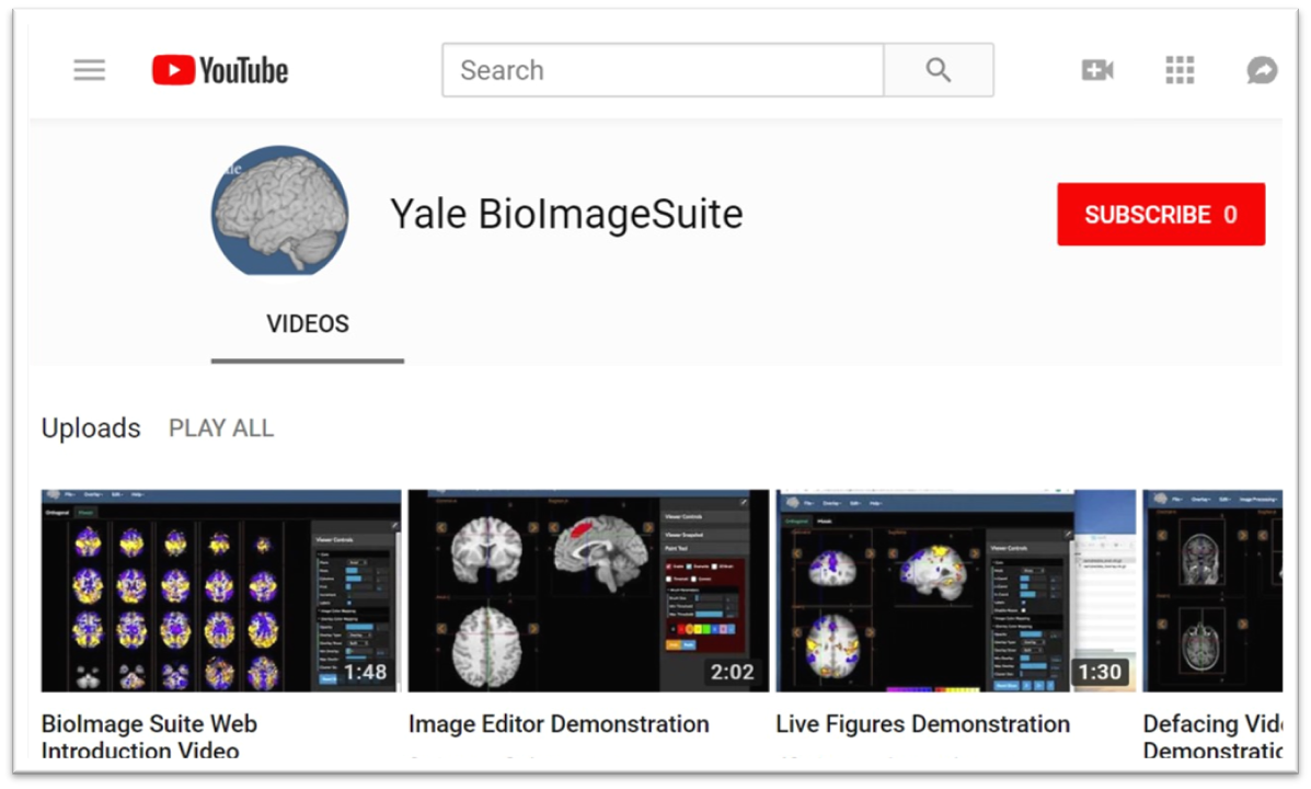
Xenophon Papademetris is a Professor of Biomedical Informatics & Data Science and Radiology & Biomedical Imaging at Yale School of Medicine. He serves as Associate Director of Biomedical Imaging Data Sciences at Yale Biomedical Imaging Institute and directs the Medical Software and Medical Artificial Intelligence Certificate Program. PhD in Electrical and Information Sciences from Yale University (2000) BA from Cambridge University (1994) Postdoctoral Fellowship at Yale University (2002) His research focuses on medical image analysis, machine learning, and biomedical software development. He has developed tools like BioImage Suite Web and contributed to standards committees at the Association for the Advancement of Medical Instrumentation (AAMI). His work spans modalities including MRI, CT, PET, and optical imaging. Recent publications emphasize neuroimaging analysis, explainable AI in healthcare, and multimodal data integration across species. He leads NIH-funded research under the BRAIN Initiative (R24 MH114805) and has authored a textbook on Medical Software published by Cambridge University Press. IEEE Senior Member Yale Brown-Coxe Postdoctoral Fellowship Harding Bliss Prize for Excellence in Engineering He directs the BioImage Suite Project, creating web-based image analysis tools using JavaScript and WebAssembly. His teaching includes both academic courses and a Coursera program on Medical Software with over 14,000 enrollments.
David A. Muller serves as the Samuel B. Eckert Professor of Engineering in the School of Applied and Engineering Physics at Cornell University and co-directs the Kavli Institute at Cornell for Nanoscale Science. His research group focuses on developing quantitative electron microscopy methods to understand materials properties at the atomic scale, with particular emphasis on sustainable energy applications and quantum materials. Muller's laboratory utilizes some of the world's highest resolution electron microscopes housed in specially designed, environmentally isolated rooms. Muller received his undergraduate education at the University of Sydney and earned his Ph.D. in Physics from Cornell University in 1996. Between 1997 and 2003, he was a member of the technical staff at Bell Laboratories, where he applied his expertise in imaging single atoms and atomic-scale spectroscopy to determine the physical limits of transistor miniaturization. In 2003, he returned to Cornell as a faculty member, where he has since established himself as a leader in advanced electron microscopy techniques. Muller's research spans multiple frontiers in materials science, with particular focus on understanding how electronic-structure changes at the atomic scale control macroscopic behavior in diverse systems like turbine blades, fuel cells, and transistors. His current work emphasizes the physics of renewable energy materials, atomic-scale control of materials to create electronic phases that cannot exist in bulk, and developing hardware and algorithms for 'big data' acquisition from high-bandwidth pixelated electron microscope detectors. His group's work bridges theoretical physics and experimental techniques, requiring researchers who can think in both real and reciprocal space while considering both fundamental principles and practical applications. Analysis of Muller's recent publications reveals a strong trend toward advancing electron ptychography and 4D-STEM techniques for atomic-scale imaging. His group has pioneered methods for 3D atomic-scale metrology, strain mapping, and imaging of radiation-sensitive materials. The research spans applications from semiconductor technology to quantum materials and energy storage systems, demonstrating the versatility of his microscopy approaches across multiple scientific domains. Top 100 Young Innovator by Tech Review Magazine (2003) Burton Medal from Microscopy Society of America (2006) Ernst Ruska Prize of German Society for Electron Microscopy (2021) John Cowley Medal from International Federation of Societies for Microscopy (2023) Fellow of American Physical Society Fellow of American Association for the Advancement of Science Fellow of Microscopy Society of America Muller has mentored an extensive group of students and postdocs who have gone on to successful careers in academia and industry. His former students hold faculty positions at institutions including Rice University, University of Southern California, Seoul National University, Colorado School of Mines, and the University of Michigan, among others. His research has been supported by substantial grants, including a $22.5M NSF grant that accelerates materials discovery. The Muller lab maintains close collaborations with the Kavli Institute at Cornell and PARADIM (Platform for the Accelerated Realization, Analysis, and Discovery of Interface Materials). The Muller lab operates at the forefront of electron microscopy, housing specialized instrumentation including high-resolution transmission electron microscopes in environmentally isolated rooms. The group collaborates extensively with other research teams at Cornell and worldwide, focusing on understanding materials atom by atom. Current research directions include applying machine learning to electron microscopy data analysis, developing cryogenic techniques for studying low-melting-point materials, and exploring quantum phenomena in engineered materials systems.
Jakoah Brgoch is an Assistant Professor in the Department of Chemistry at the University of Houston. His research focuses on leveraging machine learning to design inorganic compounds for applications in LED-based lighting and superhard materials. Key areas include phosphor development, sparse data handling, and predicting material formation. He leads the Brgoch Group, which emphasizes interdisciplinary approaches combining computational modeling and experimental synthesis. Research interests span luminescent materials, crystal chemistry, and defect engineering, with a particular emphasis on optimizing phosphors for solid-state lighting and high-performance materials under extreme conditions. His work bridges data science and traditional materials discovery to accelerate innovation in optoelectronics and mechanical materials. Recent publications highlight advancements in cyan-emitting nitridation processes, machine learning-guided phosphor discovery, and understanding oxidation resistance in silicides. His team has developed novel phosphors like Na2CaZr2Ge3O12:Cr³⁺ for NIR bioimaging and explored luminescent properties of Sr-based solid solutions. Active in translational research, Dr. Brgoch collaborates on applications like smartphone-readable diagnostic platforms using nanophosphors and point-of-care testing. His lab emphasizes open science practices and has pioneered methods like Single-crystal automated refinement (SCAR) for structural determination.
Daniel Sage is a Lecturer and Scientific Advisor at École polytechnique fédérale de Lausanne (EPFL) , affiliated with the Biomedical Imaging Laboratory (LIB) under the College of Engineering (STI) and School of Life Sciences (SV) . He specializes in bioimage informatics , structured-illumination microscopy , and deep learning applications for biomedical imaging. His work spans algorithm development for single-molecule localization microscopy (SMLM) , fluorescence imaging , and 3D reconstruction . His research group has developed open-source tools like FlexSIM for light inhomogeneity correction, DeepImageJ for integrating deep learning in ImageJ, and Steer'n'Detect for orientation-accurate template detection. His publications focus on correcting multiple-blinking artifacts in PALM, optimal transport metrics for SMLM evaluation, and contextual feature analysis for xenograft cell classification. He mentors PhD students and contributes to interdisciplinary education through courses such as Bioimage Informatics and Fundamentals of Image Analysis , emphasizing practical software solutions and Java programming for bioimage processing. His collaborations include institutions like Howard Hughes Medical Institute and Centre National de la Recherche Scientifique (CNRS) .
Ueli Grossniklaus is an Ordinary Professor at the University of Zurich within the Faculty of Mathematical and Natural Sciences , affiliated with the Department of Plant and Microbiology . His work focuses on plant developmental biology, particularly epigenetic and genetic mechanisms governing reproduction and adaptation. Key Courses: Epigenetics, Plant Biology Workshop, Group Seminars on Current Research Laboratory Techniques: Advanced methods in plant cell mechanics, transcriptomics, and genome editing Research Interests span plant epigenetics, reproductive biology, and the interplay between environmental stress and genetic regulation. He investigates: Mechanistic control of gametogenesis and fertilization Epigenetic contributions to plant adaptation Evolutionary implications of asexual reproduction Biophysical forces in plant cell growth Publication Trends (2025–2018) reveal expertise in: Arabidopsis and fern model systems Epigenetic regulation (DNA methylation, histone dynamics) Apomixis and hybrid seed failure mechanisms Biomechanics of pollen tubes and carnivorous plants Genome editing tools (CRISPR) and long-read sequencing Scientific Collaborations include interdisciplinary projects on: Microfluidic devices for plant cell analysis Gene drive ecology and ethics 3D imaging of plant reproductive structures Advising and Grants focus on mentoring through research internships in developmental biology, genetics, and systems biology. His lab engages in: Epigenetic response to environmental stress Cell wall mechanics in reproduction Computational modeling of plant growth Laboratory Teams integrate plant biologists, bioengineers, and computational scientists to study: Mechanistic gene regulation Evolutionary developmental biology Microrobotics for cellular force measurement
Privatdozent Dr. Andreas Faust is a leading researcher at the European Institute for Molecular Imaging (EIMI) at the University of Münster, where he heads the Chemical Targeting Lab. His work focuses on developing innovative imaging agents for medical diagnostics, particularly in radiopharmaceutical chemistry and molecular imaging. He maintains strong affiliations with the Department of Nuclear Medicine at the University Hospital Münster and participates in the "Cells in Motion" excellence cluster, contributing to cutting-edge research at the intersection of chemistry, medicine, and imaging technology. Dr. Faust completed his chemistry studies at the University of Münster, earning his Diploma in 1999, followed by his doctoral degree (Dr. rer. nat.) in 2003 with research on artificial caffeine receptors. His academic journey continued with positions at the Department of Organic Chemistry and the Department of Nuclear Medicine before becoming head of the chemistry group at EIMI in 2011. Dr. Faust's research centers on organic and medicinal chemistry with specialization in radiopharmaceutical chemistry . His team develops novel tracers for diagnostic molecular imaging using positron emission tomography (PET), single-photon emission computed tomography (SPECT), optical imaging, and photoacoustic imaging. A significant portion of his work focuses on creating specific ligands for the alarmins S100A8/S100A9 and bacteria-specific tracers based on complex carbohydrates or siderophores. His research has important applications in inflammation imaging, infection diagnostics, and cancer theranostics, with emphasis on improving metabolic stability and target specificity of imaging agents. His publication record demonstrates consistent contributions to molecular imaging, with recent work emphasizing bacteria-specific PET tracers, inflammation imaging targeting S100 proteins, and novel optical imaging probes. The research shows a clear trajectory toward developing clinically applicable imaging agents with improved specificity and metabolic stability, particularly in the areas of infection diagnostics and inflammation monitoring. 2017: Best Poster Award at Symposium "Molecular Imaging Agents in Medicine," Groningen 2009: Young Investigator Award at Deutscher Röntgenkongress, Berlin 2005: Best Scientific Poster Award at 4th Annual Meeting of the Society of Molecular Imaging, Köln Dr. Faust leads multiple significant research projects, including as Coordinator of a project on immune cell distribution imaging (2019-2024) and as Principal Investigator for CRC-project A03 "Targeting of S100A8/A9 for imaging of inflammatory disorders" and research on vascular graft infections (both 2021-2024). His Chemical Targeting Lab comprises a multidisciplinary team working at the intersection of chemistry, microbiology, and medical imaging, securing substantial funding from the Innovative Medicines Initiative and DFG Collaborative Research Centre. The Chemical Targeting Lab maintains state-of-the-art facilities for chemical synthesis, radiochemistry, and biological testing. The lab collaborates extensively with microbiologists, clinicians, and imaging specialists to translate basic research into clinical applications. Current research directions include optimizing bacterial imaging probes for clinical diagnostics and developing new inflammation-specific tracers for early disease detection, with particular focus on S100A9-targeted imaging and siderophore-based bacterial detection systems.