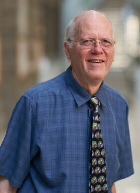
David Van Essen is the Alumni Endowed Professor of Neuroscience at Washington University School of Medicine in St. Louis. His primary affiliation is with the Department of Neuroscience within the School of Medicine. He is renowned for his leadership in the Human Connectome Project (HCP), focusing on advanced neuroimaging and brain mapping techniques. His research spans cerebral cortex structure, function, connectivity, and evolution in humans and nonhuman primates. Education: BS (Honors) in Chemistry from California Institute of Technology (1967), PhD in Neurobiology from Harvard Medical School (1971), followed by postdoctoral research at Harvard and the University of Oslo. Research Interests: Dr. Van Essen’s work emphasizes neuroimaging innovations, cortical parcellation, and the integration of multimodal data. His lab develops computational tools for brain mapping and neuroinformatics. Key projects include the HCP, studying cortical folding, and analyzing connectivity in human and nonhuman primate brains. Publications: Over 200 peer-reviewed articles, including seminal works on cortical parcellation, functional connectivity, and neuroimaging methodologies. Recent trends focus on advancing MRI techniques, connectomics, and understanding brain aging. Awards: Multiple prestigious honors, including membership in the National Academy of Sciences and the Glass Brain Award. Recognized for contributions to neuroscience and neuroimaging. Labs/Teams: The Van Essen Lab at Washington University, collaborating with global institutions on neuroimaging and connectome projects.
Salomi Asaridou is a Lecturer at the University of Oxford's Department of Experimental Psychology and a Stipendiary Lecturer in Psychology at St Anne's College. She holds a PhD from the Max Planck Institute for Psycholinguistics and Donders Institute for Brain, Cognition and Behaviour, supported by a Max Planck Society fellowship. Her research focuses on individual variation in language learning and its neural correlates, particularly in children with developmental language disorder (DLD). She investigates how brain structure and function relate to language abilities in monolinguals, bilinguals, and clinical populations. Education: Bachelor’s in Psychology (Aristotle University of Thessaloniki) Master’s in Neuroscience and Education (Columbia University, Fulbright Scholar) PhD in Psycholinguistics (Max Planck Institute) Research Groups: Brain, Speech, & Language; MRC-funded BOLD study. Her work explores neural mechanisms underlying language disorders and brain reorganization in cases of congenital hemispheric defects. She has contributed to NIH-funded projects at the University of California, Irvine, and uses neuroimaging techniques to study cortical and subcortical structures in language development. Awards include the Fulbright Scholarship and Max Planck IMPRS Fellowship. Salomi teaches Experimental Design and Methods at Oxford and actively collaborates with interdisciplinary teams to advance understanding of language neurobiology.
Dr. Qianqian Yang is a Senior Lecturer in applied and computational mathematics at the School of Mathematical Sciences, Queensland University of Technology (QUT). She holds a PhD in computational mathematics from QUT (2010) and has been recognized with prestigious awards such as the QUT Outstanding Doctoral Thesis Award (2010) and an ARC DECRA fellowship (2014). Her research focuses on fractional differential equations, numerical methods, and their applications in medical imaging, particularly diffusion MRI. She has secured major grants, including an ARC DECRA fellowship (2015–2022, part-time) and an ARC Discovery Project (2019–2022). Dr. Yang’s work bridges computational mathematics and biomedical applications, aiming to model brain tissue microstructure using fractional models. She teaches computational mathematics units and has supervised 2 postdocs, 3 PhDs, and numerous Honours and VRES students. Her research is published in top journals like NeuroImage and SIAM Journal on Scientific Computing. Expertise: Fractional PDEs, Numerical Analysis, MRI Modeling Grants: ARC DECRA, ARC Discovery Project
Juan Arellano is a Research Scientist in the Department of Neuroscience at Yale School of Medicine. His research focuses on cortical development, hippocampal neurogenesis, and neurodegenerative mechanisms in primates. Education: PhD in Neuroscience (Complutense University of Madrid) Current Research: Cortical Development, Tau Protein Pathology, Calcium Signaling in Aging Research Themes: Structural and functional organization of the cerebral cortex Mechanisms of tau hyperphosphorylation in aging primates Role of calcium signaling in cognitive decline Comparative neurobiology of hippocampal neurogenesis Genomic regulation of cortical development Neurodegenerative disease modeling Recent Collaborations include work with the Arnsten Lab and Rakic Lab, focusing on primate neuroanatomy and molecular mechanisms of cognition.
Professor Kevin Keay is a Professor of Neuroscience and Chief Anatomist at the University of Sydney's School of Medical Sciences, part of the Faculty of Medicine and Health. He serves as Deputy Head of School and Theme Leader for Neuroscience, with membership in the Brain and Mind Centre. His academic qualifications include a BSc(Hons), GradDipScMed(Pain Management), and PhD. Research focuses on pain mechanisms, neuroimmunology, and anatomical education. Key interests include chronic neuropathic pain, glial-neuronal interactions, and translational studies across species. He investigates placebo/nocebo effects, brainstem pain modulation, and the role of PACAP/VIP neuropeptides in nervous system disorders. Recent work explores sex differences in pain responses and the impact of spinal cord injury on neuroinflammation. Publications span 40+ years, emphasizing neuroanatomical imaging (7T fMRI), rodent behavioral models, and medical education innovations. Notable contributions include studies on body donor programs, cadaver-based learning, and global health initiatives through international anatomy partnerships. Research also addresses pandemic-era student perceptions and cultural competency training. Grants include NHMRC funding for pain mechanisms (2020), equipment grants for advanced imaging (2002-2013), and educational innovation grants. Advises current PhD students on topics like viral gene therapy for genetic disorders (Christianson Syndrome) and trigeminal neuropathic pain mechanisms. Labs/groups include collaborations with Brain and Mind Centre researchers, neuroimaging teams, and international anatomy departments. Active in promoting interdisciplinary research through interprofessional learning and global student exchanges.
Todd Vanderah is a Regents Professor at the University of Arizona , affiliated with multiple departments including Pharmacology , Anesthesiology , Neurology , and Neuroscience . He serves as Co-Director of the MD/PhD Dual Degree Program , Department Head of Pharmacology , and Director of the Comprehensive Pain and Addiction Center . His research focuses on pain mechanisms, opioid tolerance, endogenous opioid systems, and neuroinflammatory pathways. Leadership Roles : Pharmacology Executive Committee, Dean's Council on Faculty Affairs, Director of Medical Pharmacology Graduate Program Key Research Areas : Chronic pain, cannabinoid-opioid synergy, blood-brain barrier integrity, neuroimmune signaling Recent publications highlight his work on opioid-cannabinoid interactions , sphingosine-1-phosphate pathways , and novel pain models (e.g., red light exposure, DAGLα inhibition). His studies frequently employ rodent models to explore pharmacological interventions for neuropathic pain, traumatic brain injury, and opioid-induced dysregulation. Findings from Vanderah's lab have advanced understanding of CRMP2 phosphorylation , NHE1 regulation , and neuroinflammatory mediators in pain states. Collaborations span pharmacology , neuroscience , and oncology , with implications for drug discovery and clinical pain management.
Lijing Xin is an Assistant Professor at the Department of Physics and a research staff scientist at the Center for Biomedical Imaging (CIBM) at Ecole polytechnique fédérale de Lausanne (EPFL), Switzerland. She teaches courses on Biomedical Imaging and Translational MR Neuroimaging, while also contributing to academic administration. PhD in Physics (2010, EPFL) Master's Project (2002-2005) on MRI instrumentation Her research focuses on high-field magnetic resonance spectroscopy (MRS) and MRI for studying brain function and neurological diseases. She develops novel acquisition and quantification methods for 1 H, 13 C, and 31 P nuclei, particularly on 7T clinical platforms . Her work bridges preclinical and clinical research , with collaborations in psychiatry to explore pathophysiology and biomarkers for disorders like schizophrenia and mood disorders. Recent publications include studies on epilepsy , Alzheimer's disease , brain energy metabolism , and neurochemical profiling using advanced MRS and deep learning for psychosis classification. She has contributed to RF coil design , macromolecule suppression , and metabolic pathway analysis across multiple disciplines. She advises PhD students and collaborates on interdisciplinary projects involving neuroimaging hardware , metabolic disease research , and psychiatric biomarker identification . Her lab at EPFL CIBM-AIT develops cutting-edge techniques for high-resolution brain metabolism analysis and clinical translation .
Pingchuan Zhang, M.D., Ph.D., is a faculty member at the Mayo Clinic College of Medicine in the Department of Laboratory Medicine and Pathology . He is based in Rochester, Minnesota, and specializes in Renal Pathology and Cardiovascular Surgery . His work spans Kidney Transplantation , Glomerulonephritis , and Tissue Engineering for cardiac repair. Education: Ph.D. in Surgery, Peking Union Medical College (2006) Masters in Surgery, Sun Yat-sen University (2003) MD, Peking University Medical School (1997) Dr. Zhang's research focuses on Renal Immunopathology (e.g., IgG4-related nephritis, monoclonal immunoglobulin deposits), Diagnostic Accuracy in kidney biopsies (mass spectrometry vs. immunofluorescence), and Cardiovascular Regeneration using biopolymers and progenitor cells. His publications highlight collaborations with leading institutions like Mayo Clinic and University of Ottawa. Article trends show emphasis on Renal Allograft Monitoring , Autoimmune Kidney Diseases , and Advanced Diagnostic Protocols . Key subfields include Transplant Prognostication , Mass Spectrometry Applications , and Cardiac Biomaterials . Scientific Awards: 2nd place podium presentation, Texas Society of Pathologists Annual Meeting (2013) Lawrence Soloway Research Fellowship (University of Ottawa, 2009) Honorable Mention in Basic Science Research (University of Ottawa, 2008) Dr. Zhang maintains active memberships in the American Society of Nephrology , Renal Pathology Society , and United States and Canadian Academy of Pathology . His clinical work includes Kidney Biopsy Analysis and Heart Valve Surgery retrospective studies. Labs and teams: Collaborated with Mayo Clinic's Renal Transplantation Group , University of Ottawa's Cardiovascular Regeneration Lab , and SUN Yat-Sen University's Cardiac Surgery Department .
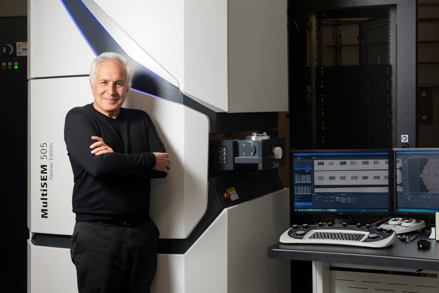
Jeff Lichtman serves as the Jeremy R. Knowles Professor of Molecular and Cellular Biology and Santiago Ramón y Cajal Professor of Arts and Sciences at Harvard University, where he leads research within the Department of Molecular and Cellular Biology in the Faculty of Arts and Sciences. His laboratory focuses on the physical basis of long-lasting memory and experience-driven neural circuit restructuring through advanced connectomic approaches. Dr. Lichtman's research program centers on connectomics and neural circuit development , utilizing transgenic Brainbow models for in vivo visualization and automated serial electron microscopy for nanometer-resolution mapping. His lab investigates synaptic competition during mammalian development, circuit rearrangement in peripheral nervous systems, and the structural underpinnings of memory formation. Key research areas include cellular and molecular neuroscience with emphasis on developmental processes, synaptic connectivity dynamics, and experience-dependent circuit refinement. Analysis of his 2021-2025 publications reveals dominant methodological innovation in machine learning-guided microscopy (e.g., SmartEM) and correlative imaging techniques , with expanding applications across model systems including zebrafish, octopus, and human tissue. Recent work increasingly addresses Alzheimer's disease pathology within connectomic frameworks while maintaining core focus on developmental circuit formation. The Lichtman Lab maintains active development of high-throughput tools for neural circuit mapping, including Fishexplorer for zebrafish circuit dissection and large-scale human cerebral cortex reconstructions, with ongoing projects examining both peripheral and central nervous system connectivity.
Timothy O'Connor serves as Professor in the Department of Cellular & Physiological Sciences at the University of British Columbia's Faculty of Medicine. He holds concurrent appointments as Member of the Program in Cell and Developmental Biology, Member of iCORD (Collaborations on Repair Discoveries), Associate Member of the Department of Zoology, and Member of the Program in Neuroscience. His academic credentials include a BSc and PhD from the University of Toronto, establishing his foundation in cellular and developmental neuroscience. Dr. O'Connor's research centers on semaphorin-mediated axon guidance mechanisms in neural development, with particular emphasis on Semaphorin 5B's dual roles as repellent cue and synapse regulator. His laboratory employs advanced in vitro models including PC12 cell systems to investigate growth cone dynamics, neurite consolidation, and proteolytic processing of guidance molecules. Key methodologies span molecular assays, live-cell imaging, and functional characterization of neuronal morphogenesis pathways. Analysis of his 2009-2015 publications reveals a cohesive research trajectory focused on repulsive guidance cues in spinal cord innervation, corticofugal pathfinding, and hippocampal circuit refinement. His work consistently bridges molecular mechanisms (e.g., Sema5B proteolysis) with functional outcomes in neural circuit assembly, demonstrating how guidance molecules orchestrate sensory afferent projection and synapse elimination. The O'Connor Lab actively recruits highly motivated graduate students and postdoctoral fellows, requiring competitive academic records and research experience. Candidates are expected to pursue external funding through studentships and fellowships to support their work in developmental neurobiology. As part of UBC's Centre for Brain Health ecosystem, the lab maintains strong collaborative ties with iCORD on neural repair initiatives while advancing fundamental understanding of axon guidance principles applicable to regenerative medicine.
Professor Marjorie Lorch is a distinguished academic at Birkbeck, University of London, holding the position of Professor of Neurolinguistics within the School of Creative Arts, Culture and Communication. She has been affiliated with Birkbeck since 1986, initially as a part-time Lecturer and Research Officer before becoming a full-time staff member in 1991. Her academic journey includes a BA in Linguistics, Anthropology, and Biology from Washington University, St. Louis (Phi Beta Kappa), a Grass Foundation Non-Clinical Fellowship in Neurology at Barnes Hospital, postgraduate studies in Linguistics at University College London, and a Ph.D. in Language Behavior (Neurolinguistics) from Boston University. BA, Washington University in St Louis PhD, Boston University Professor Lorch's research spans multiple dimensions of language processing and its neurological underpinnings. Her work integrates neurolinguistics, psycholinguistics, and historical perspectives to explore how language is organized in the brain, particularly through the investigation of neurogenic language and communication disorders. She has made significant contributions to understanding cross-linguistic comparisons and bilingual speakers' language processing, as well as conducting extensive archival research on 19th-century approaches to language function and disorder. Her interdisciplinary approach bridges linguistics, neurology, psychology, education, and history. Analysis of Professor Lorch's recent publications reveals a consistent focus on historical perspectives in neurolinguistics, particularly examining 19th-century understandings of language organization in the brain. Her work demonstrates a unique integration of historical scholarship with contemporary neurolinguistic research, with recurring themes including aphasia research history, language localization debates, and the evolution of speech and language pathology. The publications also highlight her expertise in cross-linguistic studies and her interest in the relationship between language, thought, and other cognitive domains like music. Silver Gilt Award, Member of the Embroidered Minds Collective, Epilepsy Garden, Chelsea Flower Show, Royal Horticultural Society (April 2018) President, International Society for the History of the Neurosciences (February 2010) Professor Lorch has supervised numerous doctoral and master's students across a wide range of topics related to language disorders, multilingualism, and historical linguistics. Her supervision portfolio includes both principal and second supervisory roles, demonstrating her collaborative approach to academic mentoring. She has served on the editorial boards of prestigious journals including Cortex (2003-2025), Historiographia Linguistica, and Journal of the History of the Neurosciences, and has held significant roles such as Associate Editor of Cortex and membership on the executive boards of the Henry Sweet Society and the International Society for the History of the Neurosciences. Her extensive international collaborations reflect her standing in the academic community. Professor Lorch maintains active international research collaborations and has held numerous visiting positions at institutions including Emory University, Brown University, Duke University, and Loyola University Chicago. Her research program involves interdisciplinary work with medical practitioners, psychologists, speech and language therapists, educators, and historians, reflecting her commitment to bridging theoretical and applied perspectives in language sciences.
Dr. Laura Parkes is a Professor of Neuroimaging at The University of Manchester, specializing in neuroimaging techniques such as MRI and MEG. She holds a MPhys (Oxford), MSc (UCL), and PhD (UCL) in neuroimaging and cerebrovascular physiology. Her research focuses on quantifying neurophysiological properties of the human brain, particularly cerebral blood flow (CBF) and oxygen metabolism using arterial spin labeling (ASL). Key areas include aging, cerebrovascular disease, Parkinson’s, Alzheimer’s, and mechanisms of brain plasticity. She has received a Medical Research Council New Investigators Award (2006). Teaching roles: Neuroimaging Techniques (MSc), Physics of Medical Imaging (MPhys), Advanced MRI (MSc), Neuroscience tutoring (BSc) Collaborations: External with UCL, Lancaster, Nottingham, Liverpool, Leeds; Internal with over 15 Manchester researchers Professional memberships: International Society for Magnetic Resonance in Medicine, Society for Neuroscience Her work addresses cerebrovascular dysfunction in neurodegeneration, blood-brain barrier integrity, and sensory-motor plasticity. Recent advancements include MRI-based blood-brain barrier permeability measurements in rat models and human studies of oxygen extraction fraction in aging populations. Research outputs include 116 peer-reviewed articles (2025 highlights: ATRT tumor analysis, FEXI BBB measurements, and ENIGMA-PD neuroimaging collaboration). Projects funded by MRC and industry partnerships focus on vascular dysfunction in Parkinson’s, stroke, and dementia.
Dr. Christoph Lerche is a researcher at the Institute of Neuroscience and Medicine (INM) , specifically in the Physics of Medical Imaging (INM-4) department at Forschungszentrum Jülich GmbH . His research focuses on advanced medical imaging technologies, particularly positron emission tomography (PET) and hybrid imaging systems like PET-MRI. Key areas include improving PET detector design, developing novel reconstruction algorithms, and applying these techniques to neurodegenerative diseases (e.g., Alzheimer’s) and oncology (e.g., glioma diagnosis). His work emphasizes interdisciplinary collaborations, such as optimizing BrainPET insert compatibility with ultra-high field (UHF) MRI systems and analyzing sleep-related effects on synaptic density. He contributes to clinical applications, such as distinguishing glioma relapse from treatment effects using FET PET radiomics, and advancing multimodal imaging (e.g., simultaneous PET/MR/EEG). Notable projects include enhancing dead-time correction for accurate BrainPET quantification, designing dipole antenna arrays for hybrid systems, and developing photon-counting CT-based PET attenuation maps. His innovations aim to bridge gaps between imaging hardware, data processing, and clinical neuroscience.
Lena H. Ting is a Professor of Biomedical Engineering at Georgia Institute of Technology, holding an adjunct position in the Woodruff School. Her research focuses on neuromechanics of movement, combining experimental, computational, and robotic approaches to study locomotion and balance in humans and animals. She leads efforts in rehabilitation technologies and robotics, with implications for prosthetics and physical therapy. Her work bridges biomechanics and neuroscience, addressing how neural systems interact with musculoskeletal dynamics. Education includes a B.S. in Mechanical Engineering from UC Berkeley (1990), M.S.E. in Biomechanical Engineering from Stanford (1993), and a Ph.D. in Mechanical Engineering from Stanford (1998). She completed postdoctoral fellowships in neurophysiology, including one in Paris. Research interests explore neuromechanical principles of movement control, including: Neuromechanical modeling of muscle coordination Neural control of posture and gait Robotics inspired by biological systems Rehabilitation engineering for spinal cord injury and cerebral palsy Her work integrates robotics, biomechanics, and clinical applications to develop innovative treatments and prosthetic devices. Awarded the American Physiological Society Arthur C. Guyton Award (2007), UC Berkeley Miller Visiting Professorship (2007), and multiple teaching honors including the Georgia Tech CETL/BP Junior Faculty Teaching Award (2006). Her contributions to biomechanics and education are widely recognized. Graduate students from her lab pursue careers in academia, industry (surgical robotics, biomechanics), and consulting. Notable alumni work in rehabilitation robotics and prosthetics development. Her lab collaborates across disciplines to advance neuromechanics research, including projects developing compliant robots for balance studies and analyzing motor control in impaired populations.
Nereo Kalebic is a Research Group Leader at the Centre for Neurogenomics, focusing on molecular and cell biological mechanisms underlying human neocortex development, its implications for neurodevelopmental disorders, and brain cancers. He holds a BSc in Molecular Biology from the University of Zagreb (2007) and a PhD from the European Molecular Biology Laboratory (EMBL) and the University of Heidelberg (2012). His postdoctoral research (2013-2019) at the Max Planck Institute explored neocortex development and evolution. Research interests include neural stem cell biology, glioblastoma stem cell mechanisms, and evolutionary aspects of cortical expansion. His group employs CRISPR/Cas9, advanced live imaging, and cerebral organoids to study these topics. Key projects involve identifying therapeutic targets in glioblastoma and understanding how developmental pathways contribute to neurodevelopmental disorders like Down syndrome. Publications highlight breakthroughs in mechanisms driving tumor growth, neurogenesis differences between humans and Neanderthals, and serotonin receptor roles in progenitor proliferation. The group also develops innovative imaging techniques, such as SOLIST for nanoscale tissue analysis. Advising includes mentoring PhD students (Carlotta Barelli, Ilaria Bertani, Emanuele Capra, Nikola Cokorac, Stefania Faletti) and postdoctoral researchers. Collaborative projects utilize ferret models and human primary samples to bridge basic research and clinical applications. Future work aims to dissect cancer-cell proliferation parallels with neural development and uncover evolutionary mechanisms shaping human cognitive abilities through neocortex expansion.