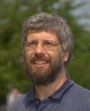
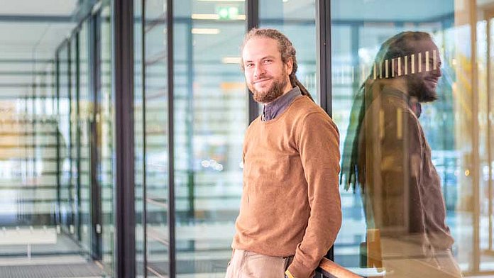
Nasir M. Rajpoot is a Professor in the Department of Computer Science at the University of Warwick, UK. His research focuses on computational pathology, medical image analysis, and deep learning applications in histology. He leads interdisciplinary projects integrating artificial intelligence with healthcare, particularly in cancer diagnostics and pathology workflows. Rajpoot’s work emphasizes developing robust algorithms for histology image analysis, including nuclear segmentation, tumor classification, and domain generalization in computational pathology. His contributions include the TIAToolbox, an open-source framework for tissue image analytics, and the CoNIC Challenge to advance nuclear detection and counting in histology images. He collaborates with clinicians and biologists to translate AI models into clinical practice, addressing challenges like tumor heterogeneity and staining variability. Rajpoot’s research spans colorectal, lung, and oral cancers, with a focus on predicting clinical outcomes via histological features and genomic data integration. Notable projects include the development of Handcrafted Histological Transformer (H2T) for unsupervised representations of whole slide images and the SAFRON framework for histology image synthesis. His work addresses domain adaptation, robustness evaluation, and explainability in AI-driven pathology systems.
Prof. Dr. Karsten Niehaus serves as Head of the Proteome and Metabolome Research Group at the Center for Biotechnology (CeBiTec) and Faculty of Biology, University of Bielefeld. His research focuses on proteomics and metabolomics applications in plant-microbe interactions, bacterial stress responses, and disease model systems. His laboratory employs advanced mass spectrometry imaging and cell phenotyping technologies to investigate molecular responses in crops like sugar beet and grapevines under abiotic stress conditions, as well as in cancer models where differentiation therapy impacts tumor malignancy. The group also explores microbial biotechnology through Xanthomonas campestris studies on xanthan production and stress adaptation. Selected publications highlight innovations in 3D microfluidics for biomarker detection and bioinformatics platforms like MetHoS for metabolomics data analysis. His work appears in journals covering Frontiers in Plant Science , Scientific Reports , and Journal of Experimental Botany . Contact: kniehaus@cebitec.uni-bielefeld.de | Office: UHG W7-117
Dr. Kurt Schmoller is a Group Leader at Helmholtz Munich, heading the Schmoller Lab within the Institute of Functional Epigenetics (IFE). His research focuses on understanding how cells regulate their size, maintain organelle homeostasis during growth, and adjust protein composition according to cell size. The lab employs interdisciplinary approaches combining quantitative biology, live-cell microscopy, AI-based image analysis, and mathematical modeling. Dr. Schmoller received his Diploma in Biophysics from TU München (2004-2008), followed by PhD studies at TU München in Andreas Bausch's lab (2008-2012), where he studied the mechanics of in vitro reconstituted cytoskeletal networks. He then pursued postdoctoral research at Stanford University with Jan Skotheim (2012-2017), developing his interest in cell size regulation. Since 2017, he has led the 'Cell Size and Organelle Control' research group at Helmholtz Munich. His research spans four main areas: Maintenance and Adaptation of Cell Size, Histone Homeostasis, Mitochondrial DNA Maintenance, and AI for live-cell imaging data analysis. Using model organisms including budding yeast (S. cerevisiae) and green algae (C. reinhardtii), his lab investigates fundamental cellular processes that are broadly conserved across eukaryotes. His work has significant implications for understanding diseases like cancer where cell size regulation is often disrupted. Analysis of his recent publications reveals a strong focus on the relationship between cell size and various cellular processes, with particular attention to histone regulation, mitochondrial DNA homeostasis, and the development of AI tools for image analysis. His work bridges molecular biology, biophysics, and computational approaches to address fundamental questions in cell biology. Dr. Schmoller leads a diverse research team including postdocs, doctoral researchers, and technicians working across multiple projects. His lab has developed notable tools such as Cell-ACDC, an open-source software for bioimage analysis that has been adopted by multiple laboratories. The lab maintains strong collaborations across institutions and participates in the broader 'Epigenetics at Helmholtz Munich' initiative. The Schmoller Lab operates from the Neuherberg Campus (Building 35.26 / Room 014) and maintains an active presence through their lab website, BioRxiv, and GitHub repositories. Their work receives support from Helmholtz Munich's infrastructure and resources, including access to advanced microscopy facilities and computational resources for AI-based image analysis.
Prof. Dr.-Ing. Tim Wilhelm Nattkemper leads the Biodata Mining Group at the Faculty of Engineering , Universität Bielefeld , while holding affiliations with the Center for Biotechnology (CeBiTec) and the Institute for Bioinformatics Infrastructure . His work bridges bioinformatics with marine environmental monitoring , focusing on machine learning and computer vision applications. The group specializes in multivariate bioimage analysis , developing platforms like BioIMAX for web-based high-dimensional data exploration. Research spans from MALDI imaging to deep-sea megafauna classification , integrating information visualization and web technologies . Recent projects address seafloor macrolitter monitoring , coral stress response analysis , and self-supervised learning for diatom classification. Their 15 most recent publications (2023-2025) highlight advancements in marine imaging , automated annotation systems , and AI-driven biodiversity assessment , particularly in polymetallic nodule fields. The group also tackles technical challenges like data imbalance in marine image classification and FAIR data principles implementation. As module responsible for courses like Information Visualization and Introduction to Bioinformatics , Nattkemper contributes to academic training in bioinformatics and data science . His interdisciplinary collaborations span physics , chemistry , and ecology within Bielefeld's Material World strategic research area.
Pasquale Cervero is a Research Fellow affiliated with the Institute of Medical Microbiology, Virology and Hygiene at the University Medical Center Hamburg-Eppendorf (UKE) , part of the University of Hamburg . His research focuses on podosome biology , cellular invasion mechanisms , and endocytic pathways . He holds a PhD (Dr. rer. nat.) and has developed computational tools like Poji for analyzing podosome structures. Key research areas include podosome dynamics in cancer metastasis, mechanosensory signaling in cells, and actin cytoskeleton regulation. His work bridges molecular mechanisms with cellular behaviors, contributing to understanding pathological processes such as tumor invasion. Publications highlight advancements in podosome lifecycle analysis, cargo-specific endocytosis, and computational methods for microscopy data. Collaborations with groups like Linder S. and Condeelis J. underscore his interdisciplinary approach to cell biology.
Professor Ingo Roeder is a Professor for Medical Statistics and Biometry and Head of the Institute for Medical Informatics and Biometry (IMB) at the Medical Faculty Carl Gustav Carus, Technical University of Dresden since 2010. His research group focuses on systems biology with applications in medicine, stem cell and developmental biology, developing mathematical models to understand biological systems across multiple scales. Professor Roeder's educational background includes: Diploma in Mathematics from TU Dresden (1994) PhD in Theoretical Biology from University Leipzig (2003) Habilitation in 'Medical Bioinformatics, Statistics and Epidemiology' (2009) Professor Roeder's research interests center around mathematical modeling of biological systems at multiple scales. His group develops and applies mathematical models and simulation strategies of regulatory processes on the molecular, cellular, and tissue levels. Major research foci include theoretical stem cell biology, investigation of stem cell-niche interactions, analysis of clonal competition processes, modeling of aging-related phenomena in the hematopoietic system, and understanding gene regulatory principles. In bioimage informatics, the group specializes in developing automatic single cell tracking algorithms and model-based segmentation methods for analyzing cellular genealogies, cell migration, and cell-cell contacts. Professor Roeder's publication record demonstrates consistent contributions to systems biology, stem cell research, and computational medicine. His work shows progression from fundamental mathematical modeling of stem cell organization to increasingly complex applications in cancer treatment and advanced imaging technologies. The research spans multiple disciplines including mathematics, computer science, biology, and medicine, reflecting the interdisciplinary nature of modern systems biology. His notable scientific contributions include: Mathematical modeling approaches for characterizing clonal heterogeneity among hematopoietic stem cells Dynamic modeling of imatinib-treated chronic myeloid leukemia with clinical implications Development of automated single-cell tracking methods for studying hematopoietic stem cell behavior Mathematical analysis of Nanog variability in pluripotency regulation of embryonic stem cells High-speed panoramic light-sheet microscopy techniques for studying endodermal cell dynamics As Head of the Institute for Medical Informatics and Biometry, Professor Roeder oversees research activities that bridge mathematics, computer science, and medical applications. His group maintains active collaborations across disciplines, working with experimental biologists to validate mathematical models and with clinicians to translate research findings into potential medical applications. The group participates in interdisciplinary graduate programs such as the Dresden International Graduate School for Interdisciplinary Life Sciences. The research group operates at the intersection of mathematical modeling and experimental biology, with particular focus on developing computational methods to address complex biological questions. Their work on single cell tracking and modeling represents a significant technical contribution to the field of bioimage informatics, enabling detailed analysis of cellular behavior that would be impossible through manual methods alone.
Dr. Markus Ankenbrand is a Group Leader in BioMedical Data Science at the Center for Computational and Theoretical Biology (CCTB) at Julius-Maximilians-Universität Würzburg. He holds a PhD in Biology and Informatics from the same university, completed at the Graduate School of Life Sciences. His research focuses on developing algorithms and tools for biomedical data analysis, with a current emphasis on multi-modal methods and machine learning model interpretability. Positions: 2020–present: Group Leader, BioMedical Data Science, CCTB 2019–2020: Group Leader, Computational Cardiology, Universitätsklinikum Würzburg 2018–2019: PostDoc, MIT, Boston Research Interests: His work spans bioinformatics, computational biology, medical imaging analysis, and machine learning applications in healthcare. Key projects include deep learning models for cardiac MRI segmentation and integrative genomics in ocean ecosystems. Publications: Over 25 peer-reviewed articles since 2013, with recent contributions on prostate cancer detection via PET/CT, influenza RNA visualization, and cardiac function modeling in large animal studies. Education: Bachelor of Biology (2009–2012), University of Würzburg Master of Biology (2012–2014), University of Würzburg Bachelor of Informatics (2011–2015), University of Würzburg PhD (2014–2017), Graduate School of Life Sciences, University of Würzburg Bachelor of Computational Mathematics (2020–2023), University of Würzburg Labs/Teams: Leads the BioMedical Data Science group at CCTB, collaborating with institutions worldwide on projects like myocardial pathology segmentation benchmarks (MyoPS) and ecological trait data standards.
Prof. Dr. Karl Duderstadt is a Professor of Experimental Biophysics at the Technical University of Munich (TUM) and a group leader at the Max Planck Institute of Biochemistry. His research focuses on understanding the dynamic processes of chromosome duplication using single-molecule imaging techniques. He holds a B.A. in Physics from Oberlin College and a Ph.D. in Biophysics from UC Berkeley. Prior to his current roles, he conducted postdoctoral research at the University of Groningen under the Human Frontier Science Program. His academic career includes leadership of the Max Planck Research Group "Structure and Dynamics of Molecular Machines" since 2016. Key research areas include replisome dynamics, DNA replication stress, and the interplay between replication and transcription. He has received prestigious awards, including an ERC Starting Grant (2019) and an HFSP fellowship (2012–2015). Publications highlight advancements in single-molecule technologies, such as the Mars data analysis suite, and discoveries about replication fork protection mechanisms. His work bridges biophysics, structural biology, and molecular genetics, with applications to cellular and epigenetic stability.
Constantin Pape is a Junior Professor at Georg-August University Göttingen in the Institute of Computer Science, where he leads the Computational Cell Analytics research group since March 2022. His work focuses on developing deep learning and AI methods for biology and medicine, with particular emphasis on biomedical imaging applications. His research spans two main areas: building foundation models for biology and medicine, and protein structure analysis in cryogenic electron microscopy and super-resolution microscopy. Dr. Pape's group is dedicated to open source and open science, contributing to projects like the bioimage.io modelzoo, ome.ngff image data format, and MoBIE Fiji plugin. His research is funded by the DFG through a Sachbeihilfe, the SFB1286 on Quantitative Synaptology, and the Multiscale Bioimaging Cluster of Excellence (MBExC). Dr. Pape's work on foundation models includes Segment Anything for Microscopy (Nature Methods, 2025) and extensions to histopathology and medical imaging. His group develops segmentation models for various applications, including SynapseNet for automatic synapse reconstruction. He has published extensively in top venues including Nature Methods, Cell, Science, and ECCV. His teaching includes a lecture on Deep Learning for Computer Vision and seminars on Deep Learning in Biology and Medicine at the University of Göttingen, as well as co-organizing the Deep Learning for Image Analysis course at EMBL and teaching advanced image analysis through the Hertha Sponer College.
Markus D. Herrmann is an Assistant Professor of Pathology at Harvard Medical School and serves as Director of Computational Pathology and Assistant Computational Pathologist at Massachusetts General Hospital (MGH). His lab operates from the Computational Pathology unit at MGH's Longfellow Building in Boston, MA. His research transforms qualitative histopathology into quantitative science using microscopy imaging, proteomics, and machine learning to develop image-based clinical diagnostics. Key interests include cancer biomarker discovery, computational diagnostic tests, and analyzing cellular interactions during tumorigenesis and metastasis through spatial molecular feature extraction. His 2014-2020 publications reveal consistent focus on computational pathology infrastructure (DICOM standards), multiplexed protein mapping, and AI-driven image analysis across oncology. Work spans quantitative cancer imaging informatics, digital pathology standardization, and high-content cell/transcriptome profiling, establishing him as a leader in translating computational methods to clinical pathology. As faculty, he mentors trainees within Harvard's ecosystem though specific students aren't listed. His research aligns with MGH Pathology's $19 million annual research portfolio, indicating substantial institutional support for computational pathology initiatives. He leads the Computational Pathology lab collaborating across MGH, Harvard, and clinical networks to develop multimodal imaging methods (immunofluorescence, electron microscopy, OCT) and clinical validation frameworks for AI-based diagnostic tools, emphasizing real-world implementation and performance monitoring.
Peter Ludewig is a Professor at the University Medical Center Hamburg-Eppendorf (UKE), affiliated with the Department of Experimental Medicine and the Medical Faculty. His research focuses on neurovascular mechanisms, stroke pathophysiology, and advanced imaging techniques like Magnetic Particle Imaging (MPI). He leads interdisciplinary projects combining experimental medicine with clinical applications, particularly in cerebral perfusion analysis and neuroinflammation. Key areas of expertise include: stroke therapy development, immune responses in brain injury, and translational imaging technologies. His work bridges basic science and clinical practice, with contributions to understanding neutrophil dynamics, cytokine regulation (e.g., IL-17A), and novel drug delivery systems using nanobodies. Recent studies address cerebral microvascular remodeling, nanopore sequencing for lymphoma diagnosis, and optimizing MPI tracers for real-time perfusion assessment. He collaborates across departments including Neurology, Radiology, and the Cardiovascular Research Center (CVRC).
Prof. Dr. Ullrich Köthe is an Associate Professor and group leader in the Visual Learning Lab Heidelberg at the University of Heidelberg . He focuses on Explainable Machine Learning , leveraging Invertible Neural Networks to enhance transparency and utility in image analysis and medical applications . He also maintains the widely used VIGRA image analysis library. Education: PhD in Informatics, University of Hamburg, 2000 Habilitation in Informatics, University of Hamburg, 2008 His research interests center around machine learning , image analysis , and scientific computing , particularly the development of robust algorithms for medical imaging , computer vision , and life sciences . His work on invertible neural networks and parameter-free segmentation has led to significant advancements in the field. Recent publications highlight his contributions to Bayesian inference , neural network interpretability , and stochastic modeling , with applications ranging from disease outbreak dynamics to connectomics . Key trends include generative models , parameter-free segmentation , and likelihood-free inference . Scientific Awards: DAGM 2003 Main Prize DAGM Best Paper Award 2008 He has supervised numerous Master and Bachelor theses in machine learning and image analysis , with teaching roles in Advanced Machine Learning and Explainable AI . His collaborative research grants include funding from HARMAN International (2024). He leads the Explainable Machine Learning subgroup and has contributed to open-source software projects like ilastik and VIGRA , which are critical tools in bioimage analysis .
Peter Horvath is a Principal Investigator at AI for Health, Helmholtz Munich, Germany, and serves as Director of the Institute of Biochemistry at the Biological Research Centre in Szeged, Hungary. He is a Visiting Scientist at FIMM-EMBL, Helsinki, Finland. D.Sc. in Single-cell Analysis (2023) Ph.D. in Digital Image Analysis (2007) M.Sc. in Software Engineering (2003) His research lies at the intersection of biology, engineering, and computer science , focusing on single-cell analysis using image analysis and machine learning . He combines wet-lab experiments with advanced computational techniques to study molecular processes and develop personalized therapies for cancers and other diseases. The publications highlight his work in bioimage analysis (2023), single-cell proteomics (2022), and intelligent cell isolation (2018). These span fields like computational biology, biomedical imaging, and machine learning , emphasizing single-cell technologies and quantitative methods . Szent-Györgyi Talentum Prize (2019) Pfizer Research Award (2016) Bolyai plaquette (2018) Marie Curie Fellowship (2007) Horvath has held leadership roles in institutions across Germany, Hungary, and Finland. He is actively involved in networks like the Human Cell Atlas and the Society of Biomolecular Imaging and Informatics , and has contributed to advancing deep visual proteomics and bioimage segmentation methodologies.
Prof. Dr. Marc Thilo Figge serves as Professor (W3) for Applied Systems Biology at Friedrich Schiller University Jena and Head of the Applied Systems Biology Research Group at the Leibniz Institute for Natural Product Research and Infection Biology (Leibniz-HKI) since 2011. His career spans theoretical physics, computational biology, and infection research, with significant leadership roles in national research initiatives including NFDI4Bioimage, DFG Research Training Groups, and the Excellence Cluster 'Balance of the Microverse'. His research focuses on three interconnected pillars: Host-pathogen interactions of human pathogenic fungi Automated image analysis through the open-source JIPipe platform Spatiotemporal computer simulations of infection dynamics These areas combine experimental microbiology with computational modeling to address fundamental questions in infection biology. Analysis of his 2025 publications reveals strong interdisciplinary output spanning microbiology, computational biology, and biomedical engineering. His work demonstrates consistent methodological innovation in image analysis (particularly through JIPipe) and computational modeling of fungal infections, with increasing applications in rapid diagnostics and organ-on-chip technologies. The publications show growing international collaboration and methodological transfer between fungal pathogenesis and broader infection biology contexts. His scientific recognition includes: medac Research Award (2023 and 2024) Adjunct Fellowship at Frankfurt Institute for Advanced Studies (2011-2017) Editorial roles at PLOS Complex Systems, Biological Imaging, and Scientific Reports Prof. Figge maintains an active supervision program with approximately 14 students across multiple graduate schools, supported by substantial funding from DFG, BMBF, and Leibniz Association initiatives. His group participates in the International Leibniz Research School and Jena School for Microbial Communication, with strong connections to the Microverse Imaging Center's advanced microscopy facilities. He regularly organizes the biennial international symposia on Image-based Systems Biology (IbSB) and Systems Biology of Microbial Infection (SBMI), fostering global collaboration in the field. The Applied Systems Biology group operates at the intersection of physics, computer science, and infection biology, developing computational tools that have gained international adoption in bioimage analysis. Current projects emphasize translation of fundamental research into diagnostic applications, particularly through microfluidics and organ-on-chip technologies.
Dr. Robert Haase is a Lecturer and Training Coordinator at the Center for Scalable Data Analytics and Artificial Intelligence (ScaDS.AI) under Leipzig University , with prior leadership roles at the DFG Cluster of Excellence 'Physics of Life' at TU Dresden . He specializes in Bioimage Analysis , GPU-Accelerated Image Processing , and Large Language Models (LLMs) for life sciences. His research focuses on democratizing bioimage analysis through open-source tools like CLIJ , clesperanto , and bia-bob , aiming to bridge microscopy with data science . Recent projects explore LLM-driven code generation for image analysis and interactive workflow design in platforms like napari . He leads initiatives such as the NFDI4BioImage consortium for research data management in Germany and GloBIAS , a global society for bioimage analysts. Funded by organizations including the Chan Zuckerberg Initiative (CZI) and DFG , his work emphasizes reproducibility , open science , and interdisciplinary collaboration in bioimaging. As an educator, he conducts training programs like "Large Language Models for Bioimage Analysis" and "Collaborative Working with Git" , and contributes to workshops at institutions such as EMBO , Institut Pasteur , and ScaDS.AI Summer Schools .