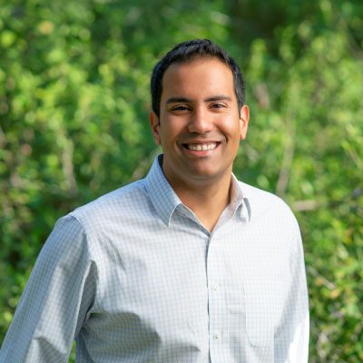
Andrew T Crane is an Assistant Professor at the University of Minnesota's School of Medicine , affiliated with the Transplant and Cellular Therapy Division of the Department of Medicine. His work bridges stem cell biology , gene editing , and neuroregenerative medicine . Institution: University of Minnesota Rank: Assistant Professor Division: Transplant and Cellular Therapy Research Interests Dr. Crane focuses on exosome-based therapies for stroke and traumatic brain injury (TBI), brain organoid engineering to model neurodegenerative diseases, and human-animal chimeras for organ regeneration. His CRISPR-mRNA platforms enable precise cell engineering for transplantation, while his blastocyst complementation studies explore interspecies organogenesis. Scientific Contributions Co-investigator on exogenic organ transplantation projects (NIH-funded) Led 3D brain organoid research for chronic TBI and CTE Key publications on stem cell guidelines for human embryos and chimera research Collaborative work with Professor Andrew Grande on canine TBI models Grants & Collaborations Dr. Crane has secured grants from the NIH and Minnesota Office of Higher Education for projects involving immunosuppression-free transplantation and exosome therapy . Collaborations span neuroscience , stem cell research , and biomedical engineering .
Pamela VandeVord is a Professor in the Department of Biomedical Engineering and Mechanics at Virginia Tech's College of Engineering. She serves as Director of the Center for Injury Biomechanics (CIB) and leads the Traumatic Nerve Technologies Lab. Her research focuses on traumatic brain injury (TBI), neurotrauma, and the molecular mechanisms of blast-induced injury. Ph.D., Biomedical Engineering, Wayne State University (2002) M.S., Biomedical Sciences, Wayne State University (1996) B.S., Physiology, Michigan State University (1992) Her work investigates the cellular and molecular effects of blast-induced neurotrauma, emphasizing injury biomechanics, neuroinflammation, and behavioral outcomes like anxiety and cognitive deficits. She develops realistic injury models and explores therapeutic strategies such as hemostatic nanoparticles and tissue engineering approaches. Recent publications highlight advancements in understanding astrocyte reactivity, glial activation, and chronic behavioral impairments following traumatic brain injury. Her studies utilize rodent and minipig models to bridge preclinical and clinical gaps. Fellow, Biomedical Engineering Society (2020) Virginia Tech Engineering Dean's Award for Excellence in Service (2020) Fellow, American Institute for Medical and Biological Engineering (AIMBE) (2017) Presidential Early Career Award for Scientists and Engineers (PECASE) (2009) Dr. VandeVord’s lab collaborates on combat-related TBI models and traumatic epilepsy research, with grants supporting nanoparticle therapies and biomarker discovery. Her team works on developing standardized animal models and translational tools for injury prevention and treatment.
Martin Lauritzen is a Professor in the Department of Clinical Medicine, specifically within the Neurology Center for Healthy Aging Damage and Repair, and also holds a Professor position in the Department of Neuroscience focusing on Neuronal Signalling at the University of Copenhagen's Faculty of Health and Medical Sciences. With an extensive publication record of 223 research outputs, his work spans multiple disciplines within neuroscience and neurology, with particular emphasis on brain aging, neurovascular coupling, and cerebral blood flow regulation. His research has garnered significant attention, including media coverage of his work on brain fluid dynamics during sleep. Professor Lauritzen's primary research interests focus on the intricate relationship between brain function and vascular dynamics. His work explores how neurovascular coupling changes with age, the mechanisms underlying cortical spreading depression in migraine, and the role of the blood-brain barrier in neurological disorders. He has made significant contributions to understanding how sleep affects brain fluid dynamics and waste clearance, with his 2019 research on 'brain rinse cycle' during sleep receiving widespread media attention. His investigations into cerebrovascular responses in early Alzheimer's disease, particularly regarding amyloid-β accumulation and its effects on cerebral blood flow, represent a critical frontier in dementia research. Lauritzen's laboratory employs advanced techniques including two-photon microscopy, in vivo imaging, and electrophysiological recordings to study neuronal and vascular function in awake animal models. Analyzing his recent publication trends (2023-2025), Lauritzen's research demonstrates a clear trajectory toward understanding age-related changes in brain vasculature and their cognitive implications. His work bridges basic neuroscience with clinical applications, particularly in the areas of migraine pathophysiology, Alzheimer's disease biomarkers, and brain aging. A significant portion of his recent work focuses on the role of precapillary sphincters and pericytes in regulating cerebral blood flow, especially in aging brains. His research also explores cognitive reserve factors in longitudinal population studies, connecting vascular health with cognitive outcomes in aging populations. The interdisciplinary nature of his work is evident in publications spanning from instrument development to clinical studies of cognitive aging in the Danish Metropolit 1953 male cohort. Professor Lauritzen has been involved in significant collaborative research efforts, particularly evident in his work with S. Grubb on sleep-related brain fluid dynamics, which generated substantial media coverage across multiple outlets. His work with the Metropolit 1953 Danish male cohort indicates involvement in long-term population studies examining cognitive aging and vascular health. Based at Blegdamsvej 3B in Copenhagen, Lauritzen leads research teams studying neurovascular function, particularly focusing on the aging brain. His work with two-photon microscopy and in vivo imaging suggests a well-equipped laboratory capable of advanced neuroimaging techniques. The breadth of his collaborations, evident from co-authorship patterns across numerous publications, indicates an active research group with connections across neuroscience, neurology, and gerontology disciplines.
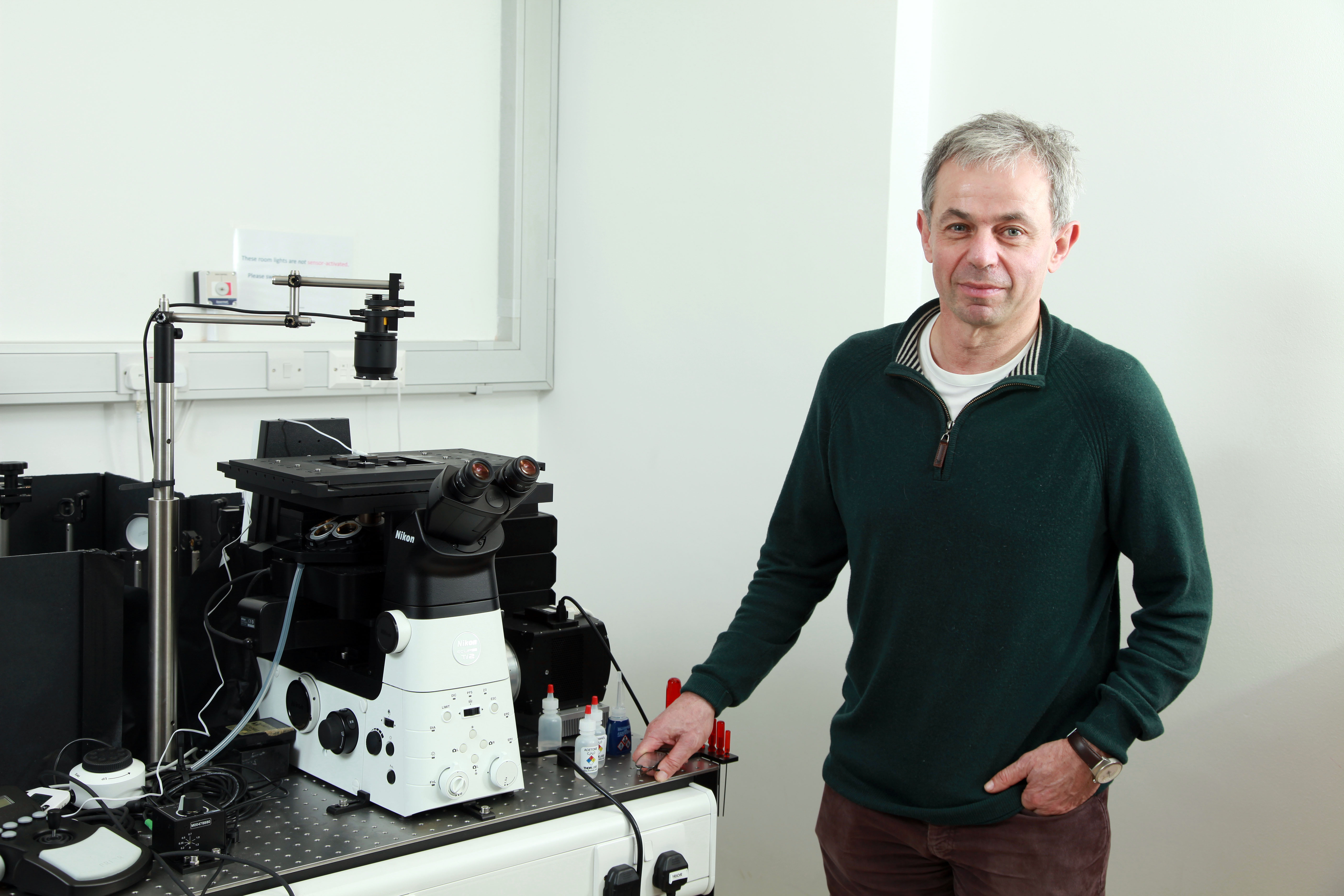
Professor Sir David Klenerman , FMedSci FRS at the Yusuf Hamied Department of Chemistry , University of Cambridge, develops quantitative biophysical methods using single-molecule fluorescence and scanning probe microscopy to address unresolved biological questions. His work bridges physical sciences and biomedical research , focusing on neurodegenerative diseases (Alzheimer's, Parkinson's) and immune signaling via T-cells and Toll-like receptors. Research Highlights : Imaging protein aggregates in test-tubes and living cells Developing 20nm-resolution imaging techniques Co-inventor of next-generation DNA sequencing technology Investigating immune receptor triggering mechanisms Part of the Cambridge Dementia Research Centre Scientific Awards : Fellow of the Royal Society (FRS) Fellow of the Academy of Medical Sciences (FMedSci) Royal Society GSK Research Professor Todd-Hamied Fellowship Team Leadership : Supervises 12 PhD students working on topics ranging from Tau aggregation to chromatin organization , with collaborators across biochemistry, neuroscience, and clinical medicine.
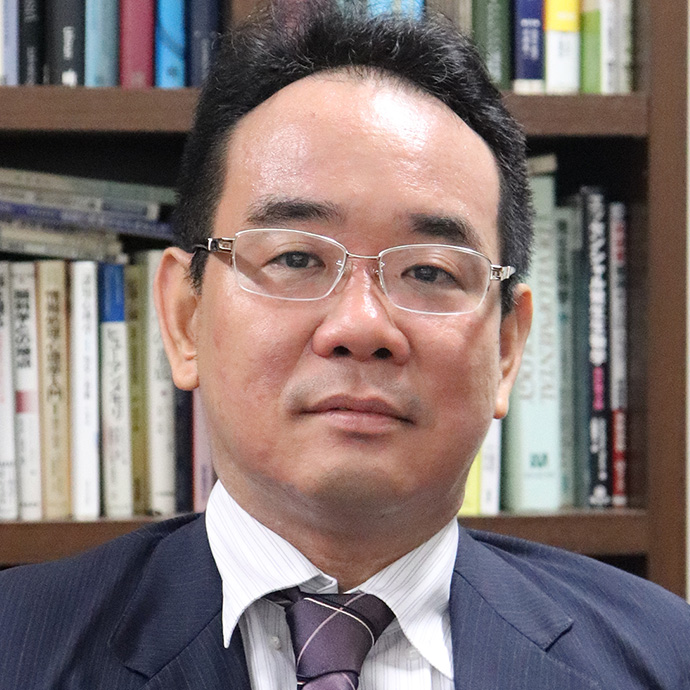
Kenichi Ohki is a Professor in the Department of Physiology at the Graduate School of Medicine, The University of Tokyo, and serves as Deputy Director and Principal Investigator at the International Research Center for Neurointelligence (IRCN). His research focuses on visual neuroscience and functional brain mapping, with particular emphasis on understanding how the visual cortex processes information at cellular resolution. Education: 1990-1996: Medical degree (MD) from Faculty of Medicine, The University of Tokyo 1996-2000: PhD in Medicine from Department of Physiology, The University of Tokyo 1996: Visiting scholar at Department of Brain and Cognitive Sciences, MIT Dr. Ohki is renowned for developing single-cell resolution functional mapping with two-photon calcium imaging in 2005, which revolutionized the understanding of visual cortex architecture. His work has revealed fundamental principles of how orientation selectivity emerges in visual neurons and how developmental programs interact with neural activity to shape cortical function. His laboratory continues to investigate the interplay between innate developmental circuits and neuronal activity in determining cortical function using advanced imaging and computational techniques. Analysis of Dr. Ohki's publications shows a consistent progression from foundational work on visual cortex organization to understanding developmental mechanisms and more recently, integrating neuroscience with artificial intelligence approaches. His recent work demonstrates increasing sophistication in analyzing neural circuits, with growing emphasis on computational modeling and cross-disciplinary approaches that bridge neuroscience and AI. Scientific Recognition: Multiple high-impact publications in Nature, Nature Neuroscience, and other top journals Development of innovative techniques for functional brain mapping Leadership as Deputy Director of IRCN, a major international research center Selection for Beyond AI Institute's mid- to long-term research project Regular recognition of laboratory members with research awards Dr. Ohki actively mentors a diverse research team including project assistant professors, postdoctoral researchers, and graduate students. His laboratory has received significant institutional support for research on visual neuroscience and neural circuit development. Several of his students have received prestigious awards at major neuroscience conferences in Japan, demonstrating his effective mentorship and the quality of research conducted in his laboratory. The Ohki Laboratory operates within the Department of Physiology at the University of Tokyo and is affiliated with the International Research Center for Neurointelligence (IRCN). The lab utilizes advanced techniques including in vivo two-photon calcium imaging, optogenetics, and computational modeling to investigate visual information processing in the mammalian brain. The research team consists of scientists with diverse expertise in neuroscience, imaging technology, and computational analysis, creating a highly collaborative and interdisciplinary research environment focused on understanding the fundamental principles of visual processing in the brain.
Flavie Lavoie-Cardinal is an Associate Professor in the Department of Psychiatry and Neuroscience at Université Laval and holds the Canada Research Chair in Intelligent Nanoscopy of Cellular Plasticity. Her research combines optical nanoscopy , machine learning , and molecular neurosciences to develop intelligent microscopes for studying synaptic plasticity. Education: Bachelor’s, Master’s, Ph.D. in Chemistry, University of Siegen (Germany) Postdoctoral Fellow, Stefan W. Hell Lab (2011–2014), Paul De Koninck Lab (2014–2017) Research Focus: She pioneers super-resolution microscopy and deep learning frameworks to analyze molecular interactions in synaptic communication. Her work emphasizes neurophotonics and quantitative imaging , addressing challenges like data scarcity and automated optimization. Article Trends: Recent publications highlight her innovations in STED microscopy , GAN-based resolution enhancement , and AI-driven image analysis , applied to studying stress effects on barriers, synaptic dynamics, and mitochondrial transport. Scientific Awards: Canada Research Chair in Intelligent Nanoscopy of Cellular Plasticity Labs & Collaborations: Leads the FLC-Lab, collaborating with CERVO Brain Research Centre and international institutions. Her team integrates biophotonics , neuroscience , and machine learning for cutting-edge biomedical imaging.
Martin Lévesque is a Professor in the Department of Psychiatry and Neuroscience at Université Laval. He serves as Director of the Integrative Neuroscience and Experimental Therapies Research Axis, focusing on Parkinson’s disease mechanisms and experimental treatments. His research explores dopaminergic circuits, axon development, mitochondrial function, and neurorestoration strategies. Education: Ph.D. in Neurobiology, Université Laval (2003) Postdoctoral Fellowship, IRCM, Montreal (2006-2008) Postdoctoral Fellowship, National Institute for Medical Research, London (2008-2011) His work has identified key transcription factors like Lmx1a and Lmx1b in dopaminergic neuron survival, linking mitochondrial dysfunction to Parkinson’s pathology. Recent publications highlight advancements in light-sheet microscopy , cell replacement therapy , and synucleinopathy models . Scientific Awards: Research Scholar, Fonds de recherche du Québec – Santé Lévesque mentors a multidisciplinary team including Ph.D. students Axelle Dovonou, Anne-Marie Castonguay, Charles Gora, and Victoria Soto, alongside postdoctoral fellows and research assistants. His lab maintains collaborations with international institutions and receives funding from organizations like the Michael J. Fox Foundation.
Caroline Ménard (PhD) is an Associate Professor in the Department of Psychiatry and Neurosciences at Université Laval's Faculty of Medicine. She holds the Sentinel North Junior Research Chair and leads research at the CERVO Brain Research Center's Neurophotonics Centre. Her work focuses on neurovascular and neuroimmune mechanisms in stress, depression, and Alzheimer's disease, particularly through blood-brain barrier permeability and gut-brain axis interactions. 2024-05: FRQNT team grant for "Intelligent cages and photometric methodology for anorexia model" 2024-04: FRQS Inserm collaborative grant on neurocognitive aging 2023-02: Washington Post citation for inflammation-depression connection Research interests include: Sex-specific neurovascular adaptations in mood disorders Chronic stress effects on blood-brain barrier and gut microbiota Immune system's role in neurodegeneration and depression Translational models bridging mouse and human studies Neuroplasticity mechanisms in aging and disease Her recent publications (2023-2025) emphasize sex differences , neurovascular integrity , and gut-brain bidirectional interactions . Key awards include the 2024 CAN New Investigator Award, 2022 FRQS Relève étoile, and 2020 CCNP Young Investigator Award. She supervises doctoral students and collaborates with multidisciplinary teams across Canada and France.
Michael Hastings is a Research Leader at the MRC Laboratory of Molecular Biology, University of Cambridge, where he investigates the molecular neurobiology of circadian rhythms. His work focuses on the suprachiasmatic nucleus (SCN) as the central circadian pacemaker in mammals, utilizing advanced techniques including real-time in vivo imaging, genomics, and molecular genetics to dissect clock mechanisms. Research Interests: Dr. Hastings explores how clock genes and proteins assemble into functional oscillators, how astrocytes and neurons interact to sustain circadian rhythms, and how central/peripheral clocks coordinate physiological processes. His group also studies evolutionary conservation of timing mechanisms in marine organisms. Publication Trends (2022-2025): Recent articles emphasize astrocyte-mediated regulation of SCN circuitry, dynamic protein behavior (e.g., PER2, CRY1), and genomic/environmental control of circadian timing. Key themes include glial-neuronal crosstalk, molecular switches for clock resetting, and multi-omic approaches to network-level timing. Research Team: Current group members include Elena del Carmen Gómez García, Olivia Johnson, Andrew Oliphant, Andrew Patton, Chee Sia, and Nicola Smyllie.
James Connor is a Distinguished Professor at Penn State University, holding appointments in both the Department of Neurosurgery and Department of Pediatrics . He serves as Vice Chair for Research and leads the Penn State Neuroscience Institute and Penn State Cancer Institute initiatives. With over 383 research outputs and 30 grants, his work bridges neuroscience, iron metabolism, and oncology. Research Interests: Dr. Connor's research focuses on Brain iron regulation and its role in neurodegeneration Iron metabolism in glioblastoma and other cancers Sex-based differences in neurological diseases Genetic variants affecting iron homeostasis (e.g., HFE gene) Therapeutic strategies for iron-deficiency disorders Article Trends: Recent publications highlight iron's role in Neurodegenerative diseases (Alzheimer's, mucolipidosis IV) Glioblastoma survival and metal content Sex-specific cancer metabolism Developmental brain disorders Epigenetic regulation of iron transport Clinical implications of anemia in oncology Scientific Awards: 2022: Excellence in Career Mentoring Award Grants: Major funded projects include studies on Sex-based glioma differences (National Cancer Institute) HFE gene variants and Alzheimer's susceptibility (National Institute on Aging) Mechanisms of brain iron uptake (NINDS) Iron deficiency therapies for mucolipidosis IV (NINDS)
Lorella Maria Teresa Canzoniero is a Full Professor in the Department of Science and Technology (DST) at the University of Sannio, where she specializes in Pharmacology (BIO/14). She has held various leadership roles, including Coordinator of the PhD in Earth and Life Sciences, President of the Master's Degree in Biology, and Rector's Delegate for International Relations. She currently serves on multiple university commissions, including the Doctoral School in Environmental and Health Sciences and Technologies (STAS) and the University Library Commission (CAB). She earned her medical degree in 1986 from the University of Naples Federico II and completed her PhD in Endocrinological and Metabolic Sciences in 1993 at the University of L’Aquila–Chieti. Her early research career was based at Washington University in St. Louis (1991–2001), where she progressed from Research Associate to Research Assistant Professor in Neurology. Her research centers on the molecular mechanisms of neurodegeneration, particularly the roles of calcium and zinc in excitotoxicity and neuronal death. She investigates ion channel function, neuroprotective pathways, and proteomic changes in Alzheimer’s disease models. Her work has significant implications for understanding and treating neurodegenerative disorders. Analysis of her 15 most recent publications reveals a consistent focus on calcium and zinc signaling, neuroprotection, excitotoxicity, and Alzheimer’s disease. Her work spans cellular models, animal studies, and molecular pharmacology, with recurring themes of ion homeostasis, mitochondrial function, and synaptic integrity. Her scientific awards include: SIF (Italian Society of Pharmacology) Scholarship (1991) NATO CNR Advanced Fellowship (1992) Post-Doctoral Fellowship, University of Naples Federico II (1995, 1996) AIRIC (Italian Brain Aging Study Group) Scholarship (1999) She has advised or collaborated with numerous researchers, though formal student listings are not provided. She has been involved in international collaborations, including a Double Degree program with the University of Coimbra, Portugal. She has not held part-time academic positions and remains an active, full-time faculty member. She leads research on neuroprotective mechanisms, particularly through ion channel modulation and zinc signaling, and continues to publish in high-impact journals in neuroscience and pharmacology.
Martine Mirrione is a Professor of Biomedical Sciences at Quinnipiac University , where she also serves as Director of Graduate MLS and BMS Programs . With a PhD in Molecular and Cellular Pharmacology from Stony Brook University and postdoctoral training at Brookhaven National Laboratory and Cold Spring Harbor Laboratory, she specializes in neuronal circuitry in stress and depression. Bachelor of Science in Biology, Marist College PhD in Molecular and Cellular Pharmacology, Stony Brook University Her research focuses on neuroinflammation , microglial dynamics , and antidepressant mechanisms , particularly through studies involving rodent models of chronic stress. She has secured institutional grants and published extensively on synaptic dysfunction and cellular metabolism in mood disorders. In 2021 alone, she contributed to studies on PFKFB3 stress markers , astrocyte morphology , and CaMKII regulation in depressive states, alongside a journal article on NMDA receptor subunits in the hippocampus. Her work bridges molecular neuroscience with translational mental health research. Faculty Scholarship and Creative Works Impact Fund grant recipient Dr. Mirrione teaches courses spanning pharmacology, neuroanatomy, and research methods while leading an active laboratory dedicated to understanding and treating stress-related neurological impairments.
Paul Wolujewicz is an Assistant Professor of Biomedical Sciences at Quinnipiac University with a joint appointment in the Department of Medical Sciences at the Frank H. Netter MD School of Medicine. He holds a PhD in Physiology, Biophysics, and Systems Biology from Cornell University, alongside advanced degrees in Biophysics, Biomedical Sciences, and Biostatistics/Epidemiology from Rutgers University. BS in Biophysics MS in Biomedical Sciences MPH in Biostatistics and Epidemiology PhD in Physiology, Biophysics and Systems Biology His research focuses on leveraging computational genomics and biomedical data science to understand complex genetic disorders, particularly neurodevelopmental conditions like spina bifida and schizophrenia. His work has been published in high-impact journals including Translational Psychiatry , Genetics in Medicine Open , and Human Molecular Genetics . Recent publications highlight his expertise in neurogenomics, computational genomics, and biomedical data science. Key themes include pathogenic expansions in neural tube defects, aberrant calcium signaling in autism, and genome-wide analysis of structural anomalies like spina bifida. Recipient of the Timothy M. George Award for Excellence in Neural Tube Defect Research and Clinical Care Dr. Wolujewicz teaches courses in Biomedical Sciences, including Genetics, Biotechnology, and Computational Biomedicine. His lab at Quinnipiac University uses computational approaches to study genetic disorders of the nervous system.
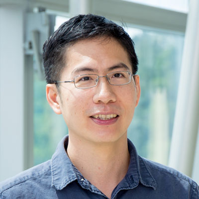
Associate Professor Pakorn Tony Kanchanawong is a Principal Investigator at the Mechanobiology Institute (MBI) and affiliated with the Department of Biomedical Engineering at the National University of Singapore (NUS). His research focuses on nanoscale cellular machinery in mechanobiology, particularly cell adhesion structures and mechanotransduction pathways. His educational background includes a Bachelor's degree (A.B. summa cum laude, 2001) in Chemistry and Biological Sciences from Cornell University, a PhD in Biophysics from Stanford University (2007), and postdoctoral training at NIH with Dr. Clare Waterman where he pioneered iPALM super-resolution microscopy. Research interests center on Nanoscale Structure-Function Relationships, Super-resolution Microscopy, and Cellular Biophysics, with emphasis on focal adhesions, cell-cell junctions, and bioimage informatics. His interdisciplinary approach combines advanced imaging, molecular engineering, and machine learning to decode cellular machine operations. Recent publications (2024-2025) reveal dominant trends in super-resolution imaging of adhesion nanoarchitecture, stem cell mechanobiology, and computational image analysis. Key themes include force transmission mechanisms in adherens junctions, Rac1-dependent cytoskeletal remodeling in pluripotent stem cells, and vortex interference techniques for 3D nanoscopy. Scientific recognition includes: HHMI Predoctoral Fellowship during doctoral studies National Research Foundation (NRF) Fellowship for establishing his lab Mentorship involves direct supervision of six PhD students across multiple cohorts (2018-2022) and leadership of a 10-member research team. His group secured a significant MOE AcRF Tier 3 grant for endomembrane aging research and maintains active collaborations with MBI labs including Li, Yu, and Ravasio groups. The Kanchanawong Lab operates advanced imaging facilities including iPALM and structured illumination systems within MBI's Molecular Mechanics of Mechanotransduction Group, driving innovation in cellular mechanobiology through cutting-edge technology development and rigorous quantitative analysis.
Ajit S Divakaruni is an Associate Professor in the Molecular and Medical Pharmacology department at the University of California Los Angeles . His research focuses on the intersection of mitochondrial metabolism and immune cell function. Key areas: Mitochondrial bioenergetics, immunometabolism, lipid signaling, and metabolic diseases. Research Trends : Recent publications highlight his work on macrophage activation mechanisms, coenzyme A as a metabolic regulator, and mitochondrial responses in neurodegenerative diseases and cancer. Articles span Cell , Nature , and EMBO journals, emphasizing metabolic flexibility and disease models. Scientific Awards : While no direct honors are listed, institutional programs like the Suzanne Eaton Memorial Prize and NIH T32 Training Grants indicate UCLA's recognition frameworks.